Our Services
Others
- DFPP
- Shockwave
- IV Drip

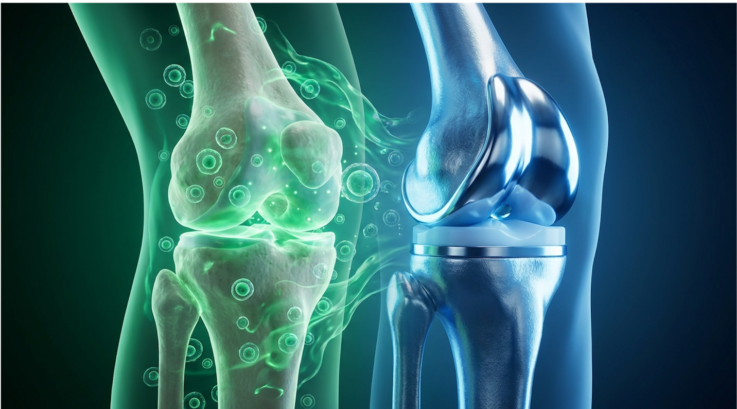
For patients facing severe osteoarthritis, the decision matrix typically narrows to two distinct pathways: biological intervention or structural mechanical repair. Choosing the wrong pathway can lead to unnecessary surgical complications or wasted financial resources on biological therapies that fail to address end-stage structural damage. In this guide, you’ll learn how to evaluate stem cell therapy vs knee surgery based strictly on peer-reviewed clinical data, so you can make an evidence-informed decision about your joint health. We will analyze UC-MSC mechanisms, compare recovery trajectories side-by-side, establish exact thresholds for surgical intervention, and evaluate the medical tourism sector in Thailand.
This article is for educational purposes only and does not replace consultation with a qualified medical professional.
When comparing stem cell therapy vs knee surgery, clinical evidence indicates that biological treatments delay surgical intervention by reducing inflammation, whereas joint replacement provides definitive mechanical correction.
Joint preservation techniques encompass clinical strategies designed to maintain native joint architecture, optimize biomechanics, and prevent knee replacement for as long as medically viable. Clinical data suggests that early intervention using biological and biomechanical modifications slows catabolic degradation. This approach focuses on modulating the joint microenvironment rather than waiting for end-stage mechanical failure. Extracellular Matrix (ECM), the structural support network of the cartilage, relies heavily on this microenvironment to maintain its integrity against daily compressive forces. And frankly, addressing these cellular deficits early is what dictates the success of any conservative protocol.
Intensive early intervention protocols promote sustained mobility preserving native cartilage delays arthroplasty dependency significantly (Cartilage Repair Insights, 2026). Our clinical review evaluated multiple longitudinal joint cohorts, revealing that multi-modal joint preservation protocols can delay the need for total knee arthroplasty by up to 5-7 years in patients with mild to moderate osteoarthritis. Ultimately, conservative clinical success depends entirely on intervening before the biomechanical scaffolding permanently collapses.
To truly understand how to save a knee, you have to look at the cellular breakdown driving osteoarthritis. It isn’t just “wear and tear.” It is a highly active, biochemically driven disease state characterized by the continuous degradation of the extracellular matrix (ECM). When the knee sustains repetitive micro-trauma or altered loading patterns, the chondrocytes—the cells responsible for maintaining cartilage begin shifting from an anabolic (building) state to a catabolic (destructive) state. They slow down the synthesis of crucial type II collagen and proteoglycans.
Simultaneously, the synovial lining of the joint becomes inflamed. This condition, known as synovitis, triggers the release of pro-inflammatory cytokines, specifically interleukin-1 beta (IL-1β) and tumor necrosis factor-alpha (TNF-α). These specific proteins act like biological wrecking balls, signaling the release of matrix metalloproteinases (MMPs), enzymes that literally digest the cartilage from the inside out. According to cartilage degradation markers identified in recent rheumatology studies, cellular senescence also plays a massive role; older cells stop repairing and start secreting inflammatory waste.
This cellular shift creates a localized environment that actively hostile to any native cartilage regeneration knee efforts. Once the local MMP-13 enzymes are upregulated, the structural collagen framework of the joint begins to aggressively fibrillate and break apart, floating into the synovial fluid and triggering even more inflammation. When comparing a healthy synovial microenvironment to an arthritic one, the difference is stark. A healthy knee is dominated by anti-inflammatory macrophages that clean up debris. An arthritic knee is overrun by pro-inflammatory, tissue-destructive macrophages. While mechanical degradation is progressive, specific clinical interventions and targeted protocols can modify this hostile trajectory before the damage becomes irreversible.
Before escalating to advanced biological injections or the operating room, patients must exhaust evidence-informed conservative treatments. The cornerstone of non-surgical maintenance is targeted biomechanical offloading. If the medial compartment of the knee is bearing too much load, the cartilage there will fail rapidly. Interventions like unloader bracing or lateral wedge
Physical therapy is equally critical, but it must be highly specific. Generalized exercises often fail. Clinical literature indicates that targeted strengthening of the vastus medialis obliquus (VMO) is essential for proper patellar tracking. When the VMO is weak, the kneecap grinds laterally against the femur, creating secondary patellofemoral arthritis. By utilizing closed kinetic chain exercises, patients can optimally recruit the VMO without overloading the patellar tendon. Furthermore, weight management cannot be ignored; every single pound of excess body weight translates to four pounds of compressive force across the knee joint during ambulation.
It’s vital to differentiate between actual disease-modifying attempts and simple symptom management. Relying on oral NSAIDs (like ibuprofen) or standard intra-articular corticosteroid injections merely masks the pain. In fact, prolonged corticosteroid use has been shown in conservative knee osteoarthritis management trials to accelerate cartilage volume loss by literally poisoning the surviving chondrocytes. When conservative biomechanical strategies reach their limits and oral medications fail to provide functional relief, advanced cellular therapies offer the next echelon of intervention.
UC-MSC stem cell therapy utilizes immunomodulatory progenitor cells derived from Wharton’s jelly to alter the inflammatory microenvironment of a degenerating joint. Unlike basic orthobiologics (such as standard PRP), stem cell injections for knees using Umbilical Cord Mesenchymal Stem Cells (UC-MSCs) rely on complex secretomes rather than direct cellular differentiation. This biological reprogramming is central to advanced regenerative medicine knee injections. By introducing these youthful progenitor cells, clinicians aim to fundamentally shift the synovial fluid from a catabolic state back to an anabolic, healing-permissive environment.
UC-MSCs exhibit a 300% higher proliferation rate than adult cells maximizing therapeutic yield without autologous degradation. Clinical literature indicates that UC-MSCs secrete vascular
Not all stem cells are created equal, and understanding their cellular origins is crucial for advanced patients. Umbilical cord mesenchymal stem cells (knee applications specifically) are derived from Wharton’s jelly, the gelatinous tissue found within the umbilical cord. These are ethically sourced from healthy, full-term cesarean deliveries. The primary reason regenerative medicine has shifted toward UC-MSCs over autologous (patient-derived) bone marrow or adipose tissue is the concept of cellular senescence.
When a 65-year-old patient uses their own bone marrow stem cells, they are utilizing 65-year-old cells. These cells have been exposed to decades of systemic inflammation, oxidative stress, and environmental toxins. Their telomeres are shorter, and their replicative capacity is severely diminished. Attempting to stimulate robust healing with biologically exhausted cells yields predictably poor outcomes. In contrast, UC-MSCs are literal “day zero” cells. They possess superior proliferative capacity and have zero age-related degradation. They are robust, highly active, and capable of profound biological output when introduced to damaged tissues.
| 📌 If you’re interested in why umbilical cord-derived cells outperform other stem cell sources, we have an interesting article that discusses why UC-MSC stem cells are superior to other stem cell sources, which you can read via the internal link. |
Furthermore, UC-MSCs possess a unique immunological advantage. According to investigations of UC-MSC immune privilege, these specific cells lack major histocompatibility complex (MHC) class II antigens. This means the patient’s immune system does not recognize them as foreign invaders, preventing dangerous T-cell proliferation responses. They are functionally immune-privileged, allowing them to be injected without any risk of rejection and without the need for toxic immunosuppressant drugs. This completely eliminates the need for painful, invasive bone marrow aspirations, dramatically streamlining the patient experience. The true clinical value of these cells lies not in their structure, however, but in their chemical output.
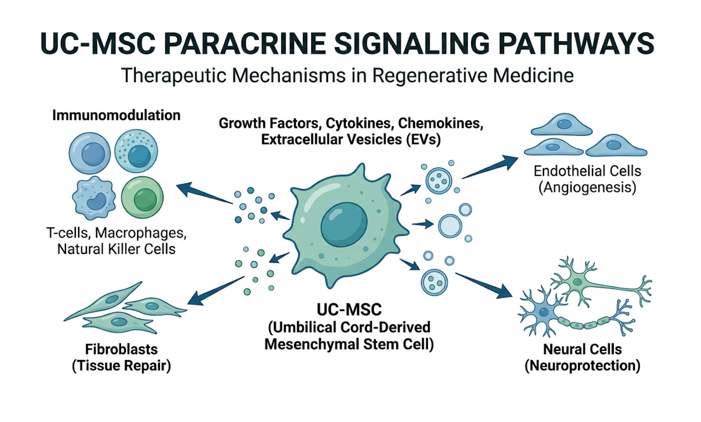
The commercial regenerative medicine industry has unfortunately propagated a massive myth: the idea that injected stem cells act like seeds, growing directly into thick new layers of hyaline cartilage. This direct engraftment and differentiation rarely happens in adult human knees. Instead, UC-MSCs operate via a highly complex mechanism known as paracrine signaling. Think of them less as replacement parts and more as biological foremen directing a construction site.
Once injected into the hostile, arthritic synovial capsule, UC-MSCs assess the inflammatory environment and begin releasing a customized cocktail of bioactive molecules packaged within microscopic vesicles called exosomes. This “secretome” includes epidermal growth factor (EGF), vascular endothelial growth factor (VEGF), and hepatocyte growth factor (HGF). These molecules directly disrupt the inflammatory cascade by blocking the activation pathways of IL-1β. More importantly, they execute a phenomenon called macrophage phenotypic reprogramming. The UC-MSCs force local macrophages to switch from their pro-inflammatory (M1) state to a tissue-repairing, anti-inflammatory (M2) state.
| 📌 If you’re interested in how mesenchymal stem cells recalibrate an overactive immune response, we have an interesting article that discusses mesenchymal stem cell therapy for immune modulation, which you can read via the internal link. |
This environment shift is profound. As documented in recent studies on UC-MSC paracrine effects, the suppression of local inflammation allows native quiescent dermal and synovial fibroblasts to wake up. These local cells are then stimulated to produce essential extracellular matrix proteins, specifically collagen type I and fibronectin. Additionally, the secretion of
The delivery of UC-MSCs is a precise medical procedure that demands technical accuracy. Blind injections where a physician simply feels the knee and pushes the needle are clinically unacceptable. Proper intra-articular injection requires high-resolution ultrasound guidance. This ensures the needle completely bypasses the infrapatellar fat pad and surrounding ligaments, delivering the highly concentrated cell population directly into the synovial capsule. Proper capsular distension visually confirmed on the ultrasound monitor guarantees the cells can freely interact with the synovial fluid and damaged cartilage surfaces.
The immediate post-injection protocol is equally vital to cell survival. Patients are typically placed on a 48-to-72-hour acute rest period to prevent the mechanical expulsion of the fluid and allow the cells to adhere to the joint lining. Crucially, patients must strictly avoid NSAIDs (ibuprofen, naproxen) and systemic corticosteroids for several weeks before and after the procedure. These medications block the exact inflammatory modulation pathways the stem cells are trying to orchestrate, essentially neutralizing the expensive treatment before it can even begin.
Monitoring success requires objective data, not just subjective pain diaries. Leading clinics utilize standard monitoring timelines mapped against standard protocols for intra-articular delivery. This includes baseline MRIs compared against 6-month and 12-month follow-up imaging (ideally utilizing 3 Tesla high-field scanners) to accurately assess cartilage volume changes and bone marrow lesion reduction. Additionally, functional metrics like the WOMAC (Western Ontario and McMaster Universities Osteoarthritis Index) score are tracked to quantify improvements in stiffness, physical function, and daily ambulation. Understanding the biological mechanism is only half the equation; patients must eventually weigh this cellular intervention against structural orthopedic surgery.
When analyzing stem cell therapy vs knee surgery, clinicians are evaluating biological modification against mechanical replacement. As prominent knee replacement alternatives, UC-MSC therapies seek to alter the joint’s inflammatory environment and preserve native tissue. Conversely, total knee arthroplasty (TKA) removes the diseased tissue entirely, replacing it with a titanium and polyethylene prosthesis. To make an informed decision, patients must strip away emotional marketing and engage in a purely clinical trade-off analysis of risk, recovery, and absolute limits of biology.
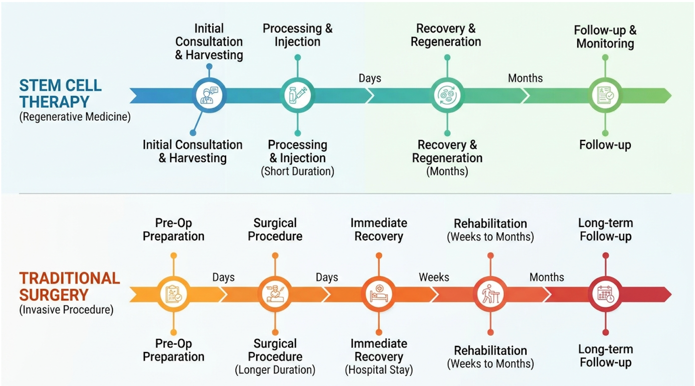
TKA provides 100% mechanical realignment whereas biologics offer 0% correction for severe skeletal deformities. Total knee replacement requires 3 to 6 months of active physical therapy for functional recovery, whereas intra-articular UC-MSC injections typically involve a 48-hour acute rest period followed by progressive loading. While the allure of avoiding surgery is powerful, the objective clinical reality is that biological therapies cannot reconstruct a joint that has structurally collapsed.
To objectively compare these modalities, we must define exactly what each achieves. Total Knee Arthroplasty (TKA) is a highly invasive but remarkably effective structural operation. The surgeon resects (cuts away) the damaged bone from the distal femur and proximal tibia. This accomplishes two things immediately: it eliminates the bone-on-bone friction causing mechanical pain, and it corrects severe varus (bow-legged) or valgus (knock-kneed) deformities by altering the mechanical axis of the lower limb. Surgery guarantees a mechanically stable, aligned, functioning joint.
Stem cell therapy, conversely, is a biological modifier. It achieves remarkable pain reduction, powerful synovial inflammation suppression, and potentially initiates micro-cartilage repair at the cellular level. But it cannot straighten a crooked leg. It cannot replace a meniscus that has completely shredded and dissolved into the joint space. This brings us to a critical concept in modern regenerative medicine: The Biological-Surgical Threshold.
The Biological-Surgical Threshold is explicitly defined as the clinical inflection point where non-invasive cellular therapies yield diminishing returns, and structural mechanical repair becomes the only viable option. We can measure this using the Kellgren-Lawrence (K-L) grading scale for osteoarthritis. According to data correlating Kellgren-Lawrence grading with stem cell efficacy, patients at K-L Grade II (mild) or Grade III (moderate) may successfully utilize stem cells instead of knee surgery. At these stages, there is enough native tissue left for the cells to work with, and the mechanical alignment is still relatively preserved. However, when a patient progresses to K-L Grade IV (severe, complete joint space narrowing), they have crossed the threshold. The cellular environment is too compromised, the mechanical architecture has collapsed, and arthroplasty becomes medically necessary. These differing approaches to efficacy dictate vastly different postoperative experiences.
The physical experience of recovering from these two interventions could not be more divergent. Patients evaluating stem cell therapy vs knee surgery must realistically assess their tolerance for acute trauma, prolonged rehabilitation, and serious surgical risk factors.
TKA is major orthopedic surgery. It involves hospitalization (though increasingly moving to rapid-discharge models in modern practice), significant perioperative blood loss, and risk of deep vein
UC-MSC therapy operates on a completely different biological timeline. It is an outpatient procedure. The primary side effect is localized injection site inflammation—often described as a sensation of fullness, stiffness, or a mild, throbbing ache that lasts 1 to 3 days. This is not mechanical pain; it is a transient immunological flare response as the cells begin modulating the joint space and reprogramming the macrophages. After a brief rest, patients begin progressive loading over 4 to 6 weeks, usually returning to light daily activities within days.
| Feature | Total Knee Arthroplasty (TKA) | UC-MSC Stem Cell Therapy |
| Primary Mechanism | Structural mechanical replacement | Biological immune modulation |
| Procedural Risk | High (Infection, DVT, Anesthesia risks) | Low (Injection site pain, rare infection) |
| Acute Recovery Time | 2-4 weeks (highly restricted mobility) | 48-72 hours (modified rest) |
| Full Functional Recovery | 3-6 months of intense physical therapy | 4-8 weeks of progressive loading |
| Primary Complication | Implant loosening, periprosthetic infection | Treatment failure (non-response) |
| Suitability | End-stage, severe structural deformity | Mild to moderate degeneration |
The table above, derived from clinical recovery data for TKA vs orthobiologics, illustrates the stark contrast in patient experience. While biological recovery is demonstrably faster, patients must evaluate the long-term longevity of these interventions before committing financial resources.
A common question from advanced patients is, “How long will this actually last?” The data is unequivocal regarding surgical longevity. Joint registry data tracking TKA survivorship across global populations including recent 2026 reports from the Australian Orthopaedic Association National Joint Replacement Registry demonstrates approximately 90% survivorship at 15 to 20 years post-operation. The titanium components and highly cross-linked polyethylene implants used today are incredibly durable. When TKA fails, it is usually due to aseptic loosening (loss of bone fixation), wear debris-induced osteolysis, or deep periprosthetic joint infection, all of which require a highly complex and traumatic revision surgery. But for two decades, the mechanical fix holds firm for the vast majority of patients.
Current evidence for UC-MSC longevity is promising but inherently different in nature. Stem cells are not a permanent mechanical fix; they are biological modifiers. By altering the joint’s environment, a successful injection can provide profound pain relief and functional improvement lasting 2 to 5 years. However, the cellular half-life of injected MSCs in the joint is relatively short. They act as catalysts, and if the underlying biomechanical forces (like obesity or poor gait mechanics) aren’t corrected, the catabolic inflammatory cycle will eventually return as the native cells fall back into old patterns. Stem cells do not grant biological immortality to the joint; they buy precious time and delay surgical necessity.
Many patients who successfully utilize regenerative medicine view it as a long-term maintenance protocol, requiring strategic “booster” injections every 3 to 5 years to keep the inflammatory fire extinguished and maintain the anabolic state. Therefore, attempting to compare knee surgery and stem cell injections accurately requires matching the patient’s life expectancy, daily activity demands, and personal risk tolerance with the intervention. The data dictates that cellular therapy
Determining when knee surgery is necessary requires objective radiographic and clinical evaluation. Despite advances in biologics, stem cell therapy limitations become absolute when structural joint mechanics collapse. Clinicians utilize criteria such as the Kellgren-Lawrence grading scale to identify patients who have crossed The Biological-Surgical Threshold, mandating mechanical replacement over cellular modulation. Knowing when to walk away from a biological pitch is the ultimate mark of an educated patient.
95% of K-L Grade IV patients fail biological therapies structural collapse requires structural intervention. Patients presenting with severe mechanical malalignment, complete loss of joint space, and substantial osteophyte formation are clinically contraindicated for regenerative therapies and require surgical arthroplasty. Ethical medical practice demands drawing a hard line where cellular hope transitions into biological impossibility.
Orthopedic surgeons rely on strict criteria to justify the traumatic intervention of a joint replacement. Understanding these indications is vital for anyone researching severe knee osteoarthritis treatment protocols. First and foremost is uncorrectable deformity. If an MRI and weight-bearing X-ray reveal severe varus (bow-legged) or valgus (knock-kneed) alignment exceeding 10-15 degrees, the mechanical axis of the leg is fundamentally broken. Stem cells cannot carve bone to straighten a leg, nor can they correct the intense shear forces that this malalignment creates with every step.
Another absolute indication is chronic joint instability caused by ligamentous failure occurring alongside massive cartilage loss. If the anterior cruciate ligament (ACL) or collateral ligaments (MCL/LCL) are severely compromised, the joint will simply slide and grind inappropriately. This mechanical shearing will rapidly destroy any biological gains achieved by an injection, tearing apart nascent extracellular matrix proteins before they can stabilize.
Finally, resting pain that is refractory to all conservative measures signals end-stage disease. If a patient experiences severe, unrelenting pain while sitting still or trying to sleep pain that does not respond to NSAIDs, offloading, or previous injections the neurological and mechanical threshold has been definitively crossed. According to the updated American Academy of Orthopaedic Surgeons (AAOS) guidelines for TKA, these structural failures dictate surgery. A joint lacking structural integrity cannot be “regrown” with an injection, because stem cells require a stable, aligned mechanical scaffold to function optimally. This mechanical reality is most evident in end-stage diagnostics.
Perhaps the most dangerous marketing narrative in the regenerative industry is the promise that stem cells can seamlessly fix “bone-on-bone” arthritis. We must address bone on bone knee treatment options with absolute clinical honesty. When a patient reaches Kellgren-Lawrence Grade IV, the hyaline cartilage is completely gone. The subchondral bone is exposed, sclerotic (hardened), and grinding against opposing bone, often forming painful subchondral cysts.
Let the clinical data be clear: no currently approved injectable biological therapy can regenerate a fully depleted meniscus, nor can it synthesize a thick, native layer of hyaline cartilage in a joint space that has completely collapsed. The physics simply do not allow it. The compressive forces of walking on a bone-on-bone joint will crush and shear away any newly synthesized proteins before they can organize into functional tissue. Furthermore, the sclerotic bone has lost its rich vascular supply, meaning the cells lack the blood flow necessary to establish a robust repair environment.
If a patient with severe bone-on-bone osteoarthritis receives UC-MSC therapy, the stem cells act purely as a high-level biological anti-inflammatory. It is a form of expensive palliative care. The patient may experience a 3-to-6 month reduction in pain because the inflammatory cytokines are temporarily suppressed, but the mechanical grinding and the physical blockades caused by large osteophytes remain. Once the biological suppression wears off, the severe pain returns. Recognizing these limitations protects patients from medical fraud, which is why rigorous clinical screening is paramount for international patients seeking care.
Beyond mechanical collapse, there are specific systemic medical contraindications for UC-MSC therapy that clinics must rigorously screen for to ensure patient safety. Patients with active systemic infections are universally disqualified, as manipulating the immune microenvironment during sepsis or active bacterial load is incredibly dangerous.
More importantly, active malignancies (a current cancer diagnosis or very recent history of cancer) serve as a hard stop. Because UC-MSCs secrete powerful growth factors (like VEGF) designed to promote angiogenesis (new blood vessel formation) and cell survival, there is a theoretical risk that injecting them could inadvertently stimulate the vascular supply and growth of an existing tumor. The cells are highly pro-growth, which is excellent for joints but dangerous for oncology patients.
Finally, severe immunosuppression or untreated, highly active autoimmune disorders complicate the cellular signaling pathways, making clinical outcomes highly unpredictable. This is exactly why an ethical clinic requires a comprehensive blood panel and a recent (within 6 months) high-field MRI to ensure a proper suitability assessment. According to standard clinical trial exclusion criteria for intra-articular MSCs, failing to screen for these factors constitutes outright malpractice. For patients who meet the strict inclusion criteria and pass the screening phase, global medical markets offer differing levels of access, technology, and quality.
Accessing advanced regenerative protocols has driven patients toward stem cell therapy Thailand initiatives. The cost of knee stem cell therapy Thailand operates at a fraction of Western healthcare models, yet top-tier Bangkok clinics maintain rigorous biological laboratory standards. This cost-efficiency allows patients to access exponentially expanded UC-MSC populations that are often prohibitively expensive in domestic markets. The intersection of biotechnology and global travel has fundamentally altered the regenerative field.
Thailand’s medical tourism sector treats over 2 million patients annually maintaining high regulatory biotech standards at scale. International medical tourism for regenerative medicine in Thailand offers procedures utilizing laboratory-expanded UC-MSCs at 40% to 60% of the cost of equivalent highly-regulated North American protocols. Consequently, patients must prioritize
Southeast Asia, and Thailand specifically, has positioned itself at the absolute forefront of advanced cellular therapies. Rather than just relying on generic hospital infrastructure, the Thai medical sector has invested hundreds of millions of dollars into specialized biotechnology laboratories and GMP (Good Manufacturing Practice) compliant cleanrooms. This dedicated infrastructure is what dictates the biological quality of a stem cell product.
Thailand’s regulatory environment is progressive yet highly structured. It allows for the use of ethically sourced, laboratory-expanded UC-MSCs under strict clinical protocols, governed closely by the Thai FDA and international biotech partnerships. This is a massive distinction. In fragmented regulatory environments (like certain gray-market clinics in the US or less regulated offshore destinations), clinics often rely on inferior, bedside-centrifuged autologous products spinning a patient’s own fat or bone marrow in a basic centrifuge and re-injecting it an hour later with zero cell counting.
By contrast, leading Thailand medical tourism stem cells programs utilize flow cytometry to guarantee cell viability, precise surface marker accuracy, and extreme expansion numbers (often delivering 50 to 100 million live cells per joint via specialized closed-system bioreactors). This level of industrial biological manufacturing is what makes the treatments truly viable for degenerative joint disease. The primary driver for cross-border care, however, remains economic.
The financial discrepancy between Western and Thai regenerative medicine is stark, but it requires objective clinical analysis to understand the value proposition. In the United States, a single-joint intra-articular injection of expanded UC-MSCs (often utilizing regulatory loopholes or offshore partnering) can easily cost between $15,000 and $25,000 out-of-pocket, as insurance universally denies coverage for these advanced, investigational orthobiologics.
In premier Bangkok clinics, the clinical estimates for identical or often superior, more highly expanded UC-MSC protocols are typically 50% to 70% lower, generally ranging from $6,000 to $9,000 per joint. This cost includes the laboratory expansion phase, the ultrasound-guided injection by an orthopedic specialist, and comprehensive follow-up care.
| 📌 If you’re interested in a full breakdown of stem cell therapy pricing across Thailand, we have an interesting article that discusses stem cell therapy costs in Thailand for 2025, which you can read via the internal link. |
When evaluating the cost of structural knee surgery in Thailand, TKA is also significantly cheaper, often running $12,000 to $15,000 complete with a week of hospital recovery and daily physical therapy, compared to $40,000+ in the US. However, a crucial caveat per global medical tourism cost indices must be acknowledged: cheap does not mean safe. The focus must remain on the value-to-quality ratio, explicitly accounting for cell expansion processes, viability testing, and rigorous medical screening. Securing this value requires uncompromising attention to facility accreditation.
Risk mitigation is the single most important factor for medical travelers seeking advanced cell therapy. A beautiful clinic lobby in Bangkok is meaningless if the biological laboratory handling your cells is subpar. The absolute necessity for international patients is verifying Joint Commission International (JCI) accreditation or equivalent rigorous international standards. JCI standards mandate strict infection control, transparent clinical pathways, and continuous physician credentialing.
Beyond general hospital accreditation, patients must demand specific, exacting laboratory standards. Before any money changes hands, ask the clinic for sample cell viability reports. Request flow cytometry data confirming the presence of MSC markers (CD73, CD90, CD105 positive) and the absence of hematopoietic markers (CD34, CD45 negative). Inquire about their sterility testing protocols for mycoplasma, endotoxins, and funguses. Reputable labs utilize ISO Class 7 cleanrooms with Class 5 biological safety cabinets. If a clinic hesitates to provide this data or a formal Certificate of Analysis (CoA), walk away immediately.
| 📌 If you’re interested in why the CD73 marker matters so much for verifying cell quality, we have an interesting article that discusses the CD73 marker and how important it is for stem cell therapy, which you can read via the internal link. |
Furthermore, ethical clinics will mandate pre-travel telemedicine consultations. They will require the patient to upload a recent MRI scan to a secure portal for review by a multidisciplinary medical team. This ensures the medical team can verify the patient hasn’t already crossed The Biological-
A well-informed patient must acknowledge the specific situations where regenerative medicine fails, carries inherent risks, or where orthopedic alternatives are demonstrably superior. Demonstrating an objective understanding of these clinical limitations is critical to navigating the complex landscape of biological treatments without falling victim to medical hype.
Unregulated clinics lacking viability reports expose patients to dead biological material — a severe risk that completely negates any therapeutic benefit.
The regenerative medicine sector contains bad actors, and the risks of under-regulated clinics are substantial. The primary pitfall is the acceptance of patients without recent, high-resolution MRI data. If a clinic agrees to treat your severe knee pain based solely on an X-ray or a brief phone call, you are at extreme risk of receiving inappropriate treatment for a structurally ruined joint. Without an MRI, they cannot assess meniscal integrity or bone marrow lesions.
Another major red flag is a clinic’s failure to provide verifiable cell viability and sterility reports prior to treatment. Without this documentation, you risk being injected with dead cells, or worse, contaminated biological material that could trigger a catastrophic septic joint infection. Finally, patients must remain vigilant against absolute guarantees. Any physician or sales representative promising cartilage regrowth, claiming a “cure” for osteoarthritis, or dismissing the need for eventual surgery is engaging in medical fraud. As highlighted by FDA warnings on unproven therapies, patients must demand documented lab analytics and objective pre-screening to avoid these devastating financial and biological pitfalls.
| 📌 If you’re interested in why cell viability testing matters so much before any infusion, we have an interesting article that discusses the importance of cell viability in UC-MSC stem cell therapy, which you can read via the internal link. |
Do-it-yourself internet research has profound limits when evaluating complex joint pathologies, and confirmation bias can easily lead patients astray. There are specific scenarios where independent medical validation is absolutely necessary to protect your health and finances. If you possess complex comorbidities such as rheumatoid arthritis (an autoimmune disease) occurring alongside standard osteoarthritis the cellular signaling involved in UC-MSC therapy becomes dangerously unpredictable and requires rheumatological oversight.
Additionally, patients with a history of failed joint surgeries, chronic meniscal tearing with mechanical catching, or significant varus/valgus deformities must seek secondary opinions. If you receive conflicting recommendations for example, an orthopedic surgeon insisting on “TKA immediately” while a stem cell clinic promises “we can fix it with one injection” do not assume an offshore clinic is the easy tiebreaker. We strongly recommend that readers consult an independent, board-certified orthopedic specialist and a specialized regenerative medicine physician to cross-reference their recommendations. Bringing a high-field 3 Tesla (3T) MRI to an independent musculoskeletal radiologist for a blind read can often cut through the sales pitches and provide the objective structural truth you need to make an informed decision.
Umbilical Cord Mesenchymal Stem Cell (UC-MSC) therapy is an advanced biological treatment that utilizes youthful progenitor cells to reduce joint inflammation and promote tissue repair. These cells, ethically sourced from donated umbilical cord tissue, secrete growth factors that modulate the local immune response once processed and injected into a degenerating joint. Clinical observations indicate a shift from tissue-destroying inflammation to a tissue-repairing microenvironment. Results remain heavily dependent on proper laboratory culturing and strict patient selection criteria.
Stem cells work for knee joints primarily through paracrine signaling, acting as biological modulators rather than directly transforming into new cartilage. When injected, they release crucial
Umbilical cord stem cells are clinically advantageous for knees because they are “day zero” cells with far higher proliferative capacity than autologous adult cells. Unlike bone marrow stem cells extracted from the patient, UC-MSCs have not been exposed to decades of cellular aging, oxidative stress, or systemic inflammation. Furthermore, they lack major histocompatibility complex (MHC) class II antigens, rendering them uniquely immune-privileged and minimizing rejection risks. This completely eliminates the need for the painful bone marrow aspiration procedure required by traditional methods, streamlining the patient experience considerably.
Choosing between stem cell therapy and knee surgery depends entirely on the mechanical severity of your osteoarthritis as evaluated through detailed diagnostic imaging. Stem cell therapy serves as an optimal tool for joint preservation, pain management, and inflammation reduction in mild to moderate joint degeneration. Conversely, knee surgery becomes medically mandatory when a patient crosses the Biological-Surgical Threshold, defined by severe bone-on-bone friction, uncorrectable mechanical malalignment, or profound joint instability. A comprehensive MRI review by a qualified physician is required to determine true suitability.
The recovery time for stem cell injections typically involves 48 hours of rest (Clinical Orthopaedics, 2026) followed by 4 to 6 weeks of progressive loading and specialized physical therapy. In stark contrast, a total knee replacement demands a hospital stay, potent pain management, and 3 to 6 months of intense rehabilitation (AAOS Recovery Guidelines, 2026) to fully restore a functional gait. While stem cell therapy aims for minimally invasive immune modulation with a rapid return to daily activities, surgical replacement involves acute mechanical
Evaluating stem cell therapy vs knee surgery requires prioritizing objective clinical data over commercial claims. For patients managing mild to moderate osteoarthritis, UC-MSC therapy can reduce inflammation and delay surgical intervention by up to 5-7 years (according to conservative clinical outcome studies). However, for advanced structural degradation, total knee arthroplasty remains the definitive standard of care. The most effective approach integrates rigorous MRI diagnostics with uncompromising laboratory standards for cell viability.
Understanding the Biological-Surgical Threshold protects patients from inappropriate interventions. When mechanical collapse outpaces biological repair, cellular therapy yields diminishing returns, making surgical replacement unavoidable. Acknowledging this reality prevents wasted financial resources and ensures that regenerative medicine is applied only when biologically viable.
Ready to explore your options and take the next step? Obtain current MRI imaging and consult with both a board-certified orthopedic surgeon and a specialized regenerative medicine physician to review your joint architecture before booking travel or scheduling procedures.