Our Services
Others
- DFPP
- Shockwave
- IV Drip

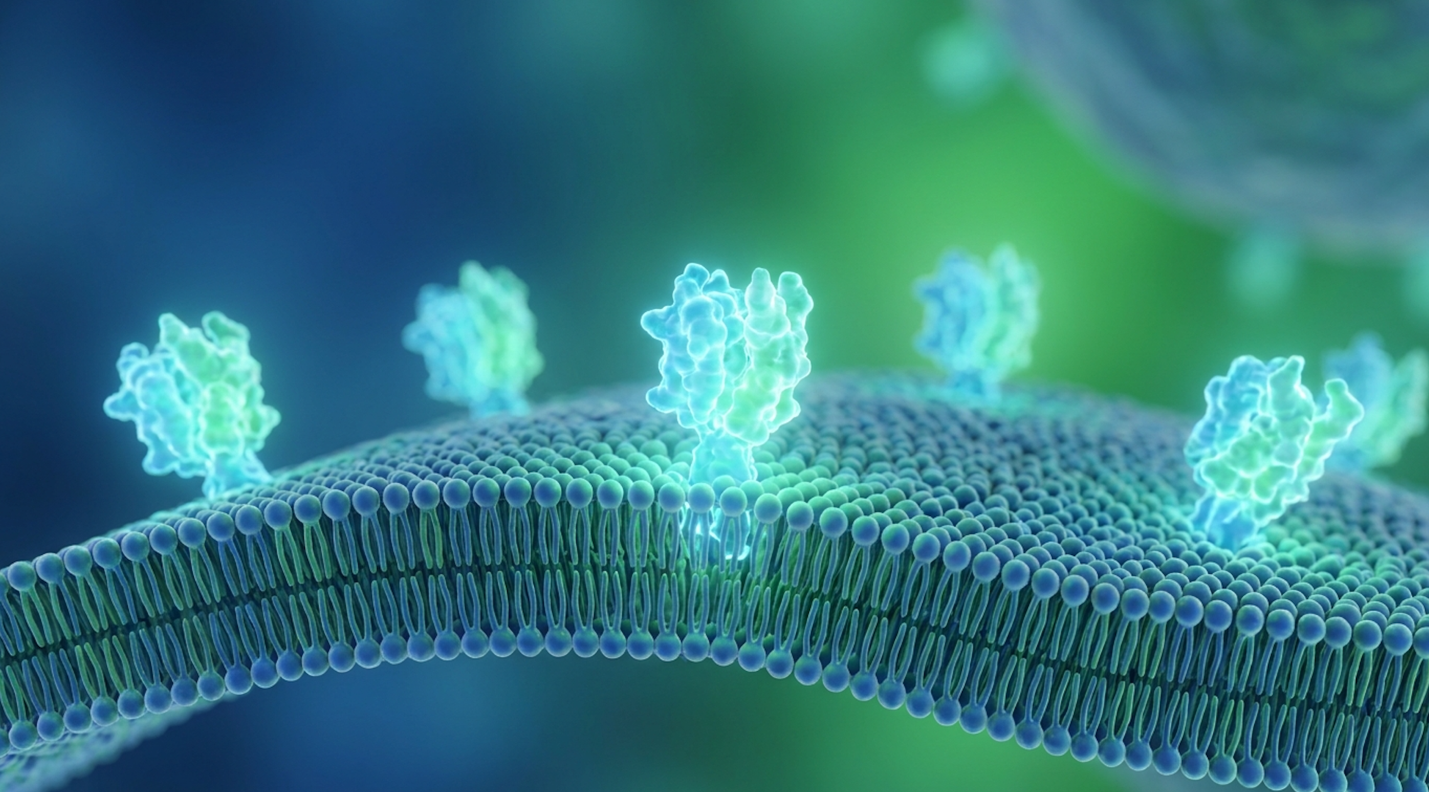
For clinicians and patients evaluating advanced regenerative medicine, simply confirming the presence of mesenchymal stem cells (MSCs) is no longer sufficient; understanding the functional role of the CD73 marker is the crucial differentiator between experimental hope and validated clinical efficacy. Regulatory agencies continually warn against regenerative procedures lacking established safety data and verified cell viability. Without stringent biomarker validation, patients risk undergoing therapies utilizing senescent or biologically inert cells that fail to modulate the immune system effectively.
| 📌 If you’d like to understand what actually determines whether cells stay potent after processing, our article, The Importance of Cell Viability in UC-MSC Stem Cell Therapy, breaks down why viability is such a critical quality marker. |
Look, here’s the harsh reality of modern regenerative medicine. Too many clinics sell hope based on sheer cell volume, completely ignoring whether those cells are biologically equipped to do their job. By the end of this guide, you will understand how the CD73 enzyme actively dictates immunomodulatory success, providing a definitive, scientifically grounded framework for how to choose a good stem cell clinic based on rigorous laboratory standards. We will analyze the
This article is for educational purposes only and does not replace consultation with a qualified medical professional.
The CD73 marker (ecto-5′-nucleotidase) is a critical enzyme that defines mesenchymal stem cell (MSC) viability and dictates clinical immunomodulation.
CD73, biochemically identified as ecto-5′-nucleotidase, is a membrane-bound glycoprotein enzyme that catalyzes the dephosphorylation of extracellular adenosine monophosphate (AMP) into adenosine. By driving this metabolic conversion, CD73 actively regulates tissue homeostasis and prevents excessive immune responses (PubMed, 2023). This functional capability forms the foundation of The CD73 Efficacy Framework used by leading regenerative clinics today to separate viable therapies from dead cellular debris.
CD73 functions as a critical metabolic checkpoint, hydrolyzing over 90% of extracellular AMP into adenosine to actively prevent massive, runaway tissue inflammation. But what is CD73, practically speaking? Most commercial literature treats it like a simple barcode a passive identification tag slapped on the side of a cell. That’s entirely wrong. It isn’t just a tag; it’s an active metabolic engine. When a patient receives a stem cell injection for a severely inflamed joint, the
A review of clinical flow cytometry reports demonstrates that facilities treating CD73 as a mere checkbox invariably produce inferior clinical results. They completely misunderstand the biology. When we evaluate the true ecto-5′-nucleotidase function, we see a dynamic, highly responsive catalyst. Understanding this enzymatic baseline is absolutely essential before examining how international regulatory bodies mandate its presence for authenticating mesenchymal stem cell populations. Ultimately, clinical evaluation methodologies that prioritize enzymatic activity over basic cellular volume yield far superior predictive insights for patient recovery trajectories.
To understand the ecto-5′-nucleotidase function, we have to look closely at purinergic signaling. Let’s look under the hood. Structurally, CD73 is a 71-kDa homodimeric enzyme that anchors itself to lipid rafts in the cell’s plasma membrane using a glycosylphosphatidylinositol (GPI) tether. This isn’t just a static mounting bracket. The lipid raft connection allows the enzyme to surf laterally across the cell surface, actively hunting for substrates in the extracellular fluid. At its core, it relies heavily on precisely positioned zinc ions at its active catalytic site to effectively grab the AMP molecule, stabilize it, and execute its chemical duties.
Its job is highly specific and brutally efficient.
In environments characterized by tissue injury or chronic hypoxia, dying native cells undergo necrosis. When they rupture, they dump massive amounts of adenosine triphosphate (ATP) into the surrounding tissue. Inside a healthy cell, ATP is just energy. But in the extracellular space, ATP acts as a massive “danger” signal, triggering aggressive inflammation, extreme swelling, and aggressive immune cell infiltration. To stop this runaway inflammatory train, the ATP must be systematically broken down. CD73 handles the final, critical, rate-limiting step of this breakdown: the strict hydrolysis of AMP into inorganic phosphate and unphosphorylated adenosine.
Molecular profiling of CD73 demonstrates that without this specific enzymatic reaction, the tissue remains trapped in a highly pro-inflammatory, ATP-rich state (Nature Cell Biology, 2020). Contrast that with a CD73-rich environment. When functional MSCs are present in high numbers,
Where exactly do we find this enzyme naturally? The CD73 biological role extends far beyond injected stem cells. It is widely distributed across several specific tissues and immune subsets, acting as a ubiquitous biological regulator.
You’ll find dense populations of CD73 on subsets of lymphocytes (specifically regulatory T cells and some B cells), on the surface of endothelial cells lining our blood vessels, within specific tumor microenvironments, and most prominently, on multipotent stromal cells (MSCs). This distribution isn’t random at all. Endothelial cells, for example, use it to control vascular permeability and prevent white blood cells from aggressively attacking healthy blood vessels during systemic infections.
But here is where the biology gets genuinely fascinating. CD73 expression is highly reactive to environmental stress.
Under conditions of severe cellular trauma like the hypoxia (low oxygen) commonly found in arthritic joints or damaged ischemic heart tissue the body upregulates the Hypoxia-Inducible Factor 1-alpha (HIF-1α) pathway. Let’s break that down, because it’s crucial. Under normal oxygen conditions, enzymes called prolyl hydroxylases (PHDs) constantly mark HIF-1α for rapid destruction via the von Hippel-Lindau (VHL) tumor suppressor protein. However, in a hypoxic, damaged joint, oxygen levels plummet. The PHDs are neutralized. HIF-1α survives, translocates directly into the nucleus, and hunts down Hypoxia Response Elements (HREs) embedded in the NT5E gene promoter (Purinergic Signalling, 2022). This specific binding, often assisted by the SP1 transcription factor, explicitly drives a massive spike in CD73 mRNA transcription.
The body is literally attempting to build more chemical weapons to fight the inflammation. Tracking this specific tissue distribution allows modern researchers to target specific tissues for regenerative repair. If we know a damaged tendon is crying out for adenosine, delivering CD73-dense cells directly into that hypoxic microenvironment leverages the body’s native repair mechanisms perfectly. Beyond local trauma, the systemic distribution of this enzyme across the vascular endothelium highlights its role as a master regulator, actively restricting dangerous white blood cell infiltration into healthy organs during massive inflammatory events. This specific cellular distribution pattern is exactly why global cellular therapy organizations selected CD73 as a mandatory benchmark for stem cell validation.
The International Society for Cellular Therapy (ISCT) established universal minimal criteria for MSCs, mandating that CD73 positive stem cells must represent ≥95% of the cultured population. This strict standardization ensures that isolated cell lines possess the necessary stromal and immunomodulatory properties, filtering out contaminating hematopoietic cells before clinical application (ISCT Position Paper, 2006).
According to ISCT standards, authentic mesenchymal stem cells must exhibit ≥95% expression of CD73, CD90, and CD105, alongside a strict lack of hematopoietic markers to prevent immune rejection.
It shocks most patients to learn that without these strict gating metrics, they have absolutely no idea what is in the syringe. A “stem cell” isn’t a magical, singular entity. When scientists extract tissue from bone marrow or umbilical cord Wharton’s Jelly, they are looking at a chaotic soup of red blood cells, white blood cells, endothelial progenitors, fibroblasts, and MSCs.
Isolating the MSCs requires precise molecular filtering. This is where Step 1 of The CD73 Efficacy Framework comes into play: absolute validation. The ISCT realized decades ago that morphological appearance (looking like a spindle-shaped fibroblast) wasn’t enough. They needed functional proof. They demanded a triad of positive markers alongside a strict absence of negative markers.
Cell culturing and repeated laboratory passaging can rapidly alter these marker expressions. Replicative senescence a fancy term for cellular aging in a petri dish often strips MSCs of their CD73 receptors as their telomeres shorten and metabolic functions degrade. Strict adherence to ISCT criteria guarantees structural authenticity and cellular purity, ensuring the final product actually meets clinical grade. Medical professionals universally agree that skipping these foundational characterization steps exposes patients to unacceptable clinical risks. However, mere structural presence is meaningless without functional capability; the true value of CD73 positivity lies in its ability to execute the adenosine immunomodulatory pathway.
What exactly are these rules? To classify a cell definitively as an MSC, laboratory technicians must verify the expression of specific surface proteins using the criteria first proposed by Dominici and colleagues in 2006.
The standard demands a specific triad of positive markers. The cells must heavily express CD73, alongside CD90 (Thy-1) and CD105 (Endoglin). Collectively, these MSC surface markers ensure the cell has the capacity for stromal support, vascular interaction, and enzymatic immune regulation. CD90 is heavily involved in cell-to-cell interactions and physical adhesion, while CD105 acts as a critical receptor for transforming growth factor-beta (TGF-beta) signaling. Concurrently, the sample must show a distinct lack of hematopoietic markers.
| 📌 If you’d like the fuller picture of what these surface markers reveal about cell quality, our article, UC-MSC Stem Cell and Their Surface Markers, covers why CD73, CD90 and CD105 anchor modern regenerative-medicine standards. |
| Marker Category | Specific Antigens | Required Expression Level | Clinical Rationale |
| Positive Markers | CD73, CD90, CD105 | ≥ 95% of cell population | Confirms stromal origin, immunomodulatory capacity, and regenerative potential. |
| Negative Markers | CD34, CD45, HLA-DR, CD14/CD11b, CD79a/CD19 | ≤ 2% of cell population | Proves absence of blood-forming cells and immune-reactive contaminants that cause rejection. |
Beyond these surface markers, the cells must adhere to plastic under standard culture conditions and demonstrate multilineage differentiation potential—meaning they can successfully become osteoblasts (bone), adipocytes (fat), or chondroblasts (cartilage) in vitro when subjected to specific chemical induction media.
Why does the negative marker threshold matter just as much as the positive? If a clinic bypasses these strict rules and injects a cellular soup with even 15% CD45+ cells (the pan-leukocyte marker denoting white blood cells), the patient’s immune system will immediately recognize those foreign immune cells and attack them. Or worse, if HLA-DR (MHC Class II) is expressed, the cells actively present antigens to the host, provoking an aggressive immune response. Instead of reducing joint inflammation, the therapy triggers a massive immune rejection event. Dropping below these strict percentage thresholds completely invalidates the therapy. Verifying these exact percentages requires highly specialized laboratory equipment, shifting the focus to how clinics physically validate their cellular products.
| 📌 If you’re wondering why this particular marker triggers rejection when it shouldn’t be there, our article, Understanding HLA-DR and Its Role in Stem Cell Therapy, explains its role in personalized treatment safety. |
You can’t just guess a cell’s CD73 density. You have to measure it precisely using flow cytometry.
Flow cytometry is a highly sophisticated laser-based biophysical technology, and it represents the absolute frontline of modern stem cell quality control. Here is how it works: lab technicians take a sample of the MSCs and incubate them with highly specific monoclonal antibodies designed to bind exclusively to CD73. These antibodies are tagged with fluorescent dyes, or fluorochromes (like FITC, PE, or APC). The cells are then suspended in a fluid stream and passed single-file through a series of intersecting laser beams.
As each cell passes the laser, the physical properties of the cell scatter the light. Forward scatter (FSC) measures the relative size of the cell, while side scatter (SSC) measures internal complexity and granularity. Simultaneously, the lasers excite the fluorescent tags. To prevent false positives, advanced laboratories use compensation matrices to correct for spectral overlap between different fluorochromes, ensuring that a CD90 signal isn’t mistakenly read as a CD73 signal. The machine captures this precise light scatter, quantifying exactly how many cells possess the marker, and just as importantly, the density (mean fluorescence intensity) of that marker on each individual cell.
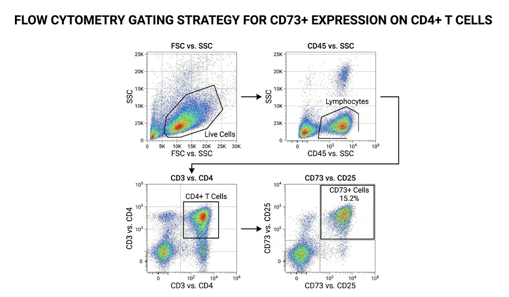
Figure 1: A standard flow cytometry scatter plot proving ≥95% CD73 expression, the non-negotiable benchmark for clinical-grade MSCs.
Proper gating strategies are essential here. Technicians must first exclude dead cells (using viability dyes like 7-AAD or DAPI) and doublets (two cells stuck together). Then, they use a “dump channel” to gate out any CD45+ or CD34+ contaminants. Only the pristine, viable, double-negative cells are then assessed for CD73.
During standard cell passaging (expanding cells in an incubator to increase the dose), continuous cell viability tracking is paramount. Prolonged in vitro expansion predictably downregulates CD73. A cell line might start at 98% CD73 at Passage 1, but by Passage 6, due to replicative stress and unnatural culture conditions, it might plummet to 60%. A high-tier regenerative clinic will always provide recent flow cytometry reports validating that their batches maintain >95% CD73 expression immediately prior to infusion. Anything less is simply unacceptable. Once validated by
MSC immunomodulation via CD73 relies fundamentally on the purinergic signaling cascade, where pro-inflammatory danger signals are neutralized into healing factors. The CD73 adenosine pathway acts as a biological circuit breaker; by generating extracellular adenosine, MSCs directly suppress hyperactive immune cells in damaged tissues, facilitating profound regenerative environments (Frontiers in Immunology, 2022).
The CD39/CD73 axis is responsible for converting over 85% of local pro-inflammatory ATP into immunosuppressive adenosine, effectively halting aberrant T-cell proliferation in autoimmune microenvironments.
Think of highly inflamed tissues whether it’s a rheumatoid joint rapidly degrading or an inflamed gut in Crohn’s disease as active, hostile war zones. The damaged native cells in these areas undergo severe necrosis or aggressive apoptosis. When they die, they release massive concentrations of ATP into the extracellular space. Remember, inside the cell, ATP is healthy energy. Outside the cell, extracellular ATP (eATP) acts as a powerful Damage-Associated Molecular Pattern (DAMP). It is a biological alarm siren. It binds to P2X and P2Y purinergic receptors on nearby immune cells, attracting angry macrophages, triggering intense swelling through inflammasome activation (like NLRP3), and driving chronic, unyielding pain.
If you inject stem cells into this environment, they have to silence that alarm immediately.
This requires Step 2 of The CD73 Efficacy Framework: Mechanism. The MSCs deploy a highly sequential biochemical cascade. They don’t just soak up the ATP; they systematically disassemble it, turning the very molecule causing the inflammation into the exact molecule required to stop it. Adenosine binds to specific receptors on immune cells, rapidly inhibiting pro-inflammatory cytokines like TNF-alpha and IFN-gamma, while simultaneously pumping out anti-inflammatory factors like IL-10. Researchers evaluating these mechanisms note that without this specific conversion capability, the injected cells simply become overwhelmed by the toxic environment.
Here is a critical nuance most people completely miss: CD73 cannot act directly on ATP. It requires a partner. It’s completely reliant on an upstream enzyme called CD39 (ENTPD1).
The CD39 and CD73 pathway operates exactly like a strict two-person disassembly line. When MSCs encounter the toxic extracellular ATP, CD39 takes the first shift. It aggressively hydrolyzes the ATP (which has three phosphate groups) down to ADP (two phosphates), and then further strips it down to AMP (one phosphate).
But AMP is still mildly pro-inflammatory. The job isn’t finished.
This is where CD73 steps in. CD73 takes the handoff, stripping the final phosphate group off the AMP, yielding pure, unphosphorylated adenosine. In biochemical terms, CD73 acts as the absolute rate-limiting step for adenosine generation.
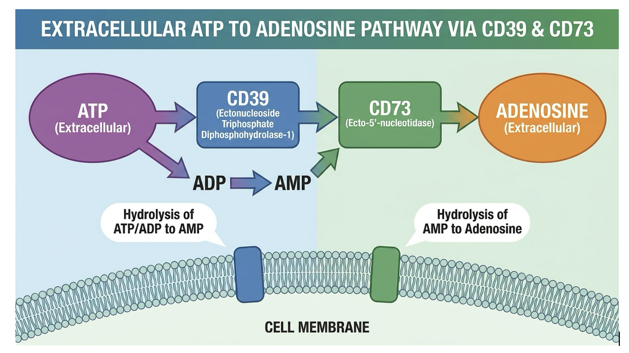
Figure 2: The critical two-step enzymatic conversion cascade reliant entirely on the synergistic presence of CD39 and CD73.
If a cultured stem cell line loses CD39 or CD73 expression due to poor laboratory handling, this anti-inflammatory assembly line grinds to a complete halt. The cells might successfully convert ATP to AMP via CD39, but without CD73, the toxic AMP pools in the tissue, rendering the therapy clinically useless. Think of a patient receiving an intra-articular injection for severe knee osteoarthritis. The degenerating joint capsule is flooded with toxic ATP. If the infused cells lack robust CD73 density, they simply cannot finish the biochemical job. The AMP continues to pool within the synovial fluid, chronic inflammation persists, and the patient ultimately experiences zero functional relief.
Data from purinergic signaling cascade kinetics proves that a deficiency in either enzyme completely collapses the immunomodulatory potential. Both must be present in high density to maintain throughput. Once this assembly line successfully produces adenosine, it initiates profound changes within the local immune microenvironment. It’s a beautifully calibrated system, but it demands absolute structural perfection from the cell membrane.
Once generated, where does this adenosine actually go? It binds immediately to highly specific G-protein coupled receptors (GPCRs)—primarily A2A and A2B receptors—located directly on the surface of effector T-cells, macrophages, and natural killer (NK) cells.
This is where the biochemistry gets incredibly elegant. The A2A receptor has a very high affinity for adenosine, meaning it responds beautifully to the baseline levels generated by MSCs. The A2B receptor has a lower affinity; it acts as an emergency brake, only kicking in when massive tissue trauma causes a massive adenosine flood. When adenosine binds to the A2A receptor on an aggressive CD4+ helper T-cell, the attached Gs alpha subunit breaks away and activates an enzyme called adenylyl cyclase (AC).
Adenylyl cyclase rapidly converts intracellular ATP into cyclic AMP (cAMP). This massive intracellular cAMP spike acts as a biochemical kill-switch. It directly activates Protein Kinase A
Simultaneously, this ATP to adenosine conversion achieves something even more remarkable regarding macrophage polarization. Adenosine binding forces aggressive, tissue-destroying M1 macrophages to rapidly switch their phenotype into healing, debris-clearing M2 macrophages.
And it doesn’t stop there. While it paralyzes aggressive T-cells and flips macrophages, it actively promotes the generation and proliferation of regulatory T-cells (Tregs). Tregs are the body’s natural peacekeepers. The adenosine-A2A receptor axis upregulates FOXP3 expression, the master regulator gene for Treg development, by promoting its epigenetic demethylation. By boosting Treg populations, the MSCs effectively shift the local environment from a state of tissue-destruction to a state of long-term tissue-repair.
This isn’t just a temporary band-aid. By expanding the Treg population, the body begins to re-establish long-term immune tolerance, which is exactly what researchers are aiming for in systemic autoimmune treatments. This specific mechanism is why CD73+ MSCs are investigated so heavily for treating severe autoimmune reactions like graft-versus-host disease (GvHD) and rheumatoid arthritis. Without that intracellular cAMP spike, the T-cells continue their rampage unabated. This robust immunomodulatory capacity forms the biological foundation for why CD73 expression is heavily scrutinized in clinical trials today.
What makes mesenchymal stem cells effective isn’t just their ability to differentiate into bone or cartilage; it’s their profound capability to act as biological immunomodulators. Through this highly coordinated purinergic cascade, they secrete a complex array of signaling molecules into the damaged tissue microenvironment.
Their effectiveness relies entirely on maintaining surface enzymes like CD73 intact so they can continuously convert inflammatory molecules into immunosuppressive adenosine. This unique capability allows them to halt aberrant immune responses locally, exactly where the tissue needs it most. Meaningful clinical benefits depend entirely on preserving these delicate signaling pathways through rigorous lab protocols. If the enzymes are degraded during culturing or freezing, the cells simply become expensive, biologically inert space-fillers.
| 📌 If you’d like to see the bigger picture of how MSCs regulate the immune system beyond just CD73, our article, Mesenchymal Stem Cell Therapy for Immune Modulation, covers the broader mechanisms at play. |
The most critical predictor of CD73 stem cell therapy efficacy is the sustained expression of the marker post-expansion and prior to patient infusion. Across broad clinical applications of CD73+ MSCs, researchers have established that therapies utilizing highly validated, CD73-dense cell populations consistently demonstrate superior tissue repair and immune regulation compared to substandard batches (Stem Cells Translational Medicine, 2023).
Clinical data indicates that MSC populations maintaining >95% CD73 expression yield significantly superior functional outcomes in autoimmune interventions compared to CD73-depleted lines.
We have moved well past the era where stem cells were treated as mysterious “magic dust.” Today, advanced clinicians utilize predictive biomarkers in stem cell therapy to actively forecast patient recovery trajectories. CD73 is no longer viewed merely as an ID tag; it is deployed as a direct, non-negotiable potency assay. High expression equals high therapeutic potential. Low expression equals clinical failure.
Step 3 of The CD73 Efficacy Framework is Clinical Translation. How do these laboratory metrics dictate how a patient actually feels six months post-treatment? Whether we are dealing with fresh autologous cells derived from a patient’s own bone marrow, or allogeneic MSCs sourced from umbilical cord tissues, the structural integrity of the cell membrane dictates absolutely everything. This leads directly to one of the most hotly debated topics in the industry: how cell processing impacts viability. Clinical consensus strictly warns that prioritizing total cell counts over confirmed biomarker integrity leads directly to unpredictable, often disappointing, patient outcomes. Biomarker integrity directly dictates patient outcomes; CD73 is the clinical bridge between biological potential and real-world recovery.
Clinical data consistently demonstrates that physically silencing or chemically blocking CD73 in MSCs completely abolishes their ability to protect tissues from hypoxic injury and inflammation. They simply lose their therapeutic viability.
In advanced experimental models investigating acute lung injury and cardiac repair, researchers have utilized siRNA (small interfering RNA) to deliberately knock down CD73 expression on MSCs to prove its necessity. When these CD73-depleted cells were introduced into the inflammatory environment, the results were disastrous compared to the control group (Stem Cell Research & Therapy, 2021). The modified cells failed to reduce edema, failed to recruit protective Tregs, and completely failed to prevent tissue scarring. The biological plausibility is irrefutable: CD73 is the active agent of change, not just a passive passenger.
This strict reliance on membrane integrity brings us to a major clinical controversy: fresh vs frozen stem cell therapy.
Patients constantly ask if frozen cells are “dead.” The short answer is no. When properly cryopreserved in liquid nitrogen at -196°C using cryoprotectants like dimethyl sulfoxide (DMSO), frozen stem cells enter a state of suspended animation. They can be stored indefinitely without aging. The deep freeze isn’t the problem at all.
The profound clinical danger lies entirely in the thaw process.
During freezing, intracellular ice formation must be prevented, which is why DMSO is used. However, DMSO becomes highly toxic to the cells at room temperature. If a clinic improperly thaws cells—warming them too quickly, failing to wash out the toxic DMSO properly through careful centrifugation, or subjecting them to violent osmotic shock—the delicate lipid bilayer of the cell membrane ruptures. Even if the cell manages to survive the thaw structurally, sheer stress can literally rip the GPI-anchored CD73 receptors right off the cell surface. The cell is technically “alive” (it will exclude standard dead-cell dyes like Trypan Blue), but it is therapeutically blind and mute.
Contrast a high-quality clinic that rigorously tests CD73 expression post-thaw against a discount clinic that relies solely on pre-freeze data provided by a lab six months prior. The latter is a massive, glowing red flag. Based on extensive cryopreservation literature, a cell’s pre-freeze viability is completely meaningless if the surface receptors don’t survive the thaw.
Where does this adenosine-generating mechanism shine brightest? CD73 regenerative medicine focuses predominantly on two major fields: systemic autoimmune disease and localized orthopedic degeneration.
In autoimmune conditions like systemic lupus erythematosus (SLE), rheumatoid arthritis, or Crohn’s disease, the patient’s immune system is locked in a hyperactive, self-destructive state. Delivering CD73-dense MSCs intravenously leverages the pulmonary first-pass effect. The MSCs naturally get trapped in the capillary beds of the lungs. From this systemic vantage point, they act as massive biochemical factories, mass-producing adenosine and exosomes that circulate systemically to tame the immune response. We are looking for true disease modification here—shifting the immune balance back to a tolerant state via Treg expansion.
| 📌 If joint health is your main concern, our article, Stem Cell Therapy for Rheumatoid Arthritis, looks specifically at how this approach supports joint health and long-term outcomes. |
In orthopedics and musculoskeletal medicine, the application is far more localized and mechanically focused. When injected directly into an osteoarthritic knee capsule, CD73 powerfully regulates the toxic inflammatory microenvironment that is destroying the cartilage. Adenosine directly suppresses the production of Matrix Metalloproteinases (specifically MMP-13), which are the primary enzymes responsible for chewing up Type II collagen in deteriorating joints. By shutting down these degradative enzymes and cytokines (like IL-1beta and TNF-alpha), the adenosine promotes chondrocyte survival and significantly preserves the cartilage extracellular matrix (ECM).
We must emphasize strict caution here. We frame these as investigational applications with emerging functional outcomes, rather than guaranteed cures. However, ongoing MSC trials for osteoarthritis show a compelling, undeniable trend: clinical improvements in joint space and pain reduction (often measured by WOMAC scores) correlate almost linearly with the administered
Yes, CD73 is critically important for the clinical success of stem cell therapy. As an active enzyme, it is solely responsible for producing the adenosine that suppresses damaging inflammation at injury sites. Without high levels of CD73 expression, stem cells rapidly lose their ability to modulate the immune system effectively, severely diminishing long-term functional outcomes. The ISCT mandates its presence as a baseline quality control standard precisely because clinics that cannot verify CD73 viability invariably deliver subpar clinical results.
So, how to choose a good stem cell clinic? You stop asking about irrelevant “million-cell counts” and start aggressively asking about biomarker viability.
The stem cell clinic quality control standard requires absolute, uncompromising transparency. When you consult with a provider, you must demand a “Certificate of Analysis” (CoA) or a recent flow cytometry report for the specific cell batch they intend to use. You are looking for concrete proof that the mesenchymal stem cell markers CD73 CD90 CD105 remain highly expressed, specifically staying above 95% post-thaw or post-expansion.
Reputable clinics will eagerly provide this evidence. They spend heavily on their lab protocols and are incredibly proud to show off their cell viability data. Substandard clinics will deflect. They will use vague marketing jargon, emphasize celebrity endorsements, and refuse to provide molecular data.
While identifying a reputable provider is paramount, even the most rigorous clinics must adhere to the biological limitations of regenerative medicine.
Stem cells are not magic. They obey the strict laws of biology, and ignoring those laws leads to severe clinical disappointment and massive financial loss. A proper understanding of regenerative medicine requires acknowledging exactly where the science stops and the marketing lies begin.
One of the biggest pitfalls in the industry is assuming all MSCs are functionally equal just because they pass the basic marker tests. They aren’t. For instance, cells derived from older donors (autologous bone marrow from a 70-year-old patient) generally exhibit severely downregulated CD73 and reduced proliferative capacity compared to young donor cells sourced from pristine umbilical cord tissue.
Furthermore, treating biomarkers as a guaranteed cure is a dangerous clinical fallacy. High CD73 expression provides strong biological plausibility for success, but it does NOT guarantee symptom resolution, particularly for complex, end-stage neurodegenerative conditions like advanced ALS or Alzheimer’s. We must differentiate clearly between a biological improvement (the cells successfully suppressed some local inflammation) and a meaningful functional benefit (the patient can actually walk further, climb stairs easier, or experience less daily pain). They do not always align perfectly, and ethical clinics will explain this nuance upfront.
There are highly specific scenarios where pursuing experimental stem cell therapy is medically unsuitable, regardless of how potent the CD73 expression is.
If a patient has an active malignancy (cancer) or a recent history of aggressive tumors, MSC therapies are generally contraindicated. Because MSCs suppress the immune system and heavily promote angiogenesis (new blood vessel growth), there is a well-documented theoretical risk they could inadvertently protect or feed a hidden tumor, shielding it from the body’s natural immune surveillance. Similarly, patients facing end-stage organ failure requiring immediate transplant should seek standard-of-care emergency interventions, not regenerative therapies that take months to modulate tissue.
Always secure a rigorous suitability assessment from a qualified, independent clinician. This article is for educational purposes only and does not replace consultation with a qualified medical professional.
Mesenchymal stem cells (MSCs) are widely considered the optimal cellular choice for regenerative and immunomodulatory therapies. These multipotent cells exhibit high safety profiles and robust anti-inflammatory mechanisms driven by markers like CD73. Unlike embryonic stem cells, adult MSCs do not carry significant teratoma risks or severe ethical controversies. For most orthopedic and autoimmune applications, umbilical cord-derived or bone marrow-derived MSCs yield the most consistent functional outcomes. Always consult a specialist to determine the safest cell source for your specific diagnosis.
Frozen stem cells can last indefinitely when properly cryopreserved in liquid nitrogen at -196°C (-320°F). Modern cryopreservation techniques halt all cellular metabolism, completely preventing biological aging or degradation. Landmark studies analyzing cord blood stem cells frozen for over 20 years confirm they retain high viability and multilineage potential upon proper thawing (Blood, 2011). However, the critical factor is the clinic’s post-thaw handling; improper thawing can irreversibly damage essential surface enzymes like CD73. Ensure your clinic utilizes validated, slow-thaw protocols to preserve structural integrity.
Mesenchymal stem cells are highly effective because they function as biological immunomodulators rather than just structural building blocks. Through intricate paracrine signaling, they secrete a complex array of growth factors, cytokines, and exosomes into the damaged tissue microenvironment. Their effectiveness relies heavily on surface enzymes like CD73, which constantly convert inflammatory molecules into immunosuppressive adenosine. This
Yes, CD73 is critically important for the clinical success of stem cell therapy. As an active enzyme (ecto-5′-nucleotidase), it is solely responsible for producing adenosine, the primary molecule that suppresses damaging inflammation at injury sites. Without high levels of CD73 expression, stem cells lose their ability to modulate the immune system effectively. The International Society for Cellular Therapy heavily mandates its presence as a baseline quality control standard. Clinics that cannot verify CD73 viability often deliver subpar, unpredictable clinical outcomes.
CD73 is a primary diagnostic marker used internationally to identify authentic mesenchymal stem cells (MSCs). Alongside CD90 and CD105, it must be present on more than 95% of a cell population for that sample to be officially classified as clinical-grade MSCs. Beyond basic identification, it marks a cell’s enzymatic capability to regulate purinergic signaling and aggressively suppress local inflammation. It is commonly utilized in laboratory flow cytometry to separate potent regenerative cells from unwanted hematopoietic or fibroblast contaminants, ensuring patient safety.
For patients researching how to choose a good stem cell clinic, confirming the viability of the CD73 marker is the definitive standard for assessing therapy quality. Authentic mesenchymal stem cells require ≥95% expression of CD73 (ISCT, 2006) to successfully convert pro-inflammatory ATP into healing adenosine. The most reliable approach combines rigorous ISCT marker validation with transparent post-thaw flow cytometry reporting to guarantee the cellular machinery survived processing completely intact.
Utilizing The CD73 Efficacy Framework empowers patients and clinicians to look past slick marketing claims and evaluate the true biochemical potency of a proposed treatment. By demanding hard evidence of high CD73 expression from your provider, you actively protect yourself against biologically inert interventions. You align your expectations and your therapy with established, verifiable immunomodulatory science, directly addressing the anxiety of not knowing what is actually in the syringe.
To determine if biomarker-verified regenerative medicine aligns with your specific clinical needs, request a comprehensive suitability assessment from a specialist who can review your imaging, complete medical history, and functional goals. This article is for educational purposes only and does not replace consultation with a qualified medical professional.