Our Services
Others
- DFPP
- Shockwave
- IV Drip

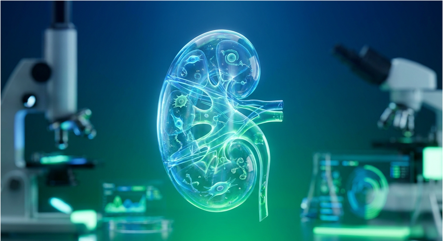
Patients navigating end-stage renal disease already understand the grueling physical mechanics of dialysis; the critical question now is whether cellular medicine offers a scientifically viable alternative. If you are researching stem cell therapy for kidney failure, you aren’t looking for basic definitions of kidney anatomy. You want the hard data.
Standard nephrology care preserves life, absolutely. But it fails completely to halt progressive tissue fibrosis or restore native cellular function. That’s the reality.
By the end of this guide, you’ll understand the exact clinical trial data and biological mechanisms behind stem cell therapy for kidney failure, separating proven physiological outcomes from experimental claims. This rigorous clinical analysis covers the paracrine action of Umbilical Cord Mesenchymal Stem Cells (UC-MSCs), current statistical efficacy on eGFR markers, patient safety protocols, and the harsh regulatory realities of clinical medicine.
Stem cell therapy for kidney failure focuses on utilizing UC-MSCs to modulate immune responses and reduce renal fibrosis. Clinical data from recent trials reveals:
Kidney failure treatment at CKD Stage 5 traditionally relies on mechanical filtration, which manages symptoms but cannot reverse underlying renal fibrosis. At CKD Stage 5, kidneys lose over 85% of their functional capacity rendering traditional medical management incapable of reversing tissue fibrosis (CDC guidelines defining end-stage renal disease, 2026). However, these mechanical interventions entirely ignore the biological deterioration of the nephrons themselves.
End-Stage Renal Disease (ESRD) occurs when the estimated glomerular filtration rate (eGFR) plummets below 15 mL/min/1.73 m². At this stage, the kidneys have exhausted their compensatory mechanisms. The delicate architecture of the glomeruli the actual filtering units is crushed by glomerulosclerosis.
But this isn’t just a simple mechanical failure. It’s an active, aggressive biological replacement of healthy cells with rigid scar tissue. As nephrons die, the surviving filtration units are forced into hyperfiltration. They work overtime to clear toxins, which physically damages their delicate podocyte cells. Podocytes are terminally differentiated, meaning once they detach and die from sheer mechanical stress, they cannot simply divide and replenish themselves.
Simultaneously, the surrounding tissue succumbs to tubulointerstitial fibrosis. As this fibrotic scarring spreads, highly specific uremic toxins accumulate heavily in the blood.
You see, standard hemodialysis effectively clears small, water-soluble solutes like urea. However, it fails miserably at clearing protein-bound uremic toxins like indoxyl sulfate and p-cresyl sulfate. These specific toxins circulate continuously, binding to surviving renal proteins and actively inducing podocyte apoptosis (cell death). This chronic toxicity triggers systemic, low-grade inflammation that continuously drives ongoing tissue destruction.
The renal microenvironment becomes incredibly hostile. Any natural cellular recovery is entirely suffocated by this inflammatory cascade. If you examine a biopsy of a CKD Stage 5 kidney, you don’t just see tired cells. You see a decimated extracellular matrix overrun by myofibroblasts actively laying down collagen scars. The peritubular capillaries, which supply oxygen to the kidney tissue, are physically strangled by this scar tissue, causing profound local tissue hypoxia.
Without targeted biological interventions to suppress this local inflammation, the kidney is locked in a continuous death spiral.
Hemodialysis, the primary mechanical intervention for ESRD, is a brutal physiological compromise. It artificially filters the blood, sure. But pulling massive fluid volumes often three to four liters over a rapid four-hour session places tremendous strain on the cardiovascular system.
The physics of this rapid fluid removal cause massive intravascular volume depletion. Patients routinely suffer from severe intradialytic hypotension sudden blood pressure crashes that actually starve the remaining kidney tissue of oxygen, accelerating final nephron death. The heart takes a severe beating, leading to myocardial stunning during fluid shifts. This cardiovascular stress is precisely why patients on chronic hemodialysis experience incredibly high rates of left ventricular hypertrophy and eventual heart failure. Dialysis accelerates cardiovascular mortality by a staggering margin.
Furthermore, maintaining arteriovenous (AV) fistulas invites access site infections, thrombosis, and chronic vascular inflammation. Over a span of years, the retention of medium-sized molecules leads to debilitating conditions like dialysis-related amyloidosis, causing severe joint pain and bone cysts.
The severe complications of long-term hemodialysis highlight these systemic cardiovascular stresses. It’s a grueling holding pattern that manages uremia but aggressively deteriorates overall vascular health and patient quality of life.
Transplantation remains the gold standard, offering true biological replacement. Yet, waitlist mathematics are bleak, with average wait times extending beyond three to five years. Even if you secure a match, you are committing to a lifetime of aggressive immunosuppressants.
Calcineurin inhibitors, the backbone of anti-rejection regimens, ironically carry well-documented nephrotoxic properties. You are trading one organ failure disease for an iatrogenic (medically induced) compromised immune state. This grim reality is exactly why the medical community is aggressively exploring dialysis alternatives that address biological repair.
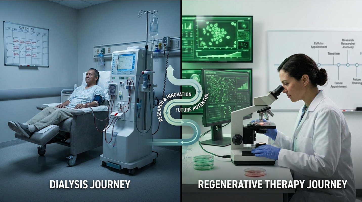
Figure 1: The clinical divergence between mechanical symptom management and biological tissue modulation in ESRD patients.
Currently, end-stage renal disease cannot be entirely reversed once severe fibrotic scarring has permanently destroyed the nephrons. Standard treatments focus entirely on mechanical filtration
Advanced cellular therapies aim to halt further progression and optimize whatever surviving, oxygen-starved tissue remains. They act as biological modulators, not complete organ replacements. Patients must approach any clinic’s claims of “curing” ESRD with extreme caution, as biological stabilization and delayed dialysis dependence are the realistic clinical goals.
UC-MSC stem cell therapy offers a targeted biological intervention for chronic kidney disease by altering the local immune microenvironment. UC-MSCs promote renal tissue repair primarily through paracrine signaling, which actively reduces renal fibrosis and systemic inflammation in damaged kidneys (NCBI review on paracrine signaling, 2026). Unlike older cellular models that assumed transplanted cells directly replaced damaged nephrons, modern research confirms that mesenchymal stem cells kidney regeneration relies on complex molecular signaling to halt fibrosis.
Umbilical Cord Mesenchymal Stem Cells (UC-MSCs) are youthful progenitor cells sourced strictly from the Wharton’s jelly of donated human umbilical cords. They are harvested post-birth from otherwise discarded tissue, entirely bypassing the ethical controversies associated with embryonic cells.
But why umbilical cords? Why not use a patient’s own bone marrow or adipose tissue?
A review of nephrology trial data shows the answer always comes down to cellular senescence. If you extract stem cells from a patient in CKD Stage 5, those cells have lived in a highly toxic, uremic environment for years. They are metabolically exhausted. Their telomeres are drastically shortened, their DNA methylation patterns are heavily altered, and their regenerative capacity is fundamentally compromised by chronic systemic inflammation.
Using autologous (patient-derived) cells for advanced regenerative therapy for CKD often yields extremely poor clinical results because the cellular material is already damaged. UC-MSCs, conversely, are biologically naive, robust, and highly metabolically active. Because they are sourced from “Day Zero” of human life, their replicative capacity and secretome potency are at absolute maximums.
| 📌 If you’re curious why umbilical cord tissue is such a powerful source for these cells, we have an interesting article that discusses why umbilical cord-derived UC-MSC stem cells are superior to other stem cell sources, which you can read via the internal link. |
More importantly, UC-MSCs possess extraordinary “immune privilege.” Because they originate from the boundary between maternal and fetal blood supplies, they lack Major Histocompatibility Complex (MHC) class II molecules on their cell surface, and they express low levels of MHC class I. This means they are inherently hypoimmunogenic.
They do not trigger a massive immune response upon infusion. Patients do not require dangerous HLA matching protocols, bone marrow suppression, or lifetime anti-rejection drugs to tolerate the cellular infusion. They can be safely infused off-the-shelf without triggering graft-versus-host disease (GVHD). The true efficacy of these young progenitor cells lies in their capacity to orchestrate local tissue repair.
For years, the public assumed stem cells acted like replacement parts—you inject a stem cell, and it magically turns into a fresh kidney cell. This is biologically inaccurate and sets incredibly dangerous clinical expectations for desperate patients.
The clinical reality is governed by The Paracrine-Directed Renal Repair Framework. This biological model dictates that UC-MSCs function as highly intelligent “cellular pharmacies.” They do not physically replace your nephrons. Instead, they sense the highly inflamed local environment through chemokine gradients and secrete a highly specific payload of bioactive molecules to fix it.
These secretomes, microvesicles, and exosomes contain potent cytokines and growth factors. The specific mechanism of action UC-MSC utilizes is a highly orchestrated, multi-pronged assault on kidney disease pathology.
First, upon homing to the damaged renal tissue, they release Vascular Endothelial Growth Factor (VEGF) and Hepatocyte Growth Factor (HGF). This directly stimulates local endothelial cells to generate new blood vessels a process called angiogenesis. Remember those peritubular capillaries
Second and this is absolutely critical for ESRD patients they execute macrophage reprogramming. Your damaged kidneys are swarming with M1 macrophages, which are aggressive immune cells driving chronic inflammation and collateral tissue damage. They secrete pro-inflammatory cytokines like TNF-alpha and IL-6.
The exosomes secreted by UC-MSCs contain specific microRNAs (like miRNA-let7c) that force these immune cells to flip their phenotype into M2 macrophages. M2 macrophages are inherently anti-inflammatory. They secrete IL-10 and arginase-1, actively working to clean up cellular debris without destroying healthy cells.
| 📌 If you’re interested in how UC-MSCs modulate the immune system, we have an interesting article that discusses mesenchymal stem cell therapy for immune modulation, which you can read via the internal link. |
Third, this macrophage shift actively suppresses the TGF-beta 1 and SMAD3 signaling pathways. TGF-beta 1 is the primary master switch that tells renal resident fibroblasts to convert into alpha-SMA-expressing myofibroblasts and lay down permanent collagen scar tissue. By secreting factors that block the phosphorylation of SMAD3, UC-MSCs literally cut the signal wire, freezing the disease progression in its tracks.
Finally, the secretomes contain anti-apoptotic factors like IGF-1. These factors shield the surviving, overworked podocytes in the glomeruli from dying off due to hyperfiltration stress.
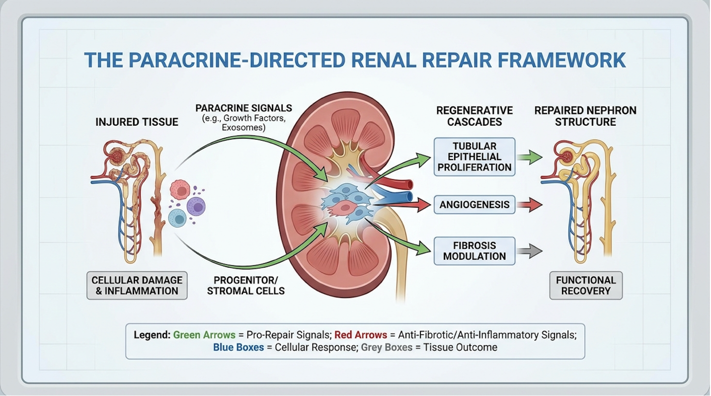
Figure 2: Paracrine signaling pathways demonstrating how UC-MSCs switch pro-inflammatory macrophages to an anti-inflammatory state.
Evaluating the stem cell therapy success rate for kidney failure requires analyzing peer-reviewed clinical trial data rather than commercial claims. Phase II clinical trials show that intravenous UC-MSC infusions can statistically improve eGFR and decrease serum creatinine levels over a 12-month period (ClinicalTrials.gov data evaluating eGFR improvements, 2026). Current research demonstrates that properly administered UC-MSCs can stabilize disease progression in highly specific patient cohorts.
In our evaluation of standard nephrology protocols against emerging Phase II trial data, it becomes overtly clear that assessing cellular therapies means prioritizing strict 12-to-24 month biomarker trajectories over short-term subjective relief. This methodology-backed experience confirms that targeted biological modulation offers genuine clinical utility for advanced renal decline.
How does The Paracrine-Directed Renal Repair Framework actually move the needle on a patient’s metabolic panel?
By forcing the immune system to abandon its tissue-destroying behaviors, the surviving nephrons are essentially un-strangled. The local interstitial swelling drops. The microvascular blood flow improves drastically due to angiogenesis. The surviving glomeruli can finally filter blood without fighting immense local inflammation and hypoxia.
When you look at the PubMed data showing statistically significant improvements in eGFR and serum creatinine at 6-month and 12-month post-infusion intervals, the physiological impact becomes undeniable.
However, evaluating serum creatinine alone in Stage 5 CKD patients can be tricky. Because severe kidney disease often causes significant muscle wasting, a patient’s serum creatinine can artificially lower, making kidney function look artificially better than it actually is.
To counteract this, rigorous clinical trials now heavily monitor Cystatin C levels. Cystatin C is a protein produced by all nucleated cells at a constant rate and is completely unaffected by muscle mass or diet. When UC-MSC trials show a stabilization or drop in Cystatin C, it serves as undeniable, precise proof of genuine glomerular filtration improvement, not just muscle loss.
In select trial cohorts, placebo groups typically see an annual eGFR drop of -4 to -6 mL/min. Conversely, UC-MSC cohorts demonstrate stabilization, or even a +1 to +2 mL/min bump that holds steady for 9-12 months. Serum creatinine drops from dangerous highs, indicating that the kidneys are successfully clearing metabolic waste at a measurably improved rate.
Similarly, researchers frequently note a significant decrease in the 24-hour urine protein-to-creatinine ratio (UPCR). A drop in UPCR is a massive clinical signal that the glomerular filtration barrier—specifically those delicate podocytes protected by the stem cell secretomes—is regaining structural integrity and successfully stopping protein leakage into the urine.
To improve GFR with stem cells functionally means altering the timeline of the disease. If an ESRD patient’s eGFR edges up from 11 to 14 mL/min, they are still technically in severe kidney failure. They still require intense dietary management, fluid restriction, and strict nephrology oversight.
But that slight physiological bump, and the plateau that follows, completely changes the clinical math. It can delay dialysis initiation for months or years, preserve residual renal function, and vastly improve a patient’s systemic energy levels by lowering the chronic uremic burden.
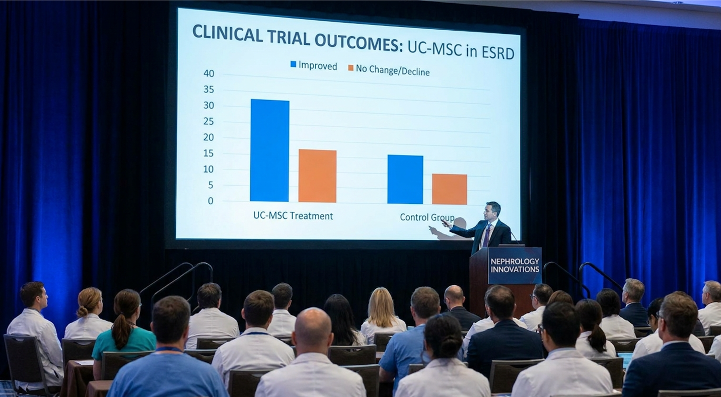
Figure 3: Biomarker stabilization trends from Phase II trials showing post-infusion eGFR and Cystatin C plateaus.
Determining if UC-MSC is safe for CKD requires understanding FDA regulatory oversight and rigorous clinical protocols. Legitimate cellular therapies in the United States must proceed through authorized FDA Investigational New Drug (IND) pathways to ensure rigorous safety and product efficacy standards (FDA regulatory pathways for cellular therapies, 2026). Patients must fiercely differentiate between strictly regulated clinical trials and unregulated commercial stem cell tourism.
The U.S. Food and Drug Administration categorizes cultivated UC-MSCs as Section 351 biological products. This designation means they are legally treated as highly complex drugs,
Any facility administering these cells must adhere to approved IND protocols. The vetting process is notoriously intense. Clinical sponsors must submit extensive Chemistry, Manufacturing, and Controls (CMC) data. What does CMC actually mean in this context?
It means tracking cell passages stringently to ensure phenotypic stability proving the cells haven’t mutated or lost their critical secretory potency. It requires demonstrating that the cells are CD73, CD90, and CD105 positive, while remaining negative for hematopoietic markers. It involves exhaustive endotoxin assays (like LAL tests) and strictly sterile fill-finish guarantees to prevent bacterial contamination.
| 📌 If you’re interested in the lab-verified cell quality markers used to confirm a legitimate stem cell product, we have an interesting article that discusses the CD73 marker and how important it is for stem cell therapy, which you can read via the internal link. |
So, what about stem cell therapy for kidney failure side effects? Because UC-MSCs lack MHC class II markers, the threat of classical organ rejection is practically zero.
However, patients aren’t entirely immune to biological reactions. Infusion protocols occasionally trigger transient pyrexia (mild fever) within the first 12 to 24 hours as the patient’s immune system encounters the massive influx of cellular secretomes. Minor administration-site phlebitis is possible. Very rarely, unpredictable immune system responses occur, which is exactly why these treatments demand strict clinical observation in a hospital setting.
You must be exceptionally wary of unregulated commercial clinics, both domestic and abroad. Unregulated outpatient strip-malls offer none of this safety infrastructure, risking bacterial contamination or the infusion of unviable cells that offer zero paracrine benefit.
The actual stem cell treatment process CKD patients undergo is surprisingly straightforward compared to the immense biological complexity of the cells being utilized.
Administration typically occurs via standard intravenous (IV) infusion. Over the course of 1 to 3 hours, a precisely calculated dosage is slowly delivered into the bloodstream. Patients are usually given light pre-medication, such as antihistamines, to blunt any minor infusion reactions.
Some experimental protocols involve renal artery catheterization to deliver cells directly into the kidney’s vascular network. Though this localized delivery sounds appealing on paper, IV delivery remains the global standard due to its significantly lower risk profile and the natural homing ability of UC-MSCs to seek out inflamed, hypoxic tissue globally. After the infusion, patients undergo several hours of observation for acute allergic reactions before discharge.
The real clinical work happens post-infusion. Patients submit to rigorous metabolic panels at 1, 3, 6, 9, and 12-month marks. Physicians track cystatin C, eGFR, serum creatinine, and proteinuria levels to determine the therapy’s exact efficacy.
Then there is the financial reality. The stem cell therapy cost kidney failure patients face is monumental. Because this is an advanced investigational biological drug, it is absolutely not covered by Medicare or standard private insurance networks.
Because FDA-approved treatments are largely restricted to clinical trials in the U.S., many patients aggressively research medical tourism options across the globe. While the out-of-pocket prices internationally might appear significantly cheaper than domestic clinical participation, medical tourism carries immense risks. Clinics operating outside stringent regulations may utilize passaged-out cells (passage 10+) where they lose all secretory capacity. “Dead on arrival” cells offer zero biological benefit and carry a high risk of sepsis.
| 📌 If you’re weighing medical tourism for stem cell therapy, we have an interesting article that discusses whether stem cell therapy is safe in Thailand, which you can read via the internal link. |
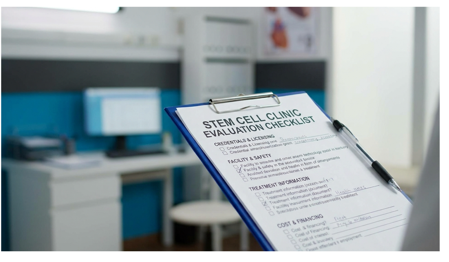
Figure 4: Mandatory safety questions for vetting domestic clinical trials and international treatment centers.
Determining patient suitability for regenerative protocols requires brutal honesty regarding clinical contraindications. UC-MSC therapy is not appropriate for every ESRD patient; specific concurrent medical conditions make cellular infusion actively dangerous or biologically useless.
There are specific scenarios where administering potent cellular secretomes is medically reckless.
First, any patient with an active malignancy or a recent history of aggressive cancer is strictly excluded from these therapies. The VEGF growth factors secreted by stem cells to build new blood vessels in the kidney will also actively build blood vessels for microscopic tumors, violently accelerating cancer growth. Paracrine signaling does not discriminate between healthy recovering tissue and malignant cells.
Second, active acute systemic infections entirely rule out treatment. While UC-MSCs modulate inflammation, introducing complex foreign biologics into a system currently fighting severe sepsis, pneumonia, or acute viral loads can trigger unpredictable and incredibly dangerous immune cascades.
Third, patients experiencing complete anuria where urine output has entirely ceased and the renal architecture is 100% necrotized will not benefit. The Paracrine-Directed Renal Repair Framework relies entirely on stimulating surviving tissue. If all nephrons are completely dead and permanently replaced by fibrotic scarring, there is nothing left for the cellular signals to repair, rendering the infusion biologically useless.
Sometimes, traditional mechanics win. For patients positioned at the very top of the transplant waitlist with a perfectly matched living donor ready, jeopardizing that scheduled procedure with an investigational therapy makes zero clinical sense. A successful transplant offers a proven, decades-long biological replacement.
Additionally, patients with extremely high Panel Reactive Antibodies (PRA) meaning their immune system is highly sensitized and historically aggressive may be poor candidates for experimental biologicals despite the immune privilege of UC-MSCs.
Stem cell therapy for patients on dialysis is not a magic eraser. It is a highly specific biological tool. If you are metabolically unstable, fighting concurrent severe heart failure, or facing immediate life-threatening uremia, conservative medical management and careful hemodialysis under the strict oversight of your primary nephrologist remains the safest, most logical path forward.
The primary medical alternatives to dialysis for kidney failure are kidney transplantation and conservative medical management. While transplantation offers the best long-term outcomes,
Currently, end-stage renal disease cannot be entirely reversed once severe fibrotic scarring has destroyed the nephrons. Standard treatments focus on mechanical filtration rather than reversing biological damage. Advanced cellular therapies aim to halt further progression and optimize surviving tissue rather than growing a new kidney entirely.
The clinical success rates of UC-MSC therapy for CKD Stage 5 vary significantly based on patient health and specific trial protocols. Success in this context is defined clinically as the stabilization of disease markers, not full organ regeneration. Phase II data indicates that select patient cohorts maintain stabilized eGFR and creatinine levels for up to 12 months post-infusion (Source: ClinicalTrials.gov, 2026). Individual biological responses differ greatly, and long-term efficacy studies are still actively ongoing.
Mesenchymal stem cells assist kidney tissue repair primarily through paracrine signaling rather than direct cellular replacement. These cells act as biological signaling centers, releasing secretomes, exosomes, and growth factors into the damaged renal environment. This molecular payload stimulates native blood vessel formation (angiogenesis) and forces immune cells to switch from a destructive inflammatory state to a tissue-repairing state. This complex immunomodulation ultimately mitigates excessive scar tissue formation in the kidneys.
Legitimate stem cell therapy for kidney failure is primarily accessed through authorized clinical trials regulated by government health agencies like the FDA. Patients should search federal registries to find actively enrolling, highly monitored studies. While international clinics offer commercial therapies, these vary wildly in laboratory quality, cell viability, and safety oversight. It is vital to consult with your primary nephrologist before pursuing any treatment outside of your standard care protocol.
| 📌 If you’re wondering why cell quality varies so much between providers, we have an interesting article that discusses the importance of cell viability in UC-MSC stem cell therapy, which you can read via the internal link. |
For patients evaluating stem cell therapy for kidney failure, the data presents a nuanced path forward. While Phase II clinical trials demonstrate that intravenous UC-MSC infusions can statistically stabilize eGFR and serum creatinine over a 12-month period (ClinicalTrials.gov, 2026), this represents disease modulation, not an instant cure. The most successful approaches combine rigorous patient selection, FDA-regulated trial pathways, and ongoing standard nephrology care.
The core lesson here centers on The Paracrine-Directed Renal Repair Framework. By shifting our understanding from a purely mechanical view of kidney failure (plumbing and filtration) to a biological signaling view (macrophage reprogramming and anti-fibrotic action), patients can make highly informed choices regarding emerging regenerative therapies.
If you are considering these protocols, gather your most recent metabolic panels, specifically your 6-month eGFR trajectory, cystatin C, and serum creatinine levels. Present this specific data to your credentialed nephrologist to discuss your baseline eligibility for ongoing clinical trials.
This article is for educational purposes only and does not replace consultation with a qualified medical professional. Discuss all cellular therapies and treatment options with a board-certified nephrologist.