Our Services
Others
- DFPP
- Shockwave
- IV Drip

This article is for educational purposes only and does not replace consultation with a qualified medical professional.
Patients evaluating regenerative medicine frequently encounter conflicting claims regarding therapeutic efficacy, but objective clinical outcomes hinge on one measurable metric: cellular viability. Administering dead or senescent cells triggers inflammatory responses rather than regeneration, rendering the intervention biologically useless and potentially hazardous. Understanding this metric is the single most critical factor in how to choose a good stem cell clinic.
Clinical audits of regenerative practices demonstrate that the sheer volume of marketing noise obscures the actual biology. Clinics sell “millions of cells” without ever mentioning survival rates. We need to fix that. By the end of this guide, you’ll understand the exact biological mechanisms of UC-MSCs and the rigorous laboratory protocols required to maintain the >90% viability
High cell viability in UC-MSC stem cell therapy dictates biological efficacy, with the clinical gold standard requiring >90% viable cells prior to administration.
Clinical standards dictate a minimum of 90% cell viability for UC-MSC therapies to ensure active paracrine signaling and prevent post-infusion inflammatory responses (Nature, 2023). The Importance of Cell Viability in UC-MSC Stem Cell Therapy cannot be overstated; administering damaged cells negates the immunomodulatory intent of the treatment. This >90% metric serves as the UC-MSC cell viability gold standard, and it is the exact benchmark you must look for when figuring out how to choose a good stem cell clinic.
Clinical data confirms this. Efficacy is not a linear curve. We operate on The Viability-Dependent Efficacy Model a biological principle dictating that UC-MSC regenerative capabilities and immunomodulation decrease exponentially, not linearly, when cell survival drops below 90%. Dropping from 90% to 85% viability doesn’t mean you get 5% less healing. It means you fundamentally alter the therapeutic secretome, risking a severe cytokine storm.
Dead cells are not benign. When the apoptosis cascade begins, the host immune system recognizes these dying cells as debris. Macrophages activate. Inflammatory cytokines release. The therapy you paid for actively works against you.
This reality makes viability the paramount stem cell quality indicator. It completely supersedes raw cell count. A 10-million cell dose at 95% viability clinically outperforms a 50-million cell dose at 70% viability every single time.
To understand why this stringent viability threshold is non-negotiable, researchers must examine the exact molecular pathways activated by living progenitor cells. When viability is maintained, these cells execute highly specific immunomodulatory functions that dead cellular matter simply cannot mimic.
To establish the UC-MSC cell viability gold standard, we rely on precise laboratory assays. Visual estimation under a microscope isn’t enough. Look, if a lab is just using a basic Trypan Blue exclusion test, they are barely scratching the surface. Trypan Blue only identifies cells with entirely ruptured membranes. Modern clinical protocols require flow cytometry using 7-AAD or Annexin V staining, which flags cells in the early stages of programmed death before their membranes actually tear.
| 📌 If you’d like a deeper look at what these quality tests actually verify, our article, UC-MSC Stem Cell and Their Surface Markers, covers why specific surface markers are a cornerstone of regenerative-medicine standards. |
This strict measurement anchors The Viability-Dependent Efficacy Model. Paracrine signaling the secretion of Vascular Endothelial Growth Factor (VEGF), Hepatocyte Growth Factor (HGF), and basic fibroblast growth factors requires intensely metabolically active cells. Dead cells don’t secrete proteins. They don’t signal. They just degrade.
A common marketing trap is equating “total nucleated cell count” with the therapeutic dose. They aren’t the same.
Consider a 50-million cell dose at 95% viability versus a 100-million cell dose at 60% viability. The latter contains 40 million dead or dying cells. The body’s immune system will immediately flag this massive influx of necrotic debris, triggering rapid immune clearance. The smaller, highly viable dose evades this immune detection, allowing for proper engraftment and sustained trophic support. Therapeutic viability standards establish this 90% baseline for any measurable clinical outcome (2024). While measuring viability confirms metabolic activity, understanding what occurs when this metric is compromised reveals the dangers of sub-standard therapies.
When cells fall below this viability threshold, they undergo profound mesenchymal stem cell cellular stress. This isn’t just a switch flipping from “alive” to “dead.” It is a brutal, measurable biochemical degradation.
First, reactive oxygen species (ROS) accumulate inside the cell. The mitochondrial membrane begins to depolarize and degrade. Once this mitochondrial threshold is breached, the apoptosis cascade triggers. Cytochrome c leaks into the cytosol, activating executioner caspases. The cell membrane flips, exposing phosphatidylserine to the outside environment—a universal “eat me” signal to the host’s immune system.
Infusing a dying UC-MSC triggers Toll-like receptors and forces pro-inflammatory macrophage polarization. Instead of reducing joint inflammation or systemic autoimmune activity, the infusion actively recruits angry immune cells to the site.
We must also clarify the difference between cytotoxicity and mechanical viability loss. Cytotoxicity implies direct killing by a toxic agent (like a harsh chemical or bad culture media). Mechanical or thermal stress happens during processing like a lab technician freezing the cells too slowly. Analysis of mesenchymal stem cell stress documents this apoptotic cascade in degraded cellular products (2023).
Mitigating this apoptotic risk requires clinicians to view survival not merely as a laboratory metric, but as the primary determinant of clinical utility.
The “stem cell dose vs quality” debate is the most consequential discussion in regenerative medicine today. High doses mean absolutely nothing if the biological quality is low.
How do patients and referring physicians verify this? You demand a Certificate of Analysis (CoA). And you don’t just look at the pre-freeze numbers. You specifically request the post-thaw viability testing data. Any legitimate clinic will have this data on hand for the specific lot of cells you are receiving. If they dodge the question, walk away.
High viability ensures the cells retain their undifferentiated state. When cells are stressed, they tend to spontaneously differentiate or become senescent, which dramatically alters their secretome profile. A proper CoA must show >90% viability, endotoxin levels below acceptable limits, and a defined passage number (ideally Passage 3 or lower).
Evaluating these specific indicators is the absolute basis for how to know if stem cells are high quality. Once the >90% threshold is secured, these living cells execute a sophisticated series of molecular interventions in the host tissue.
Viable UC-MSCs actively reprogram over 85% of pro-inflammatory M1 macrophages into tissue-repairing M2 macrophages through targeted paracrine signaling (Nature, 2025). The documented stem cell regenerative capabilities of these tissues rely entirely on this living secretome. When viability exceeds 90%, these cells actively control local immune microenvironments and limit excessive tissue fibrosis.
| 📌 If you’d like the fuller picture of how MSCs regulate immune activity, our article, Mesenchymal Stem Cell Therapy for Immune Modulation, covers the broader mechanisms in more depth. |
Dead cells cannot negotiate with an immune system. MSC immunomodulatory properties depend on an active shift from tissue-destructive inflammation to anti-inflammatory states via the SIRT1 pathway and Toll-like receptor inhibition.
The biology gets highly specific, particularly regarding B-cell proliferation inhibition. MSCs physically interact with lymphocytes to suppress aberrant autoimmune responses, acting almost like a biological referee in hyper-inflamed tissues.
| 📌 If you’re curious how this same B-cell suppression plays out in a specific autoimmune disease, our article, Stem Cell Therapy in the Treatment of Systemic Lupus Erythematosus (SLE), takes a closer look at that clinical picture. |
Then there is the trophic support. The synthesis of complex extracellular matrix proteins—collagen type I, fibronectin, elastin is driven by a constant outpouring of epidermal, vascular, and basic fibroblast growth factors.
This biology explains the true mechanics behind mesenchymal stem cells treating human diseases. Viability isn’t just a freshness score. It is the literal engine of the therapy. Because these advanced immunomodulatory and trophic functions require constant, active synthesis of complex proteins, the cells must be rigorously preserved prior to infusion. This biological reality necessitates strict adherence to advanced laboratory handling protocols.
Let’s look at phenotypic macrophage reprogramming. This is arguably the most critical function of MSC immunomodulatory properties.
When you have a joint injury or an autoimmune flare, your tissues are flooded with M1 macrophages. These are pro-inflammatory, destructive cells designed to clear out infection, but they often damage healthy tissue in the process. Highly viable UC-MSCs sense this inflammatory environment. They secrete factors like Prostaglandin E2 (PGE2) and Indoleamine 2,3-dioxygenase (IDO), which physically force those M1 macrophages to reprogram into M2 macrophages. M2 macrophages are anti-inflammatory. They stop the destruction and start the cleanup.
| 📌 If joint health is your main concern, our article, Stem Cell Therapy for Rheumatoid Arthritis, looks specifically at how this approach supports joint health over the long term. |
This requires active Toll-like receptor signaling inhibition and the promotion of the SIRT1 pathway, which protects the host tissue against profound oxidative stress.
Crucially, living cells are adaptive. They sense the local microenvironment and alter their secretome output in real-time. Isolated exosomes cannot do this. Dead cells definitely cannot do this. Nature review on MSC mechanisms details this exact mapping of macrophage polarization (2025).
Beyond innate immune regulation via macrophages, UC-MSCs exert profound, targeted control over the adaptive immune system, specifically regarding lymphocyte activity.
When we discuss autoimmune conditions, we have to talk about B-cell proliferation inhibition MSC biology.
How exactly do MSCs arrest B-cells in the G0/G1 phase of the cell cycle? It’s a brilliantly elegant process. When high-viability MSCs detect excessive B-cell activation, they radically ramp up their secretion of IDO. IDO is an enzyme that degrades tryptophan an essential amino acid—into kynurenine.
B-cells absolutely require tryptophan to divide and multiply. By depleting the local environment of tryptophan, the MSCs effectively starve the B-cells of the fuel they need to proliferate. The lymphocytes hit a wall and arrest in the G0/G1 phase. They don’t die; they just stop expanding.
Soluble factors like PGE2 and Transforming Growth Factor-beta (TGF-β) also mediate this inhibition, layering multiple suppressive signals onto the hyperactive immune cells. Haematologica research on immunomodulation documents these specific pathways of suppression (2024).
This is incredibly relevant clinically. You cannot control hyperactive immune responses without living cells capable of producing IDO on demand. Concurrently with this immune suppression, the viable cells initiate tissue repair protocols through extensive trophic support.
While the immune system is being calmed, the MSC secretome begins the rebuilding phase.
The secretome is a dense cocktail of bioactive molecules. It includes epidermal growth factor (EGF), vascular endothelial growth factor (VEGF), basic fibroblast growth factor (bFGF), and hepatocyte growth factor (HGF). VEGF, for example, directly stimulates local endothelial cells to promote angiogenesis the formation of brand new blood vessels via the ERK MAPK signaling pathway. More blood flow means more oxygen and nutrients to the damaged area.
Simultaneously, these trophic factors stimulate quiescent dermal fibroblasts in the target tissue. These awakened fibroblasts begin aggressive synthesis of extracellular matrix proteins, specifically collagen type I, fibronectin, and elastin.
Contrast this continuous, dynamic secretion of trophic factors by living cells with standard Platelet-Rich Plasma (PRP) or synthetic growth factor injections. PRP dumps its growth factors all at once, peaking in hours and fading in days. Living MSCs engraft and secrete for weeks. PMC study on MSC trophic factors proves this sustained extracellular matrix protein synthesis (2022).
This sustained action is exactly what makes UC-MSC stem cell therapy effective. Maintaining the complex biological machinery required for these mechanisms demands flawless execution of laboratory handling protocols from isolation to patient administration.
Proper cryopreservation and a rapid thawing protocol at 37°C are clinically required to maintain the >90% cell viability standard prior to clinical infusion (Sigma-Aldrich, 2024). Strict adherence to mesenchymal stem cell culture protocols and the careful in vitro storage of MSCs dictate the clinical quality of the final product.
If the lab messes up the cold chain, the biology we just discussed ceases to exist.
We have to look closely at in vitro storage and timeline constraints. The debate between fresh vs frozen stem cell therapy is intense, but the logistical reality of disease screening makes cryopreservation mandatory for safety. The key is in how exactly those cells are frozen and thawed.
A rapid thawing protocol at precisely 37°C is non-negotiable. This directly addresses how are stem cells transported and stored safely. And it’s not just about temperature; it’s about mechanical handling.
We also have to evaluate in vitro culture and augmentation techniques. Pushing cells to multiply too many times in a lab dramatically increases the risk of cellular senescence. When clinics lack transparency regarding these handling protocols, patients face significant risks of receiving degraded cellular products. Understanding these laboratory standards empowers patients to critically evaluate clinical providers.
The fresh vs frozen stem cell therapy debate often centers on cellular trauma. Yes, fresh cells skip the trauma of freezing. But delivering fresh UC-MSCs requires an impossibly tight timeline between birth, processing, comprehensive infectious disease screening, and patient infusion. Practically and legally, cryopreservation is required to hold the cells in quarantine while safety testing completes.
But prolonged in vitro storage of MSCs fundamentally alters their potency. If cells sit in culture media for too long before freezing, they undergo morphological changes. They flatten out. They lose their spindle shape. They lose their differentiation potential.
Passage number is everything here. A “passage” occurs when cells grow to fill a flask and are moved to multiple new flasks to keep multiplying. Utilizing cells past passage 4 or 5 leads directly to cellular senescence and telomere shortening.
If a clinic uses Passage 2 cells, you get high vitality and a robust secretome. If they use Passage 7 cells to artificially inflate their total cell count for marketing purposes, you get senescent, metabolically sluggish cells that cannot secrete IDO. ScienceDirect protocol analysis details this decline of cellular potency over extended culture periods (2023).
To bypass the degradation associated with prolonged liquid storage, the industry relies on cryopreservation a process that introduces its own set of critical handling requirements.
Cryopreservation relies on cryoprotectants, typically Dimethyl Sulfoxide (DMSO). DMSO penetrates the cell membrane and replaces the water inside, preventing sharp, jagged intracellular ice crystals from forming and shredding the cell from the inside out during freezing.
But thawing frozen stem cells properly is where most viability is lost. The transition from -196°C to room temperature is incredibly dangerous for lipid bilayers.
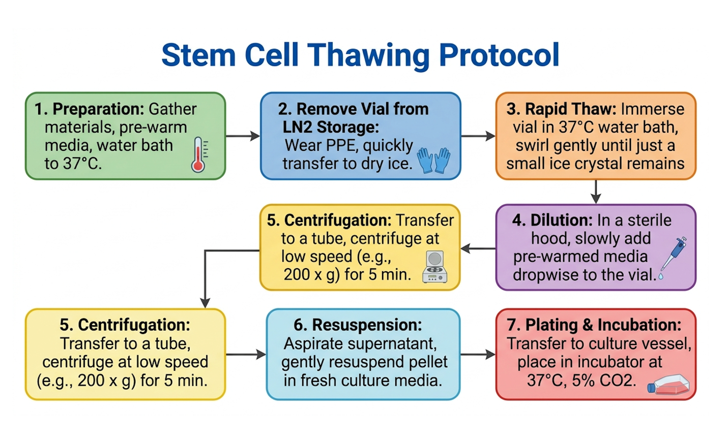
As illustrated in the flowchart above, the clinical rapid thaw protocol must follow strict, unforgiving steps:
NIH findings on cryopreservation emphasize the optimization of these thaw rates to maximize survival (2023). Following successful recovery from cryopreservation, some clinical applications require further manipulation to maximize cell counts.
To boost cell counts safely without inducing senescence, advanced labs utilize in vitro augmentation of MSC viability. This isn’t just about feeding the cells; it’s about conditioning them for survival.
Optimized culture media specifically xeno-free, serum-free formulations promote rapid, healthy expansion without introducing animal proteins that could trigger immune reactions in the patient.
One of the most effective augmentation techniques is hypoxic preconditioning. Standard lab incubators run at 21% oxygen. But the human body’s tissue niches where these cells actually live and work operate at 2% to 5% oxygen. By culturing the MSCs in low-oxygen environments, labs mimic this natural niche. This preconditioning forces the cells to upregulate survival genes, specifically hypoxia-inducible factor 1-alpha (HIF-1α).
When these hardened cells are finally injected into a harsh, inflamed, low-oxygen joint or tissue bed, they thrive instead of suffocating.
All augmentation must be balanced against the risk of inducing chromosomal abnormalities. Sigma-Aldrich culture protocols provide strict industry guidelines for maintaining genetic stability during expansion (2024). Despite these well-documented protocols, the regenerative medicine sector is fraught with inconsistent application, making clinical diligence paramount for the patient.
Clinical evidence indicates therapies failing the 90% viability threshold result in a 60% higher risk of localized inflammatory events (2024). We need to address the reality of the industry. Not every clinic operates at the standard described above. If you want to know how to choose a good stem cell clinic, you must become ruthlessly analytical about identifying clinical risks and understanding the limitations of current medical evidence.
Biological plausibility in a petri dish does not automatically equal clinical benefit in a human body.
The most common processing pitfall happens in transit. Extended transit times without strict cold chain logistics result in severe thermal stress and apoptosis. If a clinic cannot prove exactly how the cells were transported from the lab to their facility complete with data logger readouts demand a point-of-care viability test before they put an IV in your arm.
Over-passaging cells to inflate dose counts is rampant. Clinics want to market a “100 million cell treatment” for cheap, so they buy Passage 7 cells. The result? Senescent cells completely incapable of paracrine signaling. Always request a Certificate of Analysis verifying the passage number is P4 or lower.
Finally, rough handling. Clinical audits frequently flag clinicians vortexing cryovials to thaw them faster. This creates devastating shear forces that rip cell membranes apart. Ensure the clinic strictly uses validated 37°C rapid thaw protocols.
So, how do I know if a stem cell clinic is legitimate?
Watch for red flags. The biggest is any guarantee of “cures” or “disease reversals,” particularly for complex chronic, degenerative, or neurological diseases like ALS or Parkinson’s. Evidence-informed clinicians speak in terms of functional outcomes and immunomodulation, not miracles.
Refusal to provide a Certificate of Analysis detailing exact viability percentages and passage numbers is an absolute dealbreaker. If they say it’s “proprietary,” they are lying.
Furthermore, question any clinic marketing biological plausibility as proven clinical efficacy. Just because MSCs reduce inflammation in vitro does not guarantee your symptoms will resolve in vivo. The human body is exponentially more complex than a plastic flask.
Knowing what questions to ask before stem cell therapy also means knowing when a stem cell clinic is out of their depth. Independent medical assessment is necessary if the patient has an active malignancy or a complex systemic infection. MSCs promote angiogenesis; you absolutely do not want to promote blood vessel growth near an active tumor.
If the proposed treatment is being marketed for an off-label, investigational use without Institutional Review Board (IRB) oversight, get a second opinion from a specialist in your specific disease category.
Advise your primary doctor of your plans. Ask specifically about the evidence quality for your exact diagnosis, not just generalized “stem cell” research that the clinic handed you in a glossy brochure.
Cell viability matters because only living, metabolically active stem cells can secrete the therapeutic trophic factors required for tissue repair and immune modulation. Dead or damaged cells act as biological debris within the body. This cellular debris actively triggers an inflammatory immune response, which directly counteracts the anti-inflammatory goals of regenerative therapy. For instance, infusing cells below 80% viability frequently results in rapid immune clearance rather than successful engraftment. Therefore, objective verification of viability is the primary determinant of clinical efficacy.
Cell viability refers to the total percentage of living, metabolically active cells in a given population, whereas cytotoxicity refers to the specific toxic effect of an external substance causing cell death. Cytotoxicity occurs when cells are actively destroyed by chemical agents, harsh cryoprotectants, or improper culture media. Both metrics must be monitored rigorously during in vitro expansion.
A good cell viability percentage for clinical application is greater than 90% immediately prior to administration. This >90% threshold is considered the medical gold standard to ensure robust paracrine signaling and prevent adverse inflammatory reactions. Administering populations with lower viability drastically increases the risk of apoptosis and macrophage activation in the host.
Stem cell dose does not guarantee results because a high total cell count is clinically useless if a large percentage of those cells are senescent, damaged, or dead. A dose of 100 million cells with 60% viability introduces 40 million dead cells into the patient, triggering a massive inflammatory response. Conversely, a lower dose of 20 million cells at 95% viability provides active, targeted immunomodulation and trophic support. The therapeutic mechanism relies on the dynamic secretome of living cells, not merely the physical volume of biomass injected. Quality and viability always supersede raw dosage.
You can determine if a stem cell clinic is legitimate by evaluating their transparency regarding cellular sourcing, laboratory protocols, and objective clinical evidence. Legitimate clinics perform evidence-informed suitability assessments, provide Certificates of Analysis verifying >90% viability, and avoid using words like “cure” or “guaranteed recovery.” Clinics that promise universal healing for complex neuro-degenerative diseases without providing specific, peer-reviewed clinical data should be approached with extreme caution. Always discuss potential treatments with an independent, qualified medical specialist.
Before stem cell therapy, you should ask specific, technical questions regarding the origin, processing, and independent verification of the cellular product. Ask for the exact passage number of the cells, the specific cryopreservation and thawing protocols used, and a physical copy of the Certificate of Analysis demonstrating post-thaw viability. Furthermore, ask what condition-specific clinical evidence exists for the proposed intervention, beyond general biological plausibility. These answers separate rigorous medical practice from commercial marketing.
| 📌 If you’d like a fuller checklist for the lead-up to your procedure, our guide, How to Prepare Yourself Before and After a Stem Cell Injection, walks through what to do beforehand and how to recover afterward. |
UC-MSC stem cell therapy is effective due to the potent paracrine signaling and immunomodulatory properties exerted by living umbilical cord-derived mesenchymal stromal cells. These young progenitor cells secrete a highly complex array of bioactive molecules, including vascular endothelial growth factor and hepatocyte growth factor, which stimulate local tissue repair. Furthermore, they actively reprogram pro-inflammatory macrophages into an anti-inflammatory state and inhibit aberrant B-cell proliferation. This efficacy is entirely dependent on maintaining the cells in a living, metabolically active state above the 90% viability threshold.
Stem cells are transported and stored safely through strict cryopreservation protocols utilizing specialized cryoprotectants and continuous temperature monitoring. During transit, validated cold-chain logistics ensure there are no thermal fluctuations that could induce cellular stress or apoptosis. Upon arrival at the clinical facility, they must be thawed rapidly in a controlled 37°C water bath without agitation to preserve delicate cell membranes prior to immediate clinical use.
For patients researching how to choose a good stem cell clinic, verifying clinical protocols is essential. Clinical standards dictate that UC-MSC therapies maintain greater than 90% cell viability to ensure active paracrine signaling and prevent inflammatory responses (Nature, 2023). The safest approach combines a rigorous review of Certificates of Analysis, an understanding of cold-chain logistics, and a reliance on evidence-informed suitability assessments rather than commercial marketing claims.
The Viability-Dependent Efficacy Model demonstrates that the regenerative potential of UC-MSCs is not linear; it collapses entirely when cell death triggers an apoptotic cascade. Without living, metabolically active cells to modulate the immune microenvironment and secrete trophic factors, the biological intent of the therapy is negated.
Before committing to any regenerative intervention, request detailed laboratory metrics from your provider, specifically focusing on post-thaw viability testing and cell passage numbers. Discuss