Our Services
Others
- DFPP
- Shockwave
- IV Drip


“This extensive literature review delves into the molecular basis of Alzheimer’s disease, and examines the role of aberrant Amyloid Precursor Protein processing and γ-secretase mechanism in the initiation of the neurodegenerative cascade.”
Alzheimer’s disease is the leading cause of dementia, and is pathologically characterized by extracellular amyloid plaques and intracellular neurofibrillary tangles. For three decades, the Amyloid Cascade Hypothesis has been the prevailing framework for disease pathogenesis, proposing that amyloid-β peptides generated by the proteolysis of Amyloid Precursor Protein trigger a toxic cascade resulting in synaptic failure, tau pathology and cell death. Amyloid Precursor Protein is cleaved through non-amyloidogenic and amyloidogenic pathways. In the amyloidogenic pathway, amyloid-β is formed by sequential cleavage by β-secretase and γ-secretase, mainly the hydrophobic 40 and 42 amino acid isoforms. γ-secretase, comprising presenilin as a catalytic core in association with nicastrin, APH-1 and PEN-2, is responsible for processive intramembrane proteolysis. Mutations in amyloid precursor protein and presenilins affect this cleavage, shifting peptide production toward longer, aggregable forms. Soluble amyloid-β oligomers exert potent synaptotoxic effects mediated by their interaction with postsynaptic receptors, induction of calcium excitotoxicity, inhibition of long-term potentiation and activation of glycogen synthase kinase 3β signaling leading to tau hyperphosphorylation. The genetic evidence for the role of amyloid clearance in AD is strong, but clinical trials have only shown modest cognitive benefits with significant adverse events. This review integrates literature on Amyloid Precursor Protein proteolysis, γ-secretase dynamics, oligomeric synaptotoxicity, and emerging paradigms like presenilin loss-of-function and intraneuronal amyloid models.
Keywords: Alzheimer’s Disease, Amyloid Precursor Protein, γ-Secretase, Amyloid-β, Amyloid Cascade Hypothesis, Neurodegeneration
Alzheimer’s disease is a neurodegenerative disease that accounts for 60-80 % of dementia worldwide. It is marked by memory decline, executive dysfunction and loss of independence and places severe burdens. Histopathologically, the hallmarks of the Alzheimer’s brain include extracellular senile plaques (aggregates of amyloid-β peptides) and intracellular neurofibrillary tangles (hyperphosphorylated tau protein). Amyloid plaque deposition begins in neocortical areas and then spreads, whereas tau pathology begins in limbic structures and then spreads to the cortex(Kurkinen et al., 2023; Lovestone, 2000; Pimplikar, 2009).
Hardy and Higgins introduced the Amyloid Cascade Hypothesis in 1992, which has been the basis for Alzheimer’s research. This hypothesis postulates that amyloid-β accumulation in brain parenchyma is the main causative event. Downstream features include neuroinflammation, tau hyperphosphorylation, tangle assembly, synaptic loss and cell death as direct secondary consequences(J. A. Hardy & Higgins, 1992). This model was based on two findings: the first was that the main component of senile plaques was amyloid-β and the second was the discovery of mutations in the Amyloid Precursor Protein gene that lead to early-onset familial Alzheimer’s disease(Lovestone, 2000).
Amyloid precursor protein is a type one transmembrane glycoprotein with a single pass. The full-length protein and soluble cleavage products are involved in cell adhesion, neurite outgrowth, synaptogenesis and neuroprotection. The precursor protein, after post-translational modification, is transported to plasma membrane and endosomal compartments where secretases determine the processing pathway, either non-amyloidogenic or amyloidogenic. This hypothesis has unified basic science for decades(Morelli, Perry, & Tagliavini, 2012). However, recent outcomes of phase 3 trials have highlighted translational challenges(Pimplikar, 2009). This literature review summarizes evidence on amyloid precursor protein proteolysis, γ-secretase cleavage mechanics, oligomeric synaptotoxicity and emerging paradigms in Alzheimer’s research.
Amyloid Precursor Protein is enzymatically processed by two mutually exclusive pathways: non-amyloidogenic and amyloidogenic processing. Non-amyloidogenic cleavage of precursor protein, starting with α-secretase within the amyloid-β domain, accounts for most of the processing(Ricciarelli & Fedele, 2017). The activity of the α-secretase is carried out by ADAM metalloproteinases, of which ADAM10 is the main enzyme in mature neurons. α-secretase cleaves between lysine 16 and leucine 17 of the amyloid-β sequence. This cleavage disrupts the peptide sequence and prevents the formation of intact amyloid-β. This releases a soluble ectodomain (sAPP-α) into extracellular fluid, and leaves behind a membrane-bound fragment (C83) of 83 amino acids. The sAPP-α fragment is neuroprotective and promotes synaptic plasticity, learning, memory, and cell survival. γ-secretase then cleaves C83 to produce a non-toxic 3 kilodalton fragment, p3, and release of the Amyloid Precursor Protein Intracellular Domain into the cytosol(Gabuzda, Busciglio, & Yankner, 1993; LeBlanc, Koutroumanis, & Goodyer, 1998).
Conversely, the amyloidogenic pathway results in the formation of full-length amyloid-β peptides through sequential cleavage by β-secretase and γ-secretase. β-site APP cleaving enzyme one (BACE1) is an aspartyl protease that is a transmembrane protein localized in acidic endosomes and trans-Golgi networks. BACE1 cuts Amyloid Precursor Protein at Aspartate one to release sAPP-β and leave a 99 amino acid fragment, C99, in the membrane(Morelli et al., 2012). C99 contains the entire-length amyloid-β domain at its N-terminus. BACE1 can also cut at a secondary site 10 residues downstream, the β-prime site, producing a shorter C89 fragment(J. Hardy & Selkoe, 2002).
α-secretase ADAM10 and β-secretase BACE1 compete to determine whether Amyloid Precursor Protein enters the non-amyloidogenic pathway producing sAPP-α or the amyloidogenic pathway producing C99 prior to cleavage by γ-secretase (Figure 1).
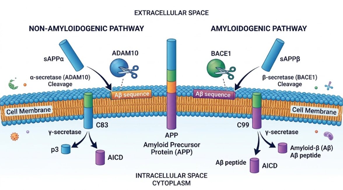
Figure 1. Processing pathways of Amyloid Precursor Protein (APP). Schematic diagram of non-amyloidogenic (left) and amyloidogenic (right) APP processing pathways at the neuronal cell membrane.
After cleavage by BACE1, C99 is then cleaved intramembranously by the γ-secretase complex. γ-secretase is a protease complex that cleaves within the membrane to hydrolyze peptide bonds within the hydrophobic lipid bilayer. The complex is comprised of 4 core subunits in a one-to-one stoichiometry: presenilin (presenilin one or presenilin two), nicastrin, anterior pharynx-defective one (APH-1) and presenilin enhancer two (PEN-2). Nicastrin has an extracellular glycosylated domain that acts as a substrate receptor for N-terminus of membrane stubs. APH-1 serves as a structural scaffold, and PEN-2 induces a conformational change in presenilin that leads to autoproteolytic cleavage into active N-terminal and C-terminal fragments(Järemo et al., 2019; Kurkinen et al., 2023; Liu, Ding, Rovere, Wolfe, & Selkoe, 2019).
Presenilin is the catalytic core of the γ-secretase complex. It is a nine-pass transmembrane protein with two conserved catalytic aspartate residues in transmembrane domains six and seven. These aspartate residues coordinate a catalytic water molecule in an internal cavity buried inside the lipid bilayer. γ-secretase binds C99 and begins cleavage at the epsilon site close to the cytoplasmic boundary. The enzyme makes carboxypeptidase-like cuts, every 3 to 4 amino acids along the peptide backbone in a processive, stepwise fashion toward the extracellular surface(Pimplikar, 2009; Volloch & Rits-Volloch, 2023).
This processive cleavage produces amyloid-β peptides of 37 to 43 amino acids. In physiological conditions, cleavage of C99 mainly occurs at position 40, yielding Aβ40, which accounts for 90% of secreted amyloid-β. 10% of cleavages stop at position 42, releasing hydrophobic Aβ42. Minor peptide species like Aβ37, Aβ38, Aβ39 and Aβ43 are also produced(Kurkinen et al., 2023; Ricciarelli & Fedele, 2017).
Aβ42 has hydrophobic residues at the C-terminus giving it different biophysical properties than Aβ40. Aβ42 has a marked propensity for self-assembly into β-sheet oligomers and fibrillar aggregates. Pathogenic mutations in PSEN1 and PSEN2 disrupt substrate-enzyme interactions in processive cleavage resulting in early substrate release and increased Aβ42 to Aβ40 ratio. Meanwhile, γ-secretase cleavage releases the Amyloid Precursor Protein Intracellular Domain. This domain translocates to the nucleus to modulate transcription(Zhang, Li, Xu, & Zhang, 2014).
Figure 2 shows that the γ-secretase complex uses the catalytic aspartate residues of presenilins within the lipid bilayer to perform the processive intramembrane proteolysis of C99, releasing Aβ40 and hydrophobic Aβ42 peptides.
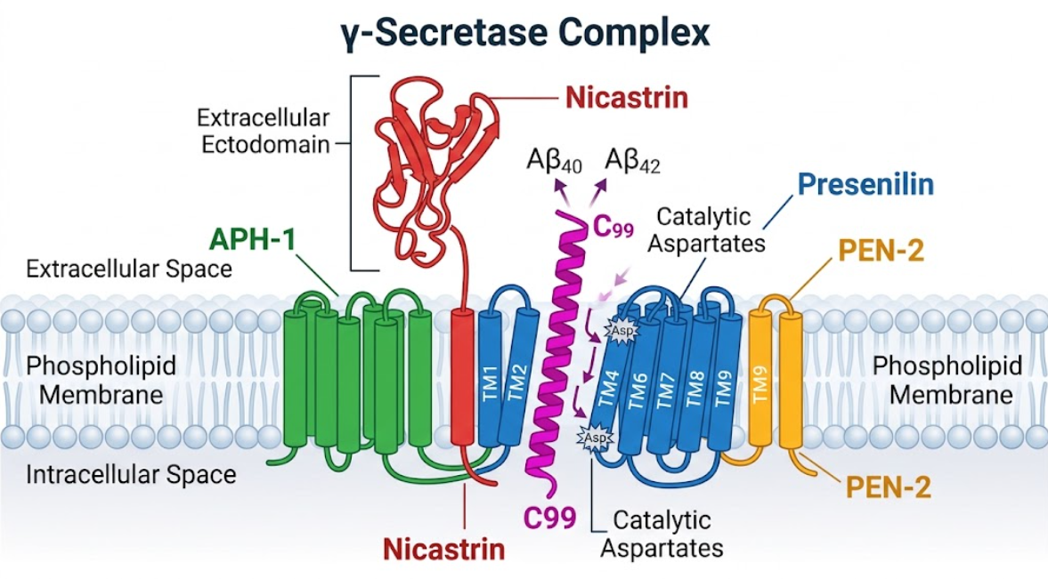
Figure 2. Organization of the γ-secretase complex structure. 3D model of the γ-secretase complex, with the 4 core subunits embedded in the membrane. C99 is cleaved processively at the presenilin active site to yield amyloid-β (Aβ40 and Aβ42) peptides.
Monomeric amyloid-β peptides self-assemble in a nucleation-dependent manner after their proteolytic release. During the nucleation phase monomers slowly aggregate into unstable oligomeric seeds. Once stable nuclei are formed, rapid elongation occurs by addition of monomers giving rise to soluble dimers, trimers, tetramers, hexamers, dodecamers and globulomers. These assemblies eventually assemble into protofibrils and cross-β sheet fibrils that deposit as parenchymal senile plaques(Lovestone, 2000; Pimplikar, 2009).
Insoluble senile plaques were initially identified in early research as the main neurotoxic entity, but clinicopathological studies showed that the volume of plaques did not correlate with the clinical severity of dementia. Cognitively normal subjects often exhibit high amyloid plaque loads and cognitive decline is closely associated with loss of synaptic density and concentrations of soluble amyloid-β oligomers. Biophysical evidence suggests that soluble oligomers are far more synaptotoxic than either monomers or insoluble fibrils, diffusing in interstitial fluid to establish a toxic penumbra around the plaque cores(Castellani, Plascencia-Villa, & Perry, 2019; Ricciarelli & Fedele, 2017).
Synaptotoxicity is evoked by soluble amyloid-β oligomers interacting with postsynaptic receptors such as α-7 nicotinic acetylcholine receptors, NMDA receptor subunits, cellular prion protein and RAGE(Volloch & Rits-Volloch, 2023). NMDA and AMPA receptor endocytosis is induced by receptor binding, which results in decreased baseline synaptic transmission and suppression of hippocampal Long-Term Potentiation. At the same time, oligomers inhibit the reuptake of glutamate by down-regulating the astrocytic glutamate transporter EAAT2, which results in excessive accumulation of synaptic glutamate and pathological intracellular calcium influx(Kurkinen et al., 2023; Ricciarelli & Fedele, 2017).
Elevated intracellular calcium activates downstream kinases, especially glycogen synthase kinase 3β (GSK-3β) and cyclin dependent kinase five. Hyperactivated GSK-3β phosphorylates tau protein at multiple residues. This causes tau to detach from axonal microtubules, resulting in microtubule depolymerization and tau aggregation into paired helical filaments and neurofibrillary tangles. Oligomers also trigger mitochondrial dysfunction, peroxidation of membrane lipids, microglial activation, cytokine release, and caspase-dependent neuronal apoptosis(Castellani, Jamshidi, Plascencia-Villa, & Perry, 2025; Josephs et al., 2008).
Soluble amyloid-β oligomers activate postsynaptic receptors to promote excitotoxic calcium influx, hyperactivation of GSK-3β, hyperphosphorylation of tau, formation of neurofibrillary tangles and loss of functional synapses (Figure 3).
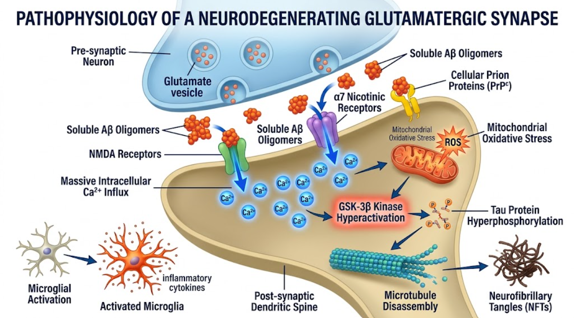
Figure 3. Pathophysiology of a Degenerating Glutamatergic Synapse. Aβ oligomers induce postsynaptic spine neurotoxicity with massive Ca2+ influx, mitochondrial stress, tau hyperphosphorylation, and microglial activation.
Biochemical models link the handling of Amyloid Precursor Protein to neurodegeneration, but results from phase 3 clinical trials targeting amyloid clearance have prompted a reassessment of the Amyloid Cascade Hypothesis. Active vaccines, passive monoclonal antibodies (bapineuzumab, solanezumab, gantenerumab, aducanumab, lecanemab, donanemab), and secretase inhibitors have been approaches for over 20 years(Cummings et al., 2021). Early γ-secretase inhibitors such as semagacestat reduced amyloid β levels, but were abandoned due to cognitive decline and skin malignancies from off-target Notch-one inhibition(Morelli et al., 2012).
Small-molecule BACE1 inhibitors (verubecestat, atabecestat, lanabecestat) could engage target and reduce cerebrospinal fluid amyloid-β by 80%. But the trials were halted because of cognitive deficits, neuropsychiatric symptoms and weight loss. Proteomic investigations have shown that BACE1 cleaves more than forty physiological substrates apart from Amyloid Precursor Protein. These include Neuregulin-one and neural cell adhesion molecules vital for myelination and synaptic plasticity(Barão, Moechars, Lichtenthaler, & De Strooper, 2016; Volloch & Rits-Volloch, 2023).
More recently, anti-amyloid monoclonal antibodies have been developed against aggregated species that can clear parenchymal amyloid plaques on positron emission tomography scans, including aducanumab, lecanemab, and donanemab. In phase 3 data from EMERGE, Clarity AD and TRAILBLAZER-ALZ, removing plaques slowed cognitive decline by 16 to 30 percent over 18 months in early symptomatic patients(Kurkinen et al., 2023; Levin et al., 2022). However, these agents do not reverse the pre-existing cognitive deficits and do not completely arrest the disease progression. Antibody therapies also have the risk of amyloid-related imaging abnormalities with edema (ARIA-E) and microhemorrhages (ARIA-H), especially in apolipoprotein E epsilon-four carriers(Castellani et al., 2025; Castellani et al., 2019).
These outcomes have prompted refined paradigms. The Presenilin Hypothesis proposes that presenilin mutations cause familial Alzheimer’s disease primarily by loss of important γ-secretase functions rather than by amyloid-β overproduction alone. Loss of normal presenilin activity impairs cell survival signaling, degrades EAAT2 regulation and disrupts synaptic function independent of plaque accumulation(Kurkinen et al., 2023). At the same time, Amyloid Cascade Hypothesis 2.0 posits Alzheimer’s disease as a two-stage disease driven by the accumulation of intraneuronal amyloid-β. In the first stage, the processes of reuptake and retention build up intraneuronal amyloid-β for decades. When a threshold is crossed, intraneuronal amyloid-β elicits a concerted stress response that involves Amyloid Precursor Protein-independent internal generation of intraneuronal amyloid-β, leading to a self-sustained second stage of tau pathology and neuronal death(Volloch & Rits-Volloch, 2023).
The research synthesis indicates that although amyloid precursor protein cleavage and amyloid-β production are critical to early disease onset, the notion that amyloid-β is the sole linear driver of late-stage clinical progression is insufficient(Pimplikar, 2009). The genetic evidence for amyloid primacy remains strong: mutations in APP, PS1 and PS2 cause early-onset disease; people with Down syndrome have 3 copies of the APP gene and all develop Alzheimer’s neuropathology by middle age; and the protective Icelandic APP mutation (A673T) reduces BACE1 cleavage efficiency, protecting carriers against both Alzheimer’s disease and age-related cognitive decline(Armstrong, 2011; Kurkinen et al., 2023; Lovestone, 2000).
However, poor correlation between insoluble plaque burden and cognitive impairment, and modest clinical impact of plaque removal in symptomatic individuals, argue that late-stage neurodegeneration becomes uncoupled from extracellular amyloid burden. Downstream cascades, including tau hyperphosphorylation, microglial dystrophy, astrocytic dysfunction, neuroinflammation, and synaptic loss, are activated once amyloid-β is outside of the cell, making the neurodegenerative process self-sustaining. Therefore, clearance of extracellular plaques in patients with significant neurofibrillary tangle load and extensive synaptic loss produces little functional improvement(Castellani et al., 2025; Pimplikar, 2009).
Furthermore, evidence suggests that monomeric amyloid-β peptides have non-pathological physiological functions. At low nanomolar concentrations, amyloid-β monomer in healthy tissue enhances presynaptic neurotransmitter release, facilitates hippocampal Long-Term Potentiation and regulates excitatory homeostasis. In innate immune defenses, peptides also function as broad-spectrum antimicrobial agents. Nonspecifically blocking the synthesis of amyloid-β or generally depleting monomeric species disrupts normal synaptic transmission and weakens immune function(Kurkinen et al., 2023; Ricciarelli & Fedele, 2017; Volloch & Rits-Volloch, 2023).
Thus, therapeutic approaches are evolving toward targeted strategies. First, anti-amyloid interventions should be initiated at preclinical or asymptomatic stages, well before widespread tau spread and structural neurodegeneration. Second, small-molecule γ-Secretase Modulators offer a promising approach that shifts γ-secretase cleavage sites to shorter non-aggregating peptides (e.g., Aβ37 and Aβ38) without inhibiting total γ-secretase activity or perturbing Notch processing. Third, a comprehensive, multi-target approach to halt disease progression is the combination of early amyloid modulation with anti-tau agents, anti-inflammatory drugs and neuroprotective compounds(Levin et al., 2022; Morelli et al., 2012).
To summarize, the proteolytic processing of Amyloid Precursor Protein by the γ-secretase complex represents a key molecular axis in the pathogenesis of Alzheimer’s disease. BACE1 cleavage of Amyloid Precursor Protein yields the membrane-bound C99 fragment which is cleaved intramembrane by presenilin within the γ-secretase complex to release monomeric amyloid-β peptides. Hydrophobic Aβ42 is strongly biophysically driven to aggregate into soluble oligomers that are potent synaptotoxins that disrupt Long-Term Potentiation, induce excitotoxic calcium influx, downregulate glutamate transporter EAAT2, and stimulate GSK-3β signaling that drives tau hyperphosphorylation, neurofibrillary tangle assembly, and apoptotic cell death. The initial genetics gave a framework for the Amyloid Cascade Hypothesis; however clinical trials show that removing parenchymal plaques in symptomatic patients leads to limited cognitive recovery. Present data suggest that amyloid accumulation is an early trigger that initiates autonomous downstream tau pathology and neuroinflammation. Successful therapeutic success in the future will require the use of disease-modifying therapies during the preclinical stages, the development of selective γ-secretase modulators that do not interfere with the cleavage of physiological substrates, and the use of multi-target therapeutic regimens that target both amyloid proteolysis and downstream neurodegenerative mechanisms.
Armstrong, R. A. (2011). The pathogenesis of Alzheimer’s disease: a reevaluation of the “amyloid cascade hypothesis”. Int J Alzheimers Dis, 2011, 630865. doi:10.4061/2011/630865
Barão, S., Moechars, D., Lichtenthaler, S. F., & De Strooper, B. (2016). BACE1 Physiological Functions May Limit Its Use as Therapeutic Target for Alzheimer’s Disease. Trends Neurosci, 39(3), 158-169. doi:10.1016/j.tins.2016.01.003
Castellani, R. J., Jamshidi, P., Plascencia-Villa, G., & Perry, G. (2025). The Amyloid Cascade Hypothesis: A Conclusion in Search of Support. The American Journal of Pathology, 195(11), 1988-1997. doi:10.1016/j.ajpath.2024.10.014
Castellani, R. J., Plascencia-Villa, G., & Perry, G. (2019). The amyloid cascade and Alzheimer’s disease therapeutics: theory versus observation. Laboratory Investigation, 99(7), 958-970. doi:https://doi.org/10.1038/s41374-019-0231-z
Cummings, J., Aisen, P., Lemere, C., Atri, A., Sabbagh, M., & Salloway, S. (2021). Aducanumab produced a clinically meaningful benefit in association with amyloid lowering. Alzheimers Res Ther, 13(1), 98. doi:10.1186/s13195-021-00838-z
Gabuzda, D., Busciglio, J., & Yankner, B. A. (1993). Inhibition of beta-amyloid production by activation of protein kinase C. J Neurochem, 61(6), 2326-2329. doi:10.1111/j.1471-4159.1993.tb07479.x
Hardy, J., & Selkoe, D. J. (2002). The amyloid hypothesis of Alzheimer’s disease: progress and problems on the road to therapeutics. Science, 297(5580), 353-356. doi:10.1126/science.1072994
Hardy, J. A., & Higgins, G. A. (1992). Alzheimer’s disease: the amyloid cascade hypothesis. Science, 256(5054), 184-185. doi:10.1126/science.1566067
Järemo, P., Jejcic, A., Jelic, V., Shahnaz, T., Oweling, M., Winblad, B., & Behbahani, H. (2019). Erythrocyte Amyloid Beta Peptide Isoform Distributions in Alzheimer and Mild Cognitive Impairment. Curr Alzheimer Res, 16(11), 1050-1054. doi:10.2174/1567205016666191010104355
Josephs, K. A., Whitwell, J. L., Ahmed, Z., Shiung, M. M., Weigand, S. D., Knopman, D. S., . . . Jack, C. R., Jr. (2008). Beta-amyloid burden is not associated with rates of brain atrophy. Ann Neurol, 63(2), 204-212. doi:10.1002/ana.21223
Kurkinen, M., Fułek, M., Fułek, K., Beszłej, J. A., Kurpas, D., & Leszek, J. (2023). The Amyloid Cascade Hypothesis in Alzheimer’s Disease: Should We Change Our Thinking? Biomolecules, 13(3). doi:10.3390/biom13030453
LeBlanc, A. C., Koutroumanis, M., & Goodyer, C. G. (1998). Protein kinase C activation increases release of secreted amyloid precursor protein without decreasing Abeta production in human primary neuron cultures. J Neurosci, 18(8), 2907-2913. doi:10.1523/jneurosci.18-08-02907.1998
Levin, J., Vöglein, J., Quiroz, Y. T., Bateman, R. J., Ghisays, V., Lopera, F., . . . Morris, J. C. (2022). Testing the amyloid cascade hypothesis: Prevention trials in autosomal dominant Alzheimer disease. Alzheimers Dement, 18(12), 2687-2698. doi:10.1002/alz.12624
Liu, L., Ding, L., Rovere, M., Wolfe, M. S., & Selkoe, D. J. (2019). A cellular complex of BACE1 and γ-secretase sequentially generates Aβ from its full-length precursor. J Cell Biol, 218(2), 644-663. doi:10.1083/jcb.201806205
Lovestone, S. (2000). Fleshing out the amyloid cascade hypothesis: the molecular biology of Alzheimer’s disease. Dialogues Clin Neurosci, 2(2), 101-110. doi:10.31887/DCNS.2000.2.2/slovestone
Morelli, L., Perry, G., & Tagliavini, F. (2012). The contribution of the amyloid hypothesis to the understanding of Alzheimer’s disease: a critical overview. Int J Alzheimers Dis, 2012, 709613. doi:10.1155/2012/709613
Pimplikar, S. W. (2009). Reassessing the amyloid cascade hypothesis of Alzheimer’s disease. Int J Biochem Cell Biol, 41(6), 1261-1268. doi:10.1016/j.biocel.2008.12.015
Ricciarelli, R., & Fedele, E. (2017). The Amyloid Cascade Hypothesis in Alzheimer’s Disease: It’s Time to Change Our Mind. Curr Neuropharmacol, 15(6), 926-935. doi:10.2174/1570159×15666170116143743
Volloch, V., & Rits-Volloch, S. (2023). The Amyloid Cascade Hypothesis 2.0 for Alzheimer’s Disease and Aging-Associated Cognitive Decline: From Molecular Basis to Effective Therapy. Int J Mol Sci, 24(15). doi:10.3390/ijms241512246
Zhang, X., Li, Y., Xu, H., & Zhang, Y. W. (2014). The γ-secretase complex: from structure to function. Front Cell Neurosci, 8, 427. doi:10.3389/fncel.2014.00427