Our Services
Others
- DFPP
- Shockwave
- IV Drip


“This study will evaluate these HLA-C*06:02 and HLA-B*27genetic codes in Psoriatic Arthritis considering that these genetic codes allow certain autoimmune cascades against the joint and skin tissues.”
Psoriatic arthritis (PsA) is a chronic inflammatory joint and skin disease. Its pathogenesis is defined by genetic predisposition, the major genetic locus being 6 p21.3 major histocompatibility complex class I region. HLA-C*06:02 and HLA-B*27 as triggers of autoimmune pathways. Mechanisms of immune activation to skin and joint by class I alleles. By contrast, HLA-C*06:02 programs epidermal (skin) T cells presenting self-peptides to CD8 + (killer) T cells driven by interleukin-23/interleukin-17 axis and producing skin plaques (and the joint needs to be infected first to get involved, very long time with minor involvement of the joint to this allele). HLA-B*27, however, causes local articular inflammation and enthesitis by its heavy chain misfolding in the peptide binding groove pockets presenting self-antigens. This resulted in axial spondylitis, enthesitis, dactylitis, and greater joint damage. These genetic factors suggest that psoriatic arthritis is a collection of several genetically different subphenotypes and not a single clinical entity. Crucially, the allele-specific pathways from genetic risk to tissue-specific autoimmunity underscore the continued potential of human lymphocyte antigen typing for future clinical subdivision and targeted therapy.
Psoriatic arthritis is a complex inflammatory disease involving the association of psoriasis with progressive musculoskeletal inflammation. It can manifest as peripheral polyarthritis, oligoarthritis, axial spondylitis, enthesitis, dactylitis, and destructive bone erosions(FitzGerald, Haroon, Giles, & Winchester, 2015; Kharouf et al., 2026). Patients also experience joint damage, pain and skin disfigurement. Therapy advances in biologics blocking the pro-inflammatory cytokines have changed the face of disease management, but much work has focused on discovering the outcome of the up-stream factors that allow tissue-specific autoimmunity to develop from a background of genetic predisposition(Maaloul et al., 2025; Zalesak, Danisovic, & Harsanyi, 2024).
Epidemiologic and familial clustering studies suggest that psoriatic disease is strongly heritable and that first-degree family members are at increased risk of joint inflammation compared with the general population. Genome-wide association studies and fine-mapping of major histocompatibility complex have identified the strongest genetic signal in the class I human leukocyte antigen on chromosome 6p21.3. These genes are responsible for the cell-surface presentation of peptide fragments to cytotoxic CD8+ T lymphocytes and for the preservation of tolerance or, in the case of a disease, the induction of autoimmune cascades(Winchester et al., 2016).
Molecular genetics and sequence-based typing have revealed psoriatic arthritis as a genetically heterogeneous disease initially perceived as a pan-inflammatory process responsible for a single disorder. Most importantly, two major class I risk alleles (HLA-C*06:02 and HLA-B*27) diversify risk for disease onset, tissue tropism, and clinical phenotype. HLA-C*06:02 is the main risk allele for psoriasis, and HLA-B*27 is an independent risk allele of musculoskeletal disease (eg subtypes like peripheral involvement, entheseal inflammation)(FitzGerald et al., 2015).
This article reviews the literature supporting immune-pathogenetic mechanisms by which HLA-C*06:02 and HLA-B*27 might drive autoimmunity to psoriatic arthritis. Mechanisms of Skin and Joint Disease Driven by Genetic Markers: Antigen Presentation, T-Cell Clonal Expansion, And Cytokine Network Activation, And Structural Tissue Response.
1. Presentation of antigen and structural features of the major histocompatibility complex class I alleles
HLA-B and HLA-C are class I major histocompatibility complex molecules that present endogenous peptide antigens to CD8+ T lymphocytes. Structurally, they are composed of a heavy chain (with 3 extracellular domains, α1, 2, 3) noncovalently associated with β2 microglobulin. The α 1 and α 2 domains form a peptide-binding groove containing polymorphic pockets (A-F), which determine the sequence motifs and chemical characteristics of bound peptides. Variations in amino acid sequence along these pockets alter electrostatic charge, 3D topography, and binding affinities for particular peptides(FitzGerald et al., 2015).
Structural analysis of HLA-C*06:02 indicates a unique peptide-binding groove favorable for presentation of hydrophobic or basic peptide motifs(Group et al., 2019). It occurs in ancient extended haplotypes (B*37:01-C*06:02, B*57:01-C*06:02) and shows very tight linkage disequilibrium across generations. HLA-C*06:02 is a perfect substrate for presentation of autoantigens or peptides with microbial mimicry to CD8+ T cells, reducing the threshold for activation of autoreactive T cells. In the skin, stress or minor trauma to keratinocytes releases endogenous peptides (e.g., the antimicrobial peptide LL-37) or autoantigens (e.g., ADAMTSL5). These peptides are processed by endoplasmic reticulum aminopeptidases and loaded onto HLA-C*06:02 to form a stable peptide–MHC complex that is recognized by specific T cell receptors (TCRs)(Costello et al., 2001; Maaloul et al., 2025).
By contrast, HLA-B*27 has a unique structure with a deep, negatively charged B pocket, due to a glutamic acid at position 45. This structure requires an arginine at peptide position 2 (P2) to be strict. Structural comparisons indicate that susceptibility alleles (e.g., HLA-B*27:05, HLA-B*39:1) favor arginine because of the negatively charged B pocket, compared to protective alleles (e.g., HLA-B*44:02), in which positively charged lysine residues exclude arginine-containing peptides. This chemical complementarity is the basis of the required repertoire of self-peptides displayed in entheseal and synovial tissues and provides an allele-specific means of selecting autoreactive T cells(Okada et al., 2014).
In addition to classical antigen presentation, HLA-B*27 has peculiar physicochemical properties that lead to cellular stress and immune activation. Newly synthesized HLA-B*27 heavy chains are slow to mature in the endoplasmic reticulum and accumulate misfolded proteins and activation of the unfolded protein response(Winchester et al., 2016). HLA-B*27 homodimers can also bind killer cell immunoglobulin-like receptors on NK and CD8+ T cells directly at the cell surface. Backup of proinflammatory signal for dual mechanism of HLA-B*27 disruption of tissue homeostasis by noncanonical and canonical peptide presentation(Macia-Villa et al., 2026; Winchester et al., 2016).
Psoriatic Arthritis: from Pathamimicry to the Clinic: Pathologies of Psoriatic Arthritis: a Cascade of Innate Stress Inflammatory Pathway and Adaptive Expansion of T Cells. In skin microtrauma and in streptococcal or ultraviolet injury of keratinocytes, signals of damage and stress are evoked. Stressed keratinocytes release self-nucleic acids in association with antimicrobial peptides, such as LL-37. Plasmacytoid dendritic cells internalize such complexes, activate toll-like receptor signaling, and produce type I interferons. Type I interferons activate myeloid dendritic cells that then phagocytose autoantigens in the skin and present them as HLA-C*06:02 complexes to CD8+ T cells(Chamian et al., 2005; Maaloul et al., 2025).
The key polarization cytokines secreted by activated myeloid dendritic cells are most importantly interleukin-12 and interleukin-23. Interleukin-23 is necessary for the differentiation, survival, and proliferation of T helper 17 cells and interleukin-17-producing CD8+ lymphocytes (Tc17). Activated T cells invade the dermis and epidermis, producing locally interleukin-17A, F and interleukin-22. Interleukin-17 causes keratinocyte hyperproliferation and secretion of chemokines recruiting neutrophils (e.g., CXCL8), thus generating the characteristic histopathological picture of psoriatic plaques. Interleukin-22 inhibits keratinocyte differentiation and features prominently to induce epidermal hyperplasia, consolidating further the cutaneous autoimmune lesion(FitzGerald et al., 2015; O’Rielly & Rahman, 2014).
A separate immunopathogenic process is taking place in the musculoskeletal tissues (i.e., entheses and synovium). The enthesis (site of tendon and ligament insertion into bone) is subjected to continuous mechanical stress and microtrauma. Mechanical stress results in microtrauma and activation of resident stromal cells and macrophages to produce interleukin 23 (IL 23). Many resident specialized immune cells at the enthesis (i.e., uncommitted RORγt CD3 CD8) express the interleukin-23 receptor and are thus responsive to the stimulus of interleukin-23 from stromal cells and macrophages. Binding of local interleukin-23 to these receptors leads to the copious production of interleukin-17, interleukin-22, and tumor necrosis factor α(FitzGerald et al., 2015; Zalesak et al., 2024).
Interleukin-22 directly affects osteoblasts and causes pathological neo-osteogenesis (spur formation and ankylosis), whereas interleukin-17 and tumor necrosis factor α recruit neutrophils and osteoclasts, resulting in erosions and marrow edema(Maaloul et al., 2025). At the same time, clonal expansion of CD8+ T cells, constrained by HLA-B27 or HLA-B*08:01, occurs in the synovial membrane, indicative of an antigen-driven response. Expression of MICA stress ligand mediates direct tissue cytotoxicity by interleukin-15-activated natural killer cells. Such inflammatory milieu results in synovial hyperplasia, vascular proliferation, and destruction of cartilages as shown in the figure 1(FitzGerald et al., 2015).
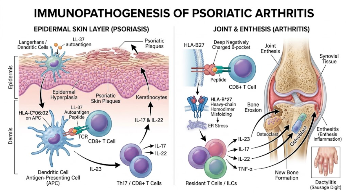
Figure 1. Immunopathogenesis of Psoriatic Arthritis. A schematic overview of the molecular and cellular mechanisms in psoriatic disease. The left panel illustrates HLA-C*06:02-driven epidermal hyperplasia via the IL-23/IL-17 pathway, leading to skin plaques. The right panel depicts HLA-B27-mediated joint and entheseal inflammation, resulting in bone remodeling, enthesitis, and dactylitis.
Genetic studies of human leukocyte antigen genotyping have revealed that psoriatic arthritis is a highly heterogeneous phenotype associated with different genetic markers. Comparisons of this kind have implicated HLA-C*06:02 and HLA-B*27 as two separate subphenotypes with distinct temporal patterns, organ involvement, and clinical courses(Winchester et al., 2016).
Classic plaque psoriasis without arthritis is strongly associated with HLA-C*06:02 (approximately 60% of cutaneous cases as opposed to around 15-20% in health). Although HLA-C*06:02 is still more common in psoriatic arthritis cohorts (28-30%), the presence of HLA-C*06:02 suggests skin disease and is not considered a primary cause for joint disease. Patients who are HLA-C*06:02 positive often develop cutaneous psoriasis at a young age, in the second or third decades, and have long delays to musculoskeletal symptoms(FitzGerald et al., 2015). In HLA-C*06:02 positive patients this arthritic phase is late to develop and occurs on average 10–15 years after cutaneous plaques. It has also been associated with a mild joint course with low rates of enthesitis, dactylitis, and severe joint deformity. In fact, these statistical associations argue for a protective role of HLA-C*06:02, as this allele spares patients of severe inflammation at entheseal sites(Winchester et al., 2016).
Conversely, HLA-B*27 is highly overrepresented in psoriatic arthritis, particularly in the presence of axial spondylitis, bilateral sacroiliitis, dactylitis and enthesitis. Unlike HLA-C*06:02, HLA-B*27 is associated with concurrent or near-concurrent onset of skin and joint disease. In about one-third of HLA-B*27 positive individuals, arthritis occurs before or concurrently with cutaneous psoriasis, reducing the latency interval seen with HLA-C*06:02. HLA-B*27 carrying subjects in clinical cohorts have predominantly symmetrical sacroiliac joint inflammation with deep bone marrow edema seen by MRI and increased systemic inflammatory activity (C-reactive protein)(Macia-Villa et al., 2026).
In more recent long-term studies, HLA-B*27 has been associated with both the new onset of dactylitis and the persistence of dactylitis: indeed, HLA-B*27/C*02 haplotype subjects are prone to dactylitis. In patients who are HLA-B*27 +, dactylitis is also more severe and tends to be unresolving in the presence of chronic inflammation (Kharouf et al., 2026). These contribute to the polygenicity of the disease and manifest with asymmetric sacroiliitis followed by deformities/fusion of joints (e.g. HLA-B* 08:01 haplotype) and synovial disease. Subphenotypes of HLA-B * 27 and HLA-C * 06.02: Problems with disease classification, immunopathogenesis, and clinical presentation.(FitzGerald et al., 2015).
| Feature | HLA-C*06:02 Subphenotype | HLA-B*27 Subphenotype |
| Primary Clinical Target | Cutaneous skin plaques (psoriasis) | Axial skeleton, entheses, and digits |
| Onset of Skin Disease | Early onset (15-25 years) | Later onset or post-articular |
| Skin-to-Joint Latency | Extended lag period (10-15 years) | Short, contemporaneous, or joint-first |
| Enthesitis Prevalence | Low / Protective effect | High / Strong positive association |
| Dactylitis Prevalence | Low / Reduced risk | High / Associated with prolonged duration |
| Sacroiliitis Pattern | Minimal or mild asymmetric | Prominent, severe, often symmetrical |
| Primary Mechanism | Epidermal antigen display to CD8+ T cells | Entheseal stress, B pocket binding, misfolding |
| Cytokine Axis Focus | Epidermal IL-23 / IL-17 axis | Entheseal IL-23 / IL-17 / IL-22 bone remodeling |
HLA-C/HLA-B27 versus HLA-C*06:02/HLA-B*27: Two sides of the same coin with new shared clues to pathogenesis of psoriatic arthritis. “Psoriatic arthritis subphenotypes” being “genetically determined and not dictated by variable expression.” So, the menu for would-be alternatives to self antigens and would-be hit anatomic site (autoimmunity)(FitzGerald et al., 2015; Maaloul et al., 2025). would be each HLA-alleles alternatives (each one’s alternative biophysics and chemical bounds and alternative peptide, as well).
This genotype-phenotype approach is most relevant in the clinic from diagnosis to treatment (Group et al., 2019). Diagnosis features: family history of disease, dactylitis, nail dystrophy, etc. (e.g., Commission on Classification Criteria for Psoriatic Arthritis) However, all these features are genetically determined and are differently represented in different ethnicities, so the diagnosis criteria may be differently applied depending on the genetic background of the authors’ study. This is especially associated with the assumption that the rheumatological diagnosis is late only in the case of strict following the diagnosis criteria (FitzGerald et al., 2015; Winchester et al., 2016), so that in the case of revealing the skin disease in strong conditions of HLA-C*06:02 values, there is the absence of nail lesions/dactylitis.
A genetic subphenotype has also emerged with biologic selection and precision medicine. For example, HLA-C*06:02 positive psoriatic patients respond well to anti-IL-12/23 (e.g., ustekinumab) versus anti-tumor necrosis factor (e.g., adalimumab) agents if HLA-C*06:02 negative with psoriatic arthritis(Group et al., 2019). In contrast, HLA-B*27 positivity is linked to axial disease with molecular etiology of psoriatic arthritis/enthesitis and good response rates to IL-23/IL-17 axis blockade. HLA-typing can be a routine germline tool to predict the most cost-effective/frequent first-line biologic choice(FitzGerald et al., 2015; Macia-Villa et al., 2026).
It remains to be answered whether HLA-C*06:02 and HLA-B*27 elicit tissue-specific responses to self-antigens. Candidate LL37 and ADAMTSL5 peptides discovered in skin lesions presented on specific HLA-B*27(FitzGerald et al., 2015; Maaloul et al., 2025). Further follow-ups of the genetic code specific for tissue destruction and development of the antigen-specific immunotherapies will require high-throughput immune peptidomics (and spatial transcriptomics and mass single cell TCR sequencing(s)).
To summarize, the genetic susceptibility based on major histocompatibility complex class I allele is the key to autoimmune attack in psoriatic arthritis. Genetic heterogeneity by two major immunogenetic codes, HLA-C*06:02 and HLA-B*27, supports the pathogenesis of separate pathogenetic and clinical diseases. HLA-C*06:02 encodes epidermal autoantigen presentation and predisposition to early-onset psoriasis with delayed and mild synovitis. Conversely, HLA-B*27 promotes entheseal and synovial inflammation via peptide presentation and misfolding of the heavy chain, resulting in axial spondylitis, dactylitis, and accelerated joint structural damage. We reinvent psoriatic arthritis as genetically defined subphenotypes, offering an alternative understanding of disease mechanisms and future consideration of human leukocyte antigen profiling for biomarker-based personalized healthcare.
Chamian, F., Lowes, M. A., Lin, S. L., Lee, E., Kikuchi, T., Gilleaudeau, P., . . . Krueger, J. G. (2005). Alefacept reduces infiltrating T cells, activated dendritic cells, and inflammatory genes in psoriasis vulgaris. Proc Natl Acad Sci U S A, 102(6), 2075-2080. doi:10.1073/pnas.0409569102
Costello, P. J., Winchester, R. J., Curran, S. A., Peterson, K. S., Kane, D. J., Bresnihan, B., & FitzGerald, O. M. (2001). Psoriatic arthritis joint fluids are characterized by CD8 and CD4 T cell clonal expansions appear antigen driven. J Immunol, 166(4), 2878-2886. doi:10.4049/jimmunol.166.4.2878
FitzGerald, O., Haroon, M., Giles, J. T., & Winchester, R. (2015). Concepts of pathogenesis in psoriatic arthritis: genotype determines clinical phenotype. Arthritis Res Ther, 17(1), 115. doi:10.1186/s13075-015-0640-3
Group, B. S., Group, B. S., Consortium, P., Dand, N., Duckworth, M., Baudry, D., . . . Stocken, D. (2019). HLA-C*06:02 genotype is a predictive biomarker of biologic treatment response in psoriasis. Journal of Allergy and Clinical Immunology, 143(6), 2120-2130. doi:10.1016/j.jaci.2018.11.038
Kharouf, F., Abarza, V. C., Mehta, P., Gao, S., Ganatra, D., Pereira, D., . . . Chandran, V. (2026). Psoriatic dactylitis: the association with HLA genetic susceptibility markers. Annals of the Rheumatic Diseases, 85(6), 1110-1116. doi:10.1016/j.ard.2026.02.011
Maaloul, M., Charfi, A., Feki, A., Gassara, Z., Hakim, F., Gaddour, L., . . . Mahfoudh, N. (2025). Association study of the HLA class I system with psoriatic arthritis in Southern Tunisia: a case–control study. Clinical Rheumatology, 44(2), 707-718. doi:10.1007/s10067-024-07290-y
Macia-Villa, C., Fernandez-Velasco, J. I., Ferre-Sanfrancisco, M., Mendoza-Bravo, D. A., Ruiz-Bejerano, A. M., Ferrez-Hernandez, C., & Morell-Hita, J. L. (2026). Exploring the phenotypic expression of HLA-B27 in psoriatic arthritis. Clinical Rheumatology, 45(6), 3399-3404. doi:10.1007/s10067-026-08144-5
O’Rielly, D. D., & Rahman, P. (2014). Genetics of psoriatic arthritis. Best Pract Res Clin Rheumatol, 28(5), 673-685. doi:10.1016/j.berh.2014.10.010
Okada, Y., Han, B., Tsoi, L. C., Stuart, P. E., Ellinghaus, E., Tejasvi, T., . . . Raychaudhuri, S. (2014). Fine mapping major histocompatibility complex associations in psoriasis and its clinical subtypes. Am J Hum Genet, 95(2), 162-172. doi:10.1016/j.ajhg.2014.07.002
Winchester, R., Giles, J., Jadon, D., Haroon, M., McHugh, N., & FitzGerald, O. (2016). Implications of the diversity of class I HLA associations in psoriatic arthritis. Clin Immunol, 172, 29-33. doi:10.1016/j.clim.2016.07.019
Zalesak, M., Danisovic, L., & Harsanyi, S. (2024). Psoriasis and Psoriatic Arthritis—Associated Genes, Cytokines, and Human Leukocyte Antigens. Medicina, 60(5), 815. Retrieved from doi:10.3390/medicina60050815