Our Services
Others
- DFPP
- Shockwave
- IV Drip


“In Alzheimer’s disease, excessive tau hyperphosphorylation causes neuronal cytoskeletal collapse, intracellular transport failure, and neurofibrillary tangle formation. These processes are discussed in detail in this literature review from a mechanistic viewpoint.”
Alzheimer’s disease is marked by extracellular amyloid plaques and neuronal neurofibrillary tangles. The pathological cascade signal begins with processing of amyloid precursor protein and increases hyperphosphorylation of tau that collapses the cytoskeleton and leads to cognitive decline. Under normal physiological conditions, tau binds to tubulin dimers to stabilize neuronal cytoskeleton for axonal transport. In Alzheimer’s disease, kinases are upregulated while phosphatases are downregulated, and there is a change to an environment conducive to hyperphosphorylation of tau on threonine and serine residues. Hyperphosphorylated tau has a low microtubule affinity and dissociates in the somatodendritic compartment. Hyperphosphorylated tau then sequesters normal microtubule-associated proteins MAP1 and MAP2 and endogenous tau, augmenting its toxic function. The depletion of a microtubule stabilizer by this sequestration leads to cytoskeletal collapse. The microtubule collapse prevents motor-mediated trafficking along the networks of axonal cytoskeletal microtubules, impeding delivery of vesicles, mitochondria and required nutrients to synaptic terminals. At the same time, tau hyperphosphorylates and aggregates into paired helical filaments of insoluble neurofibrillary tangles. Here we review recent evidence for mechanisms of tau hyperphosphorylation and disassembly of microtubules and blockage of axoplasmic transport that result in synaptic loss in Alzheimer’s disease.
Alzheimer’s disease (AD) is the most common form of dementia in the elderly. The main histopathological lesions of the AD brain are extracellular senile plaques of aggregated amyloid-β peptides and intracellular neurofibrillary tangles of hyperphosphorylated tau protein(Rawat et al., 2022). Abnormal cleavage of amyloid precursor protein and the resultant accumulation of soluble amyloid-β oligomers induce early synaptic stress, as elaborated in the preceding article. As detailed above when considering upstream pathogenic triggers, it is increasingly evident that neurofibrillary tangle density and spatiotemporal distribution are much more correlated with the clinical dementia, synaptic loss, and neurodegeneration(Boutajangout, Sigurdsson, & Krishnamurthy, 2011; Iqbal, Liu, Gong, Alonso Adel, & Grundke-Iqbal, 2009; Šimić et al., 2016). than with amyloid plaque burden.
MAPT (microtubule-associated protein tau) gene, located on chromosome 17q21, encodes tau, a normal microtubule-associated protein found in the axons of neurons. In the adult human brain, MAPT undergoes alternative splicing to produce six tau protein isoforms. According to whether they contain N-terminal inserts (0N, 1N, 2N) or microtubule binding repeats (3R or 4R)(Iqbal et al., 2009). Tau is an intrinsically disordered protein with no defined tertiary structure. It basically binds to tubulin heterodimers (repeated domains) to stabilize cytoskeleton and promote microtubule assembly. Microtubule arrays (Song et al., 2024) form tracks for trophic factor and vesicular and organellar transport in axoplasmic transport into and out of terminals.
But in diseases such as AD, tau is abnormally modified by hyperphosphorylation (Boutajangout et al., 2011). Under normal conditions, presence of 2-3 mol phosphate per mol protein, 5-9 or more mol phosphate per mol protein is observed in AD neurofibullary lesions. Advances in pathological phosphorylation remove basic charges and cause structural alterations and loss of tau binding and stabilizing associations with microtubules(Weingarten, Lockwood, Hwo, & Kirschner, 1975). This results in gradual cytoskeleton disruption, inhibition of transport routes, and neurodegeneration of Tau-affected neurons. In this review, we discuss the molecular mechanisms of tau hyperphosphorylation that result in cytoskeletal sequestration, blockage of transport pathways, and neurofibrillary tangle formation in AD(A. D. Alonso, Grundke-Iqbal, Barra, & Iqbal, 1997).
The balance between tau protein kinases and tau protein phosphatases regulates the phosphorylation status of tau. There are > 80 phosphorylation sites on tau. In AD, hyperphosphorylation is driven by increased kinase signaling and reduced phosphatase(Buée et al., 2010).
Kinases involved in tau hyperphosphorylation in AD are divided into proline-directed protein kinases and non-proline directed protein kinases. Proline-directed kinases phosphorylate tau on serine/threonine residues adjacent to proline motifs(Iqbal & Grundke-Iqbal, 2008). The main proline-directed kinases associated with tau pathology include glycogen synthase kinase-3beta (GSK-3β) and cyclin-dependent kinase 5 (CDK5). GSK-3β phosphorylates tau at Ser199, Thr231, Ser396, and Ser404 in its active form. Calpain-cleaved p35 to p25 activates CDK5 that leads to phosphorylation of tau at Ser202, Thr205, Thr212, and Ser235. Other MAP kinases, including p38, ERK1/2, and c-Jun N-terminal kinase, are also involved in response to stress(Lagalwar, Berry, & Binder, 2007).
Non-proline-directed kinases, such as protein kinase A, calcium/calmodulin-dependent protein kinase II, and microtubule affinity-regulating kinase, phosphorylate non-proline sites(Kobayashi et al., 1993). Protein kinase A and microtubule affinity-regulating kinase phosphorylate Ser262 and Ser356 in pTau repeat regions and decrease tau binding affinity to tubulin. Importantly, non-proline-directed phosphorylation is a priming event. Phosphorylation of Ser262 or Thr212 by protein kinase A or DYRK1A primes tau for secondary hyperphosphorylation by GSK-3β and CDK5, which induces conformational changes that make tau more accessible(Cho & Johnson, 2003; Wang, Wu, Smith, Grundke-Iqbal, & Iqbal, 1998).
Simultaneously, the activity of protein phosphatases is impaired in Alzheimer’s brain. The major tau phosphatase, protein phosphatase-2A, accounts for more than seventy percent of the tau dephosphorylating activity. In AD, protein phosphatase-2A activity is inhibited by 50%(Li, Sengupta, Haque, Grundke-Iqbal, & Iqbal, 2004). Endogenous protein phosphatase-2A inhibitors I1_PP2 and I2_PP2A are overexpressed and redistributed in the cytoplasm of degenerating neurons, inhibiting protein phosphatase-2A. Moreover, the hypometabolic state for glucose inhibits the antagonistic phosphorylation-induced posttranslational modification of tau by O-GlcNAcylation, worsening tau hyperphosphorylation(Iqbal & Grundke-Iqbal, 2008).
Hyperphosphorylation of tau decreases its binding to microtubules in the axons. Phosphorylation introduces negatively charged hydrophilic phosphate groups that change tau electrostatics, protein shape, and neutralize basic charges needed to move along the acidic surface of tubulin. Hyperphosphorylated tau is then cleaved from the microtubule surface into the soluble cytosolic pool and is mislocalized from axons to the somatodendritic compartment.
Hyperphosphorylated cytosolic tau is not inert but acquires a toxic function(A. C. Alonso, Zaidi, Grundke-Iqbal, & Iqbal, 1994). Hyperphosphorylated tau can bind normal tau and high molecular weight microtubule-associated proteins MAP1A, MAP1B, and MAP2 (MAP = microtubule-associated proteins). In normal neurons, MAPs are required for microtubule survival, but they are present in lesser quantities than tubulin. Hyperphosphorylated tau has higher affinity than tubulin for normal tau, MAP1, and MAP2 and sequesters them from the microtubule lattice(A. D. Alonso et al., 1997).
Hyperphosphorylation (Figure 1) initiates a cascade of structural events that remove tau from tubulin protofilaments, sequester structural proteins, and lead to depolymerization of the microtubule network.
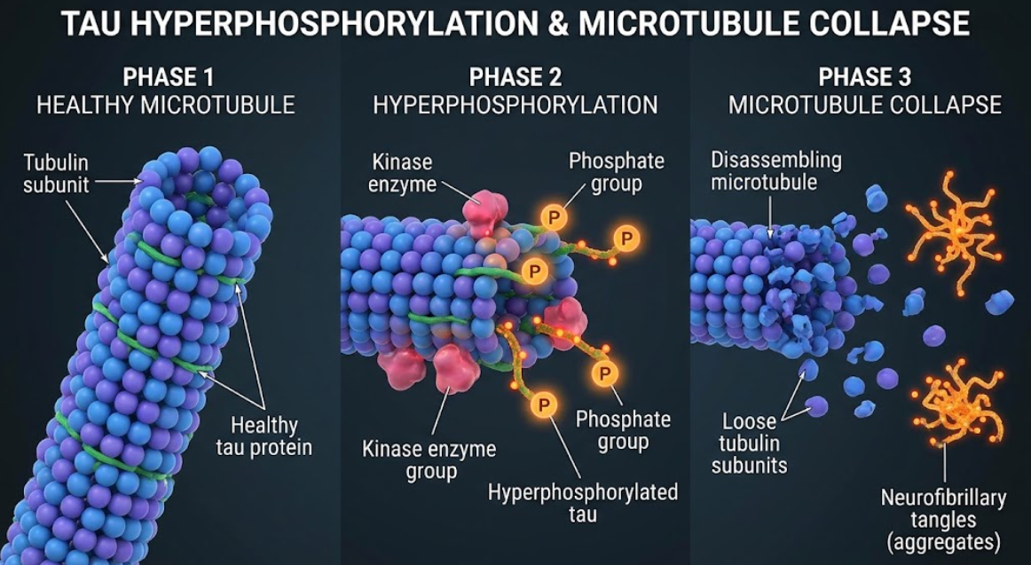
Figure 1. Microtubule collapse pathways of hyperphosphorylated Tau. Schematic of microtubule destabilization. Normal tau (left) stabilizes microtubules. Dissociation of tau causes hyperphosphorylation of tau by a kinase enzyme (center), preventing tau from promoting microtubule assembly and resulting in neurofibrillary tangle formation (right).
This sequestration deprives tubulin heterodimers of structural stabilizers, resulting in rapid depolymerization of axonal microtubules. Microtubule dissociating activity of hyperphosphorylated tau isolated from brains of patients with AD sequesters normal tubulin. Dephosphorylation of hyperphosphorylated tau with protein phosphatase-2A eliminates this sequestering activity and restores the microtubule assembling activity of tau. Thus, the direct cause of aberrant hyperphosphorylation is the destabilization of the cytoskeleton within neurons(A. D. Alonso et al., 1997).
Microtubule disassembly disrupts the intracellular transport system of neurons and jeopardizes neuron survival. These motor proteins are essential in cargo transport along the axons carrying cargo from the soma to the distal synapses. Kinesin motor proteins bind to vesicles, mitochondria, and peroxisomes, inducing anterograde flow toward nerve terminals, and dynein motor proteins do the reverse (retrograde) induced by apoptosis influx commandeering neurotrophic complexes to troop contrary to the effect back to the cell body (Futerman & Banker, 1996).
Disruption of microtubule polymerization severely handicaps axoplasmic flow and kinesin movement and thus creates traffic jams of cell cargoes on axonal shafts. The accumulated cell cargos accumulate in axonal swelling and dystrophic neurites, resulting in metabolite deficiency in presynaptic terminals (Rawat et al., 2022).
Mitochondrial transport deficits particularly affect neurons. Because kinesin facilitates anterograde transport of mitochondria, tau hyperphosphorylation impairs kinesin-dependent forward mitochondrial transport, which places mitochondria originated from distal axons at migrants retrograde to the cell body. These phenomenons result to loss of mitochondria at the terminus, problems of adenosine triphosphate stores, altered calcium homeostasis, and increased production of reactive oxygen species and oxidative stress. Hyperphosphorylated tau also binds mitochondrial fission protein Drp1, increasing the GTPase activity of the latter and leading to mitochondrial fission(Hanger, Anderton, & Noble, 2009).
Also, blocking transport precludes the contribution of peroxisomes and endoplasmic reticulum to distal neurites. Misfolded tau from the endoplasmic reticulum favors the unfolded protein response and pro-inflammatory cascades. ApoE4 aggregates via cleavage, dyspopminating oxons, and C-terminal truncation and phosphorylation, resulting in lysis of cells with starvation and demise of synaptic compartments, apoptotic death of cells with cell lysis, and “dying back” of neurites(Iqbal et al., 2009; Rawat et al., 2022).
Hyperphosphorylated tau also self-associates to form insoluble aggregates in addition to disassembling the cytoskeleton. Such tau self-association is prevented under physiological conditions by charge repulsion of basic flanking N-terminal and C-terminal domains. Hyperphosphorylation neutralizes basic charges and causes a change in tau conformation exposing hydrophobic hexapeptide motifs in microtubule-binding repeats R2 and R3. Tau self-assembly is stabilized by the β-sheet motifs and intermolecular hydrophobic interaction (Iqbal et al., 2009; Šimić et al., 2016).
The cascade of collapse includes several morphologically distinct steps:
Neurofibrillary tangles were proposed to be the major neurotoxic species in AD pathogenesis. Major paradigm shift in neuropathological evidence. Novel evidence suggests soluble tau oligomers as the most toxic species. The oligomers cause toxicity by perturbing the plasma membrane and excitotoxicity, which increases Ca2+ influx and disrupts synaptic cellular plasticity, which leads to endoplasmic reticular stress before the intermediate filaments(Song et al., 2024).
But hyperphosphorylated tau can form insoluble neurofibrillary tangles of degenerating neurons that may be protective. Hyperphosphorylated tau polymerizes to filaments in vitro and substitutes for normal MAPs, an inhibitor of MAPs active as neurotoxins in complexes with intact microtubules(Iqbal & Grundke-Iqbal, 2008). Re-expression of Tau transgene in dying neurons in transgenic mice improved cognition from pre-existing neurofibrillar tangles. However, they can form ghost tangles, even block the organelles and change nuclear cell shape in aged dying neurons(Rawat et al., 2022).
Depicted in figure 2, in their course from soluble hyperphosphorylated monomers to soluble oligomers, paired helical filaments, and dense neurofibrillary tangles, they produce traffic jams in organelles, as well as progressive synaptic loss.
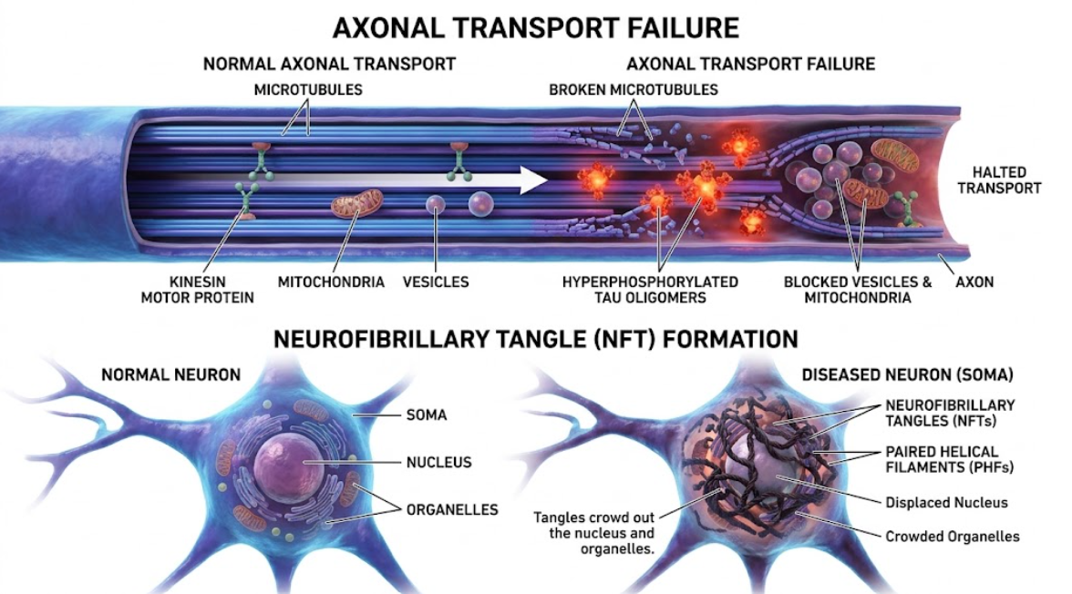
Figure 2. Cellular biology of tau pathology. Normal tau Hyperphosphorylated tau disassembles microtubules arresting axonal transport (top) and forming neurofibrillary tangles (NFTs) displacing the cell organelles (bottom).
Neurofibrillary degeneration follows a reproducible spatiotemporal cascade in networks of interconnected brain regions. A similar pattern holds for tau biology, in which one starts with tau pathology in the entorhinal cortexes, progresses onward into the hippocampus, and finally to neocortical association areas, as shown by braak neuropathological staging(Rawat et al., 2022; Šimić et al., 2016).
Tau transmission is trans-synaptic as observed for prions and is non-random. Degenerative presynaptic tau oligomer seeds are released or exocytosed into the synaptic cleft. Extracellular tau seeds reach postsynaptic recipient cells by exosome fusion as well as fluid-phase endocytosis and macropinocytosis. Once internalized, these tau seeds act as conformational templates for misfolding and aggregation of endogenous tau. Microglia also phagocytose extracellular tau and serve as a local source of undegraded tau seeds (Rankin et al., 2007; Rawat et al., 2022; Šimić et al., 2016).
Hyperphosphorylated tau transsynptically spreads and correlates with being lost from synapses. Mislocalization of tau into dendritic spines and interaction with postsynaptic density protein 95, and Fyn kinase to enhance N-methyl-D-aspartate receptor with loss of splicing and intracellular calcium overload. The cognitive decline in the network matrix of synapses progresses to dementia.(Rawat et al., 2022).
AD, tau hyperphosphorylation/neurofibrillary tangles, major cytoskeleton/organelle fall-offs, and intracytoplasmic mechanistic interference of cytoskeleton/organelle transport. Neuro-degenerative/neuro-behavioral biology loci/detections of amyloid-β oligomeric precursors at the site of pathology and tau pathology detection of synaptic stress/amyloid pore pathology, and biology of virus treatment(Rawat et al., 2022). Furthermore, soluble amyloid-β oligomers congest α7 nicotinic acetylcholine receptors and instruct cellular prion protein, Frizzled receptor(s), and pan ErbB-receptors presumably to up-regulate and regulate Wnt cascades, activate intracellular cascades of protein kinase including phosphatidylinositide 3-kinase and yeasts of Src tyrosine kinases family, and over-express glycogen synthase kinase-3β and cyclin-dependent kinase 5 activities. This double-edged cascade is believed to be associated with early pathology-related amyloid deposits and funneling down to hyperphosphorylation of tau as the progenitor of irreversible neuronal demise(De Felice et al., 2008; Iqbal & Grundke-Iqbal, 2008; Rawat et al., 2022).
Complex molecular mechanisms demonstrate the existence of several therapeutic approaches for preventing neurofibrillary degeneration and conserving neuronal architecture:
In addition, the therapeutic paradigm has dramatically shifted on the basis of this multifactorial pattern of AD process as the pure single-molecularly directed therapy is insufficient. It was found that a combined regimen that clears the toxic amyloid oligomers upstream and inhibits tau kinases downstream of amyloid and stabilizes microtubules and cellular autophagy was more effective. In this regard, development of polytherapeutic approaches in combination with support of cytoskeletal integrity in place of amyloid-clearing regimens was promising to prevent cognitive decline and neurodegeneration in AD(Iqbal & Grundke-Iqbal, 2008; Rawat et al., 2022).
Thus, hyperphosphorylation of tau is an important pathological mechanism in AD. Hyperphosphorylation by deregulated kinases and phosphatases leads to dissociation of tau from tubulin and gain of a toxic function. Hyperphosphorylated tau assembles with normal tau and MAP1 + MAP2, shattering microtubules in axons. Collapse of the cytoskeleton prevents transport of vesicles, mitochondria, and nutrients, thus starving the synapse and causing neuronal death. Concurrently, tau self-assembles to paired helical filaments and neurofibrillary tangles, which become propagating agents in neural networks trans-synaptically. Therapeutic approaches for which outlook is promising include inhibition of tau hyperphosphorylation, restoration of protein phosphatase-2A, and microtubule stabilization for preserving cytoskeletal integrity and preventing neurodegeneration in the AD brain.
Alonso, A. C., Zaidi, T., Grundke-Iqbal, I., & Iqbal, K. (1994). Role of abnormally phosphorylated tau in the breakdown of microtubules in Alzheimer disease. Proc Natl Acad Sci U S A, 91(12), 5562-5566. doi:10.1073/pnas.91.12.5562
Alonso, A. D., Grundke-Iqbal, I., Barra, H. S., & Iqbal, K. (1997). Abnormal phosphorylation of tau and the mechanism of Alzheimer neurofibrillary degeneration: sequestration of microtubule-associated proteins 1 and 2 and the disassembly of microtubules by the abnormal tau. Proc Natl Acad Sci U S A, 94(1), 298-303. doi:10.1073/pnas.94.1.298
Boutajangout, A., Sigurdsson, E. M., & Krishnamurthy, P. K. (2011). Tau as a therapeutic target for Alzheimer’s disease. Curr Alzheimer Res, 8(6), 666-677. doi:10.2174/156720511796717195
Buée, L., Troquier, L., Burnouf, S., Belarbi, K., Van der Jeugd, A., Ahmed, T., . . . Sergeant, N. (2010). From tau phosphorylation to tau aggregation: what about neuronal death? Biochem Soc Trans, 38(4), 967-972. doi:10.1042/bst0380967
Cho, J. H., & Johnson, G. V. (2003). Glycogen synthase kinase 3beta phosphorylates tau at both primed and unprimed sites. Differential impact on microtubule binding. J Biol Chem, 278(1), 187-193. doi:10.1074/jbc.M206236200
De Felice, F. G., Wu, D., Lambert, M. P., Fernandez, S. J., Velasco, P. T., Lacor, P. N., . . . Klein, W. L. (2008). Alzheimer’s disease-type neuronal tau hyperphosphorylation induced by A beta oligomers. Neurobiol Aging, 29(9), 1334-1347. doi:10.1016/j.neurobiolaging.2007.02.029
Futerman, A. H., & Banker, G. A. (1996). The economics of neurite outgrowth–the addition of new membrane to growing axons. Trends Neurosci, 19(4), 144-149. doi:10.1016/s0166-2236(96)80025-7
Hanger, D. P., Anderton, B. H., & Noble, W. (2009). Tau phosphorylation: the therapeutic challenge for neurodegenerative disease. Trends Mol Med, 15(3), 112-119. doi:10.1016/j.molmed.2009.01.003
Iqbal, K., & Grundke-Iqbal, I. (2008). Alzheimer neurofibrillary degeneration: significance, etiopathogenesis, therapeutics and prevention. J Cell Mol Med, 12(1), 38-55. doi:10.1111/j.1582-4934.2008.00225.x
Iqbal, K., Liu, F., Gong, C. X., Alonso Adel, C., & Grundke-Iqbal, I. (2009). Mechanisms of tau-induced neurodegeneration. Acta Neuropathol, 118(1), 53-69. doi:10.1007/s00401-009-0486-3
Kobayashi, S., Ishiguro, K., Omori, A., Takamatsu, M., Arioka, M., Imahori, K., & Uchida, T. (1993). A cdc2-related kinase PSSALRE/cdk5 is homologous with the 30 kDa subunit of tau protein kinase II, a proline-directed protein kinase associated with microtubule. FEBS Lett, 335(2), 171-175. doi:10.1016/0014-5793(93)80723-8
Kopeikina, K. J., Hyman, B. T., & Spires-Jones, T. L. (2012). Soluble forms of tau are toxic in Alzheimer’s disease. Transl Neurosci, 3(3), 223-233. doi:10.2478/s13380-012-0032-y
Lagalwar, S., Berry, R. W., & Binder, L. I. (2007). Relation of hippocampal phospho-SAPK/JNK granules in Alzheimer’s disease and tauopathies to granulovacuolar degeneration bodies. Acta Neuropathol, 113(1), 63-73. doi:10.1007/s00401-006-0159-4
Li, L., Sengupta, A., Haque, N., Grundke-Iqbal, I., & Iqbal, K. (2004). Memantine inhibits and reverses the Alzheimer type abnormal hyperphosphorylation of tau and associated neurodegeneration. FEBS Lett, 566(1-3), 261-269. doi:10.1016/j.febslet.2004.04.047
Rankin, C. A., Sun, Q., & Gamblin, T. C. (2007). Tau phosphorylation by GSK-3beta promotes tangle-like filament morphology. Mol Neurodegener, 2, 12. doi:10.1186/1750-1326-2-12
Rawat, P., Sehar, U., Bisht, J., Selman, A., Culberson, J., & Reddy, P. H. (2022). Phosphorylated Tau in Alzheimer’s Disease and Other Tauopathies. Int J Mol Sci, 23(21). doi:10.3390/ijms232112841
Šimić, G., Babić Leko, M., Wray, S., Harrington, C., Delalle, I., Jovanov-Milošević, N., . . . Hof, P. R. (2016). Tau Protein Hyperphosphorylation and Aggregation in Alzheimer’s Disease and Other Tauopathies, and Possible Neuroprotective Strategies. Biomolecules, 6(1), 6. Retrieved from doi:10.3390/biom6010006
Song, Z., Wang, K. W., Hagar, H. C., Chen, H. R., Kuan, C. Y., Zhang, K., & Kuo, M. H. (2024). Hyperphosphorylated Tau Inflicts Intracellular Stress Responses that Are Mitigated by Apomorphine. Mol Neurobiol, 61(5), 2653-2671. doi:10.1007/s12035-023-03689-x
Wang, J. Z., Wu, Q., Smith, A., Grundke-Iqbal, I., & Iqbal, K. (1998). Tau is phosphorylated by GSK-3 at several sites found in Alzheimer disease and its biological activity markedly inhibited only after it is prephosphorylated by A-kinase. FEBS Lett, 436(1), 28-34. doi:10.1016/s0014-5793(98)01090-4
Weingarten, M. D., Lockwood, A. H., Hwo, S. Y., & Kirschner, M. W. (1975). A protein factor essential for microtubule assembly. Proc Natl Acad Sci U S A, 72(5), 1858-1862. doi:10.1073/pnas.72.5.1858