Our Services
Others
- DFPP
- Shockwave
- IV Drip


“This literature review synthesizes multi-center clinical research and hydrodynamic models to illuminate the role of mechanical compression at the craniovertebral junction in causing cerebrospinal fluid obstruction and syrinx development in Chiari malformation type I.”
Chiari malformation type I is a complex neuroanatomical disorder characterized by the caudal displacement of the cerebellar tonsils through the foramen magnum into the upper cervical spinal canal. This structural abnormality results in spatial overcrowding at the craniovertebral junction creating a bottleneck that blocks the flow of CSF between the intracranial and spinal subarachnoid spaces. Syringomyelia is a common complication of Chiari malformation type I, which is characterized by chronic fluid-filled cavities within the parenchyma of the spinal cord. The current literature suggests that mechanical compression at the craniovertebral junction is the main physical factor for the development of a syrinx. Structural crowding, often exacerbated by osseous anomalies of the skull base (small posterior cranial fossa, short clivus, platybasia, basilar invagination) impedes subarachnoid fluid flow. During cardiac systole, herniated cerebellar tonsils act as a dynamic piston, generating pressure waves and transmedullary gradients to propel cerebrospinal fluid into spinal cord tissue through perivascular spaces. Understanding these hydrodynamic and biomechanical mechanisms is critical for early clinical identification, accurate neuroimaging assessment, and optimal surgical decision-making. Surgical decompression of the foramen magnum restores patency of the subarachnoid space, normalizes fluid circulation, relieves cervicomedullary compression and promotes syrinx regression. This review synthesizes the available evidence on the association of craniovertebral junction compression with cerebrospinal fluid obstruction and the pathogenesis of syringomyelia in Chiari malformation type I.
Keywords: Chiari Malformation Type I, Craniovertebral Junction, Syringomyelia, Cerebrospinal Fluid Dynamics, Tonsillar Ectopia, Foramen Magnum Decompression
Chiari malformation type I is a structural disorder of the craniocervical axis, characterized by caudal herniation of cerebellar tonsils below the plane of the foramen magnum > 5 mm. First described by Hans Chiari in the late 1800s, it includes manifestations ranging from asymptomatic findings to progressive neurological deficits(Chiari, 1891). The epidemiology studies suggest that the prevalence of Chiari malformation type I is approximately one percent in the general population with symptoms typically presenting in late adolescence or early adulthood(Al-Habib et al., 2020). Classical symptoms include suboccipital headache aggravated by coughing, sneezing, or Valsalva maneuvers, associated with neck stiffness, dizziness, cranial nerve dysfunctions, cerebellar ataxia, and sensory deficits in the upper extremities(Hatgaonkar, Mahajan, Hatgoankar, & Bandre, 2024).
Syringomyelia is classically associated with Chiari malformation type I and occurs in 36 to 75 percent of individuals. Syringomyelia is characterized by formation of chronic fluid-filled longitudinal cavities in spinal cord tissue, most often of cervical and thoracic segments(Fernandez et al., 2009; Fernández et al., 2009). As these intramedullary cavities enlarge, mechanical stress on traversing spinothalamic nerve fibers and anterior horn motor neurons results in dissociated sensory loss, muscle weakness, intrinsic hand atrophy, and progressive myelopathy(Alhosani & Gachechiladze, 2026).
In our previous review we addressed early embryological neural tube development, central canal occlusion, ciliary impairment and cellular signaling cascades in spinal tissue remodeling. The present review expands these premises focusing on macro-level mechanical and hydrodynamic forces at the craniocervical junction. Mechanical obstruction at the cranio-vertebral junction is the primary physical stimulus that initiates the accumulation of fluid in Chiari malformation type I, while cellular signaling controls tissue remodeling at the micro-scale(Al-Habib et al., 2020).
The craniovertebral junction is the key anatomical interface between the posterior cranial fossa and the cervical spinal canal. This transition zone is narrowed due to structural crowding or skeletal deformities and restricts the flow of cerebrospinal fluid between intracranial and spinal compartments(Mahajan, Phatak, Onkar, Ambhore, & Pantawane, 2024). This review aims to review the clinical cohorts, radiology studies, and biomechanical models that illustrate the role of mechanical compression at the craniovertebral junction in altering cerebrospinal fluid hydrodynamics, enhancing transmedullary pressure gradients, and directly contributing to the pathogenesis of syringomyelia in Chiari malformation type I(Alhosani & Gachechiladze, 2026; Fernandez et al., 2009).
The principal anatomic pathology of Chiari malformation type I is a volumetric mismatch between developing hindbrain structures and bony enclosure of the posterior cranial fossa. Morphometric studies show that underdevelopment of the paraxial mesoderm results in an abnormally small posterior cranial fossa. In a confined space the neural tissue proliferates normally leading to overpopulation of the posterior fossa and herniation of the cerebellar tonsils through the foramen magnum into the cervical spinal canal(Holder, Abbakr, Haynes, & Sanusi, 2025; Nishikawa et al., 2022).
Nishikawa et al. performed morphometric studies to measure the volume of the posterior cranial fossa and the area of the foramen magnum and classified Chiari malformation type I into definite anatomical subtypes. In some patients, there was severe underdevelopment of the occipital bone causing a small posterior fossa volume, while in other patients the posterior fossa volume was normal but focal narrowing was localized to the foramen magnum (Nishikawa et al., 2022). Caudal descent of cerebellar tonsils directly alters spatial relationships at the craniocervical border in all subtypes. The tonsils protrude into the upper spinal canal. This space normally allows fluid to circulate around the cervicomedullary junction. Clinical radiological series describe tonsillar descent from 4 to more than 22 mm with marked effacement of subarachnoid cisterns(Cogan & Barrows, 1954; Koenigsberg et al., 2005).
Osseous malformations of the skull base and upper cervical spine make this anatomical overcrowding even worse. Bony craniocervical abnormalities are present in 45% to 74% of patients with Chiari malformation type I(Fernandez et al., 2009). Al-Habib et al. 86% of patients had abnormalities of the cranio-vertebral junction. The most frequent osseous abnormality was platybasia (71%) followed by short clivus (44%) and cervical kyphosis (33%). Their results reported a significantly higher incidence of craniovertebral junction abnormalities among patients presenting with concomitant syringomyelia.(Al-Habib et al., 2020).
Radiological study of 108 consecutive Chiari malformation type I cases showed that craniocervical bony anomalies, especially skull base angle greater than 135 degrees, were significant predictors for the development of syringomyelia. Craniocervical osseous defects were associated with more than 4 times the odds of syrinx formation. Skeletal abnormalities are basilar invagination, retroflexed odontoid process, atlanto-occipital assimilation, and Klippel-Feil fusion(Gad & Yousem, 2017). A clinical case reported by Mahajan and colleagues demonstrated that severe bony anomalies involving multiple structures such as basilar invagination, platybasia, atlanto-occipital assimilation and Klippel-Feil syndrome can cause significant cervicomedullary compression, even in the absence of syringomyelia.(Mahajan et al., 2024).
These bony abnormalities and herniation of the cerebellar tonsils are called complex Chiari malformation(Vazquez et al., 2023). In complex Chiari malformations, the posterior compression by descended tonsils is associated with the anterior compression by an invaginated clivus or retroflexed dens, resulting in narrowing of the foramen magnum(Fernandez et al., 2009). Structural overcrowding correlates with neurological symptom burden and syrinx expansion as measured by assessments of the cervicomedullary compression ratio(Gad & Yousem, 2017).
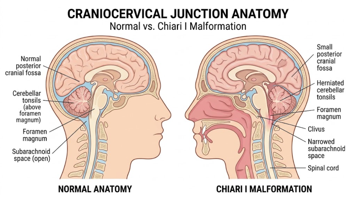
Figure 1. Normal anatomy versus Chiari I malformation of the craniocervical junction. Sagittal section demonstrating downward herniation of the cerebellar tonsils through the foramen magnum in Chiari I malformation compared with a normal craniocervical junction.
The herniated cerebellar tonsils physically crowd the foramen magnum, resulting in severe structural constriction as depicted in Figure 1 above. This anatomic crowding eliminates normal cisternal buffers, physically seals the subarachnoid space, and compresses the brainstem and upper cervical cord.
Under physiological conditions, cerebrospinal fluid is dynamically oscillating between the intracranial and spinal subarachnoid spaces in synchrony with the cardiac cycle. During cardiac systole, the influx of blood into the arteries temporarily increases the intracranial volume, causing the cerebrospinal fluid to be displaced caudally through the foramen magnum into the spinal subarachnoid space. Venous drainage allows CSF to flow back cranially into the basal cisterns during cardiac diastole. This constant movement absorbs changes in pressure and maintains pressure balance between the cranium and spine(Heiss et al., 1999; Oldfield, Muraszko, Shawker, & Patronas, 1994).
In patients with Chiari malformation type I, this hydrodynamic balance is disturbed by mechanical compression at the craniovertebral junction. Herniated tonsils and bony structures obstruct the subarachnoid space at the foramen magnum. This makes flow harder. Cine phase-contrast MR imaging reveals significant alteration of the cerebrospinal fluid flow pattern, loss of bidirectional flow and development of a high velocity turbulent jet at the craniocervical junction. The mechanical obstruction alters fluid mechanics and generates pressure forces in various hydrodynamic models(Al-Habib et al., 2020; Gad & Yousem, 2017).
Historically, James Gardner proposed the water-hammer theory, that obstruction of the foramen magnum forces pressure waves down the central canal via an open obex. Modern magnetic resonance imaging however shows that in most patients there is no communication between the fourth ventricle and syrinx and this has led to extraventricular hydrodynamic models(Al-Habib et al., 2020).
Bernard Williams developed the theory of craniospinal pressure dissociation. Williams showed that increases in intrathoracic and intra-abdominal pressure fill the epidural venous plexus and compress the spinal dural sac. The cerebellar tonsils impacted in Chiari type I malformation act as a valve that only allows fluid to move downwards, but not back up. This causes a dissociation of pressure between the spinal and cranial compartments and a suction effect that draws fluid into spinal cord parenchyma(Williams, 1969).
Edward Oldfield clarified these dynamic forces by means of his piston theory. Oldfield, using phase-contrast magnetic resonance imaging, discovered that rapid expansion of intracranial blood volume during cardiac systole forces herniated cerebellar tonsils downward like a solid piston into the obstructed foramen magnum. This displacement compresses the underlying spinal subarachnoid space and exaggerated pressure waves travel down the spinal canal and strike the cord surface with each heartbeat(Oldfield et al., 1994).
Further, Dan Greitz suggested the intramedullary pulse pressure model focusing on the Venturi effect. Hydrodynamic principles state that forcing fluid at high velocity through a narrowed subarachnoid channel at the craniovertebral junction results in a decrease in lateral fluid pressure along the cord surface. The pressure drop creates a difference in pressure between the inside of the cord and the outside subarachnoid space. This creates a localized suction that expands inner tissue(Greitz, 2006).
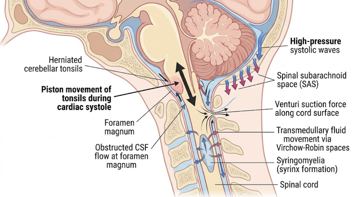
Figure 2. Pathophysiology of syringomyelia in Chiari malformation type I. Schematic representation of the fluid dynamics at the craniocervical junction. During systole, the herniated cerebellar tonsils move in a piston-like manner, obstructing normal CSF flow, and generating high pressure waves and Venturi suction forces that lead to transmedullary fluid movement into the spinal cord and eventually syrinx formation.
The mechanical squeezing of the cerebellar tonsils turns the normal flow of cerebrospinal fluid into turbulent high-velocity jets and pressure pulses that constantly act on the tissue of the spinal cord and cause fluid buildup, as shown in Figure 2 above.
Continuous mechanical compression and altered fluid dynamics at the cranio-vertebral junction clinically result in the formation and progressive enlargement of syringomyelia. Current neuro-imaging and experimental data support that the fluid within the syrinx derives mainly from the spinal subarachnoid space and interstitial fluid, and not from direct ventricular overflow(Dlouhy, Dawson, & Menezes, 2017; Koyanagi & Houkin, 2010).
The perivascular spaces (Virchow-Robin spaces) are the predominant anatomical route for fluid from the subarachnoid space to enter the spinal cord parenchyma. Perivascular spaces are fluid filled channels around penetrating blood vessels as they enter the spinal cord tissue. Under physiological conditions these channels favor the exchange and drainage of interstitial fluid. However, piston-like movement of cerebellar tonsils produces exaggerated pressure waves in the spinal subarachnoid space, establishing a strong transmedullary pressure gradient(Oldfield et al., 1994).
The increased pressure gradient leads to an inward flow of cerebrospinal fluid driven by repetitive cardiac pulsations through perivascular channels into the extracellular matrix of the spinal cord. In the presence of interstitial fluid accumulation within cord parenchyma, the compliance of tissue is exceeded. Fluid flows along the lines of least resistance in the central gray matter and microscopic fluid cavities coalesce to form a macroscopic syrinx(Rai & Rai, 2015).
As the syrinx enlarges, it exerts progressively more mechanical strain on neuroanatomical pathways. The central location of syrinx cavities initially compresses the decussating fibers of the anterior white commissure of the spinothalamic tract. The lesion results in loss of dissociated sensation over the shoulders, arms and upper chest with preservation of touch and proprioception(Kawaguchi, Fujimura, & Tominaga, 2009; Thotakura & Marabathina, 2017). Continued expansion compresses the gray matter of the anterior horn, destroys the lower motor neurons, and causes muscle weakness, atrophy, and loss of reflexes. In severe cases, lateral expansion compresses corticospinal tracts, leading to spastic paraparesis, gait ataxia, and neurogenic bladder dysfunction(Lo et al., 2026).
Surgical results of posterior fossa decompression confirm a mechanical origin of syrinx formation. Removal of the suboccipital bone and posterior arch of C1 with duraplasty or tonsillar reduction removes the physical barrier at the foramen magnum. Increasing the anatomical volume at the craniovertebral junction relieves the piston-like obstruction of the tonsils, restores patency of the subarachnoid space and reestablishes the bidirectional fluid circulation, resulting in significant shrinkage or resolution of the syrinx(Lo et al., 2026; Saenz et al., 2026).
The literature on Chiari I malformation supports a common mechanical paradigm where structural crowding at the skull base directly alters cerebrospinal fluid dynamics and leads to syrinx formation. But there are some important clinical nuances and surgical controversies that need to be evaluated in detail(Alhosani & Gachechiladze, 2026; Holder et al., 2025).
One of the main points of discussion in neurosurgery is how to define pathological compression of the craniovertebral junction precisely and identify surgical candidates(Holder et al., 2025). Diagnosis has been made on the measurement of more than 5 millimeters of cerebellar tonsillar descent below the foramen magnum on sagittal magnetic resonance imaging(Al-Habib et al., 2020). However, clinical observation has shown that the absolute degree of tonsillar herniation is not always linearly correlated with the severity of symptoms or the presence of syrinx(Holder et al., 2025). In a clinical series by Hatgaonkar et al., patients with 4 mm of tonsillar descent had extensive holocord syringomyelia, whereas patients with >22 mm descent had small syringes or minimal symptoms(Hatgaonkar et al., 2024).
This disparity has brought the attention to multi-parametric scoring systems, and volumetric overcrowd assessments. Holder and colleagues assessed the Chiari 1 Malformation Severity Classification System, and showed that the combination of classic symptoms, tonsillar descent greater than ten millimeters, and presence of syrinx, provide high diagnostic accuracy in identifying patients who benefit from surgical decompression(Holder et al., 2025).
The other big topic is surgical management of complex Chiari malformations with associated craniovertebral junction anomalies. Goel’s hypothesis suggests that central atlantoaxial instability is the primary cause of all craniovertebral junction abnormalities and that all patients should be treated with cervical spinal fusion without bone decompression(Goel, 2009; Wagner et al., 2020). Wagner et al. reviewed this hypothesis, and found no empirical evidence for universal fusion. They concluded that there is no reason to deviate from established decompression procedures to universal stabilization(Wagner et al., 2020). Vazquez et al. reviewed 449 complex Chiari cases and found that while posterior arthrodesis is important in true instability of the joint, foramen magnum decompression is very effective when structural compression is predominant(Vazquez et al., 2023).
The pediatric clinical outcomes reported by Saenz et al. strongly reinforce this conclusion. Of 41 pediatric patients with complex Chiari malformation and osseous anomalies (retroflexed odontoid, medullary kinking, or basilar invagination) 93% experienced successful isolated foramen magnum decompression with significant syrinx regression without the need for spinal fusion. This evidence indicates that posterior mechanical blockage at the foramen magnum can be successfully relieved to restore fluid pathways while maintaining mobility of the cervical spine(Saenz et al., 2026). The necessity of dural opening and duraplasty in the surgical technique of posterior fossa decompression is still a subject of discussion. Limited decompression to bone avoids opening the dura and minimizes the risk of cerebrospinal fluid leak. However, systematic reviews have confirmed that decompression with duraplasty results in significantly higher rates of syrinx resolution(Lo et al., 2026; Turk et al., 2025). In addition, Turk et al reported that arachnoid-preserving suboccipital decompression technique was successful for improving fluid dynamics and reducing syrinx volume in a study of 89 patients and showed high patient satisfaction and low postoperative complications, such as cerebrospinal fluid leakage and arachnoiditis(Turk et al., 2025). In addition, in cases of extensive syringomyelia refractory to adequate foramen magnum decompression, secondary procedures such as syringosubarachnoid shunting as described by Tanaka et al, provide effective drainage of the cavity (Tanaka, Sharma, Gori, Shohidullah, & Uotani, 2025).
To summarize, the obstruction of CSF flow and the development of syringomyelia in Chiari malformation type I is mainly due to a physical mechanism of mechanical compression at the craniovertebral junction. Anatomical crowding of the posterior cranial fossa and foramen magnum along with congenital or acquired osseous skull base anomalies lead to a bottleneck at the craniocervical junction. This structural narrowing alters the physiologic fluid mechanics and causes the herniated cerebellar tonsils to act as a piston during the cardiac systole. This results in increased subarachnoid pressure waves and transmural pressure gradients that push cerebrospinal fluid into the spinal cord parenchyma via perivascular spaces with accumulation of interstitial fluid and syrinx enlargement.
Together with previous studies of ependymal cell responses and cellular signaling pathways, these biomechanical principles provide us with a comprehensive framework for understanding syringomyelia. Surgical decompression of the foramen magnum continues to be the most effective intervention for the relief of craniovertebral junction compression, removal of abnormal pressure gradients, restoration of physiological fluid circulation and promotion of syrinx regression. Ongoing interdisciplinary research in fluid mechanics, neuroimaging and surgical outcomes will continue to refine treatment strategies.
Al-Habib, A. F., Al Abdulsalam, H., Ahmed, J., Albadr, F., Alhothali, W., Alzahrani, A., . . . Alkubeyyer, M. (2020). Association between craniovertebral junction abnormalities and syringomyelia in patients with chiari malformation type-1. Neurosciences (Riyadh), 25(4), 308-315. doi:10.17712/nsj.2020.4.20200008
Alhosani, M. S., & Gachechiladze, S. (2026). Chiari Malformation Type I: A Review of Pathophysiology, Cerebrospinal Fluid Flow Dynamics, Diagnosis, Surgical Management, and Its Relationship to Syringomyelia. Cureus, 18(1), e101226. doi:10.7759/cureus.101226
Chiari, H. (1891). Ueber Veränderungen des Kleinhirns infolge von Hydrocephalie des Grosshirns1). Dtsch Med Wochenschr, 17(42), 1172-1175. doi:10.1055/s-0029-1206803
Cogan, D. G., & Barrows, L. J. (1954). Platybasia and the Arnold-Chiari malformation. AMA Arch Ophthalmol, 52(1), 13-29. doi:10.1001/archopht.1954.00920050015002
Dlouhy, B. J., Dawson, J. D., & Menezes, A. H. (2017). Intradural pathology and pathophysiology associated with Chiari I malformation in children and adults with and without syringomyelia. J Neurosurg Pediatr, 20(6), 526-541. doi:10.3171/2017.7.Peds17224
Fernandez, A. A., Guerrero, A. I., Martinez, M. I., Vazquez, M. E., Fernandez, J. B., Chesa i Octavio, E., . . . Ramon, J. R. (2009). Malformations of the craniocervical junction (Chiari type I and syringomyelia: classification, diagnosis and treatment). BMC Musculoskelet Disord, 10 Suppl 1(Suppl 1), S1. doi:10.1186/1471-2474-10-S1-S1
Fernández, A. A., Guerrero, A. I., Martínez, M. I., Vázquez, M. E., Fernández, J. B., Chesa i Octavio, E., . . . Ramón, J. R. (2009). Malformations of the craniocervical junction (Chiari type I and syringomyelia: classification, diagnosis and treatment). BMC Musculoskelet Disord, 10 Suppl 1(Suppl 1), S1. doi:10.1186/1471-2474-10-s1-s1
Gad, K. A., & Yousem, D. M. (2017). Syringohydromyelia in Patients with Chiari I Malformation: A Retrospective Analysis. AJNR Am J Neuroradiol, 38(9), 1833-1838. doi:10.3174/ajnr.A5290
Goel, A. (2009). Basilar invagination, Chiari malformation, syringomyelia: a review. Neurol India, 57(3), 235-246. doi:10.4103/0028-3886.53260
Greitz, D. (2006). Unraveling the riddle of syringomyelia. Neurosurg Rev, 29(4), 251-263; discussion 264. doi:10.1007/s10143-006-0029-5
Hatgaonkar, A. M., Mahajan, S. M., Hatgoankar, K. A., & Bandre, G. R. (2024). MRI Insights in Chiari Malformation Type 1 and Variations With Hydrosyringomyelia. Cureus, 16(3), e55676. doi:10.7759/cureus.55676
Heiss, J. D., Patronas, N., DeVroom, H. L., Shawker, T., Ennis, R., Kammerer, W., . . . Oldfield, E. H. (1999). Elucidating the pathophysiology of syringomyelia. J Neurosurg, 91(4), 553-562. doi:10.3171/jns.1999.91.4.0553
Holder, S., Abbakr, M. A., Haynes, A., & Sanusi, T. D. (2025). Chiari 1 Malformation, Factors That May Influence Decision Making, and Introducing the Chiari 1 Malformation Severity Classification System. J Clin Med, 14(17). doi:10.3390/jcm14176113
Kawaguchi, T., Fujimura, M., & Tominaga, T. (2009). Syringomyelia with obstructive hydrocephalus at the foramens of Luschka and Magendie successfully treated by endoscopic third ventriculostomy. Surg Neurol, 71(3), 349-352, discussion 352. doi:10.1016/j.surneu.2007.08.015
Koenigsberg, R. A., Vakil, N., Hong, T. A., Htaik, T., Faerber, E., Maiorano, T., . . . Gonzales, C. (2005). Evaluation of platybasia with MR imaging. AJNR Am J Neuroradiol, 26(1), 89-92.
Koyanagi, I., & Houkin, K. (2010). Pathogenesis of syringomyelia associated with Chiari type 1 malformation: review of evidences and proposal of a new hypothesis. Neurosurg Rev, 33(3), 271-284; discussion 284-275. doi:10.1007/s10143-010-0266-5
Lo, J. J., Shiino, S., Shapiro, S. Z., Hines, B. L., Komune, N., Donofrio, C. A., . . . Tubbs, R. S. (2026). Chiari I Malformation: Review and Update of Current Treatment Options. Clinical Anatomy, n/a(n/a). doi:https://doi.org/10.1002/ca.70079
Mahajan, N., Phatak, S., Onkar, P., Ambhore, A. N., & Pantawane, P. (2024). Revealing an Uncommon Presentation of Chiari I Malformation With Diverse Craniovertebral Anomalies in the Absence of Syringomyelia and Atlanto-Occipital Subluxation: A Case Report. Cureus, 16(3), e55332. doi:10.7759/cureus.55332
Nishikawa, M., Bolognese, P. A., Yamagata, T., Naito, K., Sakamoto, H., Hara, M., . . . Goto, T. (2022). Surgical Management of Chiari Malformation Type I and Instability of the Craniocervical Junction Based on Its Pathogenesis and Classification. Neurol Med Chir (Tokyo), 62(9), 400-415. doi:10.2176/jns-nmc.2022-0078
Oldfield, E. H., Muraszko, K., Shawker, T. H., & Patronas, N. J. (1994). Pathophysiology of syringomyelia associated with Chiari I malformation of the cerebellar tonsils. Implications for diagnosis and treatment. J Neurosurg, 80(1), 3-15. doi:10.3171/jns.1994.80.1.0003
Rai, S. K., & Rai, P. S. (2015). Volume change theory for syringomyelia: A new perspective. Asian J Neurosurg, 10(4), 245-251. doi:10.4103/1793-5482.162680
Saenz, A., Zhu, H., Dhaliwal, J., Sudhakar, S., Mankad, K., Thompson, D., & Tahir, M. Z. (2026). Pediatric complex Chiari I Malformation-how complex is it? Childs Nerv Syst, 42(1). doi:10.1007/s00381-026-07325-6
Tanaka, M., Sharma, S., Gori, K. H., Shohidullah, M., & Uotani, K. (2025). A Rare Course of Chiari Malformation With Large Syringomyelia Presenting at 54 Years Old. Cureus, 17(2), e78399. doi:10.7759/cureus.78399
Thotakura, A. K., & Marabathina, N. R. (2017). Acquired Chiari I Malformation with Syringomyelia Secondary to Colloid Cyst with Hydrocephalus-Case Report and Review of Literature. World Neurosurg, 108, 995.e991-995.e994. doi:10.1016/j.wneu.2017.09.012
Turk, O., Demirel, N., Karakurt, M. T., Cetiner, R., Sahin, O. F., Karaoglu, A. C., . . . Yaltirik, C. K. (2025). Chiari Malformation Type I and Syringomyelia: Outcomes of Arachnoid-Preserving Surgical Technique. Med Sci Monit, 31, e946978. doi:10.12659/msm.946978
Vazquez, S., Dominguez, J. F., Das, A., Soldozy, S., Kinon, M. D., Ragheb, J., . . . Morcos, J. J. (2023). Treatment of Chiari malformations with craniovertebral junction anomalies: Where do we stand today? World Neurosurg X, 20, 100221. doi:10.1016/j.wnsx.2023.100221
Wagner, A., Grassner, L., Kögl, N., Hartmann, S., Thomé, C., Wostrack, M., & Meyer, B. (2020). Chiari malformation type I and basilar invagination originating from atlantoaxial instability: a literature review and critical analysis. Acta Neurochir (Wien), 162(7), 1553-1563. doi:10.1007/s00701-020-04429-z
Williams, B. (1969). The distending force in the production of “communicating syringomyelia”. Lancet, 2(7613), 189-193. doi:10.1016/s0140-6736(69)91427-5