Our Services
Others
- DFPP
- Shockwave
- IV Drip


“In this Systematic Review, we review how disruption of circumferential hoop tension in meniscal root tears results in abnormal tibiofemoral kinematics and pathologic meniscal extrusion with equally pathologic peak contact pressure and accelerated degeneration of knee joint cartilage.”
Meniscus root tears (MRTs) represent a distinct and frequently catastrophic form of knee injury, involving radial tears within 1 cm of the meniscal insertions into the tibia and/or complete bony detachment of the root. Commitment of the meniscal root to a normal meniscus converts tibiofemoral axial compressive loads into circumferential hoop tension stresses bearing and transmitting the load, thus affording an important shock-absorbing function. This requires adequate anchorage of the anterior and posterior meniscal horns to the planar surfaces of the tibial plateaus. These circumferential collagen fiber bundles of the meniscus are violated in meniscal root tears to result in a catastrophic loss of hoop tension (loss of belt effect). Destruction of the meniscal tissue within the joint compartment causes radial extrusion of the meniscus, staggering decreases in effective tibiofemoral contact area (30% to 50%) and peak contact stress (25% to >100%), biomechanically analogous to a total menisectomy. In addition, the root deficiency disturbs overall knee kinematics dramatically, with more lateral translation of the tibia, over-translation of the medial compartment, and dissociation of knee arthrokinematics during dynamic “weight-bearing” activities such as rapid walking or deep squat. In this review, we provide a synthesis of fundamental cadaveric, computational finite-element, and in vivo kinematic studies to delineate the cascade of mechanical failure from loss of hoop stress to rapid osteoarthritis.
The menisci of the knee joint are semilunar, semifibrocartilaginous structures positioned between the convex femoral condyles and the relatively flat tibial plateaus. Despite early anatomists’ belief that the menisci were vestigial and extraneous, decades of biomechanical investigation have demonstrated the key roles of the menisci in the maintenance of intra-articular equilibrium, congruence, load sharing, shock absorption, and secondary mechanical stability(M. S. Kim, In, Kim, Jeong, & Sohn, 2025). For example, during axial loading and extension of the knee during normal walking, 40-50% of the inner compartment axial load at the tibia is borne by the menisci, with 65-70% of the outer compartment axial load, as well as 90% of total axial load in deep knee flexion(Canton et al., 2026).
Meniscus root tears (MRTs) are the most common clinical form of meniscal structural compromise. An MRT is a radial tear within 1 cm (9 mm) of the meniscal bony attachment or complete bony or soft-tissue root avulsion of the meniscal terminal anchor on the tibial plateau. Unlike (1) longitudinal or complex tears, best recognized in young athletes after acute rotational trauma, (2) posterior meniscal root tears, especially of the medial meniscus, are disease entities of middle age and older patients, often following a trivial sporting or work activity, such as deep squatting or low-velocity twisting activities with the joint in 90° of flexion(Chambers, Lynch, Feeley, & Nevitt, 2023; Hantouly et al., 2024; Pache et al., 2018).
Untreated MRT is a biomechanical catastrophe in the knee. Root tears ablate the meniscal attachment to bone and its capacity to generate circumferential hoop tension in response to axial compressive loads. Without a meniscal anchor, the meniscal body extrudes outward beyond the tibial plateau, decreasing the contact surface area for axial forces. It also has a catastrophic effect on the small, mechanically overloaded, and unadapted areas of articular cartilage (point load) and leads to subchondral bone insufficiency fractures and to rapid onset osteoarthritis, cartilage degeneration, and subchondral bone marrow lesions(Banovetz, Roethke, Rodriguez, & LaPrade, 2022; Pache et al., 2018).
Complete meniscal root tears statically redistribute load but also modify tibiofemoral kinematics and increase dynamic instability in the joint. Free-floating meniscus can no longer provide the mechanical cushion or motion guidance to the joint, leading to abnormal tibia and femur translations and rotations during normal joint motion(Marsh, Martin, Harner, & Tashman, 2014). Understanding these biomechanical mechanisms is important in the clinical decision making process. Here we will review the current scientific evidence regarding hoop tension conversion, patho-mechanics of root disruption, in-vivo and dynamic kinematic change, medial-lateral differences in root tears, and mechanics of surgical repair.
Knowledge of the architecture of the native meniscus allowing for the generation of hoop tension, both on a microscopic and macroscopic level, is vital to understand the catastrophic effects of a root tear. The meniscus is primarily composed of type I collagen fibers in a dense, highly aligned fibrocartilage matrix (80–90% of dry weight of the meniscus) interspersed with water, proteoglycans, and non-collagenous proteins. The collagen fibrils in the body of the meniscus are organized in three anatomical zones(Petersen & Tillmann, 1998). The superficial zone is a fine, random meshwork of fibrils that cover the articular surfaces and reduce friction with motion. Next is the lamellar zone, in which collagen bundles are arranged in radial lines. Beneath this is the main structural core of the tissue, in which thick bundles of collagen fibrils are oriented continuously in the circumferential direction from the intertubular attachment to the tibial attachment(Y. M. Kim, Joo, An, & Song, 2022).
It is this circumferential fiber orientation that leads to the so-called hoop tension mechanism. For example, in the stance phase of the knee or during heel strike, the femoral condyle axially compresses the wedge-shaped meniscus. This compressive displacement acts to displace the meniscus radially outward, toward the outside of the joint compartment. However, any such radial outward displacement is inhibited by the resilient fibrocartilaginous meniscal root attachments, which anchor the anterior and posterior horn of the meniscus into the subchondral surface of the tibial plateau bone(M. S. Kim et al., 2025; Pache et al., 2018).
This boundary constraint allows the outward radial force to be vectorially converted into a circumferential tensile stress traveling the length of the continuous circuit of circumferential collagen fiber. This circumferential tensile resistance, like a metal hoop around a wooden barrel, inhibits expansion of the tissue laterally beneath the femoral condyle. This elastic containment allows the meniscus to yield slightly when a load is applied to absorb some of the compressive forces experienced while providing a complementary (and congruous) shape to the joint and also prevents the direct concentration of axial loads on a small area of the tibial plateau(Banovetz et al., 2022; Monson & LaPrade, 2025).
Several material and structural properties inherent to meniscus tissue also assist in maintaining hoop tension against dynamic loads, such as the circumferential alignment and root anchoring of the collagen fibers. First, lysyl oxidase crosslinks collagen fibrils covalently with tensile strength sufficient to resist rupture or slippage under maximal loading. At the same time, the negatively charged glycosaminoglycan (GAG) side chains of proteoglycan embedded in the matrix attract water molecules, which creates a hydrostatic osmotic swelling pressure(Korhonen et al., 2002). This dense, interwoven collagen matrix generates hydraulic internal pressure by providing frictional drag for compressed interstitial fluid flow, enabling dissipation of localized fiber stress concentrations. In addition, viscoelastic tissue behaviors like stress relaxation and creep add stability to the internal stress distribution under prolonged weight bearing(M. S. Kim et al., 2025).
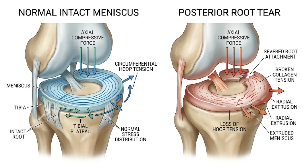
Figure 1. Biomechanical Implications of a Posterior Meniscal Root Tear. (Left) A healthy meniscus converts axial compressive forces into hoop tension. (Right) A root tear causes discontinuous fibers, loss of circumferential tension, and radial extrusion of the meniscus under load.
The meniscal root serves to tether the meniscus to the tibial plateau to transmit axial loads to hoop tension in noninterrupted collagen fibers (Figure 1) for structural integrity. In a root tear, this restraint is compromised and leads to a total failure of the laminae of hoop tension(M. S. Kim et al., 2025).
A radial tear within 1 cm of the meniscal insertion or root is a complete avulsion of the root from the bone with total failure of the biomechanical continuity of the peripheral collagen fibers. It destroys the structural loop required for maintaining circumferential tension. Absent stable root fixation, compressive loads transferred from the femoral condyle can no longer be translated into tensile hoop stress. Rather, the unanchored meniscus is extruded circumferentially out of the joint space under physiologic loads(Canton et al., 2026).
Numerous cadaveric, computational, and clinical imaging studies have characterized catastrophic load distribution patterns in the setting of meniscal root deficiency. The meniscus transmits load about the surface of the joint by enlarging the area of contact between femur and tibia. Conversely, MMPRT leads to marginalization of the body of the meniscus from the joint compartment, with an instantaneous dramatic diminishment of the total tibiofemoral contact area (often between 30% and >50%)(Allaire, Muriuki, Gilbertson, & Harner, 2008).
This is conducive to such a large increase in contact pressure (force per area, i.e., mechanical stress) across the tibiofemoral joint. Indeed, classic cadaveric studies have demonstrated that a meniscectomy or posterior root resection of the medial meniscus increases peak contact pressure in the medial compartment by 25 to 78%(Allaire et al., 2008).
Root tears also change the distribution and spatial elevate of contact pressure at the articular surface. With an intact joint, the articular cartilage within the center of the tibial plateau is designed to sustain repetitive loading. However, dynamic gait simulation studies have demonstrated that meniscal root tears lead to a very different pattern of peak pressure transmission, with flier counts posterocentral and posteromedial to the normal site. This load displacement results in excessive force-crushing of unconditioned areas of the tibial articular cartilage, which does not possess the force strength or biochemistry to tolerate heavy mechanical loads(Andriacchi & Mündermann, 2006; Banovetz et al., 2022).
This ‘point loading’ results in massive cartilage wear. Root deficiency is associated with rapid degeneration of cartilage, loss of the medial femoral condyle, and bone marrow edema and subchondral insufficiency fractures of the knee (SIFK) in animal and human models. Epidemiologic studies have shown that 95% of untreated root tears will develop osteoarthritis and 50% will develop total knee arthroplasty in 10 years(Chambers et al., 2023; M. S. Kim et al., 2025).
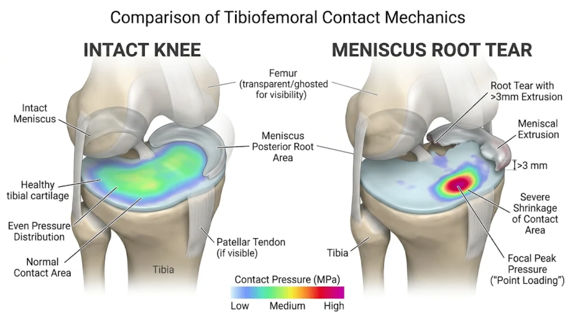
Figure 2. Tibiofemoral Contact Mechanics in Meniscus Root Tear and Intact Knees. (Left) Intact knee showing large area of contact and uniform distribution of pressure. (Right) Meniscal root tear with known >3 mm extrusion and reduced contact area with a central point of pressure (point loading) on the tibial plateau.
Meniscal root detachment permits peripheral extrusion of the meniscal body from the joint space (Figure 2) and critically decreases the contact area and precariously focuses the focal peak pressure (point loading) on unprepared articular cartilage.
Early cadaveric studies using static models showed increased contact pressures, but recent studies using high-speed biplane radiological imaging and dynamic robotic joint simulation models have shown the dramatic kinematic disruptions of meniscal root instability in vivo. The menisci serve as secondary stabilizers of the tibiofemoral joint and, in concert with the primary stabilizers of the cruciate and collateral ligaments, provide normal tibiofemoral joint kinematics and control of excessive tibiofemoral joint translation. The medial meniscus is a tightly wedged protuberance having strong meniscocapsular attachments of the posterior horn resisting the anterior translation of the tibia and stability against rotation, especially in an ACL-deficient knee(Marsh et al., 2014).
Posterior root tears significantly compromise this secondary stabilizer. Dynamic in vivo assessment of patients with unrepaired medial meniscus posterior root tears shows a clear alteration of joint kinematics even during normal-weight-bearing activities such as level walking, decline walking, and squatting. Root-deficient knees, in particular, showed significantly greater lateral tibial translation throughout the range of motion as compared with the uninjured contralateral limb(Marsh et al., 2014).
This is due to the lack of constraint of the medial meniscus, and lateral tibial translation relative to the femur is excessive. Loss of the posterior meniscal wedge also results in abnormal gliding of the medial femoral condyle on the tibial plateau and alters the rotational axis of the joint. Rather than flex and roll about physiological axes, the tibia performs complicated uncoupled motions, rolling and rotating about the intact lateral compartment with excessive excursion of the medial compartment(Marsh et al., 2014).
Critically, in vivo kinematic studies suggest that the magnitude of kinematic disruption is task-dependent, increasing with higher physical demands. Higher shear force and higher flexion angle demanding activities (e.g., use of stairs or squatting) intensify the extent of instability of such root-deficient knees. Here, the affected meniscus cannot direct the smooth motion of the joint and thus allows abnormal subluxations in the anteroposterior and coronal directions, which leads to abnormal stresses in the surrounding soft tissues and in joint capsular tissue(Marsh et al., 2014).
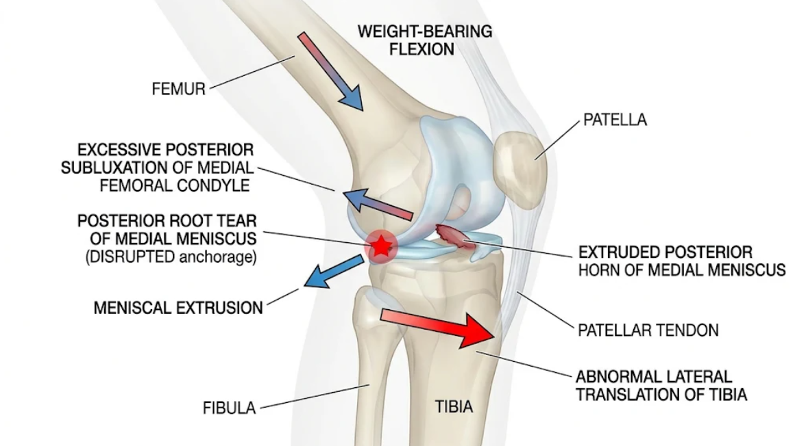
Figure 3. Abnormal Posterior Root Tear of the Medial Meniscus (Kinematics). Weight-bearing flexion with increased lateral tibial translation and medial compartment subluxation.
As in Figure 3, a model of how meniscal root loss results in abnormal tibiofemoral kinematic changes of pathologic lateral tibial translation and asymmetric medial compartment translation during the subluxation mode of active flexion.
Medial and lateral root tears of the meniscus disrupt hoop tension, but there is substantial evidence that they differ in injury patterns, as well as biomechanically and anatomically(LaPrade et al., 2015).
The majority of root injuries (approximately 75-80%) occur in the middle-aged female or obese population and involve the posterior root of the medial meniscus (MMPRT). The medial meniscus has a greater radius of curvature, is less mobile, and lacks additional ligamentous attachments to the femur. As such, MMPRT causes unrestricted meniscal extrusion, abrupt hoop stress failure, and substantial increases in medial compartment loading(Carreau, Sitton, & Bollier, 2017; Pache et al., 2018).
LMPRTs, however, are more common in young active males, secondary to acute high-energy trauma, often in combination with ACL ruptures. The lateral meniscus is also more mobile and occupies a greater proportion of the lateral tibial plateau. Importantly, the posterior horn of the lateral meniscus is often anchored to the medial femoral condyle by the meniscofemoral ligaments (MFLs), ligament of Humphry (anterior) and ligament of Wrisberg (posterior)(M. S. Kim et al., 2025).
Biomechanical analyzes conclude that an isolated LMPRT does not immediately cause a catastrophic reduction in contact area, nor a single heightened contact pressures if MFL is intact. The intact MFLs function as a biomechanical “bridge” (or secondary anchor) to counteract the lateral meniscal extrusion and maintain circumferential tension. However, compared to an LMPRT, MFL transection/disruption leads to profound disturbance of the joint-contact mechanics (similar to total lateral meniscectomy)(Sequeira et al., 2023).
Recent state of the art robotic gait simulation research has elucidated the timing of the gait cycle of maximal mechanical insult from root deficiency. While prior X-ray cadaver studies suggested peak contact pressures at deep flexion angles (≥ 30° to 90°), this has been debunked by recent 6 degrees of freedom dynamic gait simulations with physiological axial loading and shear and moment forces(Brusalis et al., 2024).
For instance, in level walking with an accompanying MMPRT (low flexion [<15°]), peak contact stress (~2 MPa) increased, while contact area decreased (~200 mm²). This time period (20-37% of gait cycle) is essentially loading response and early stance, a time of gait with peak axial compressive forces and posteriorly directed shear forces and internal torques. An important clinical observation is that root tears influence joint loading even during the near-extension phase of gait, which helps explain the failure of nonoperative treatment and provides a biomechanical rationale for rigid post-operative motion precautions to protect healing root repairs during early rehabilitation(Brusalis et al., 2024).
In conclusion, a root tear is more than just a focal, structural defect but rather the failure of the main chondroprotective mechanism of the knee joint. [28] Compared with the retained ability to translate compressive loads into circumferential hoop tension by a functioning root, a root tear converts a wide low-profile surface of the joint into a point-loaded regime exhibiting meniscal extrusion, reduced circumferential contact area, and increased peak contact stresses(Pache et al., 2018).
The paradigm of root tears, meniscal extrusion, and secondary meniscal restraints is extremely controversial in the orthopedic literature. Although historically it has been assumed that root tears lead to extrusion, recent cohort studies demonstrate that attenuation or splitting of secondary restraints, such as the meniscotibial (coronary) ligament, often occurs before frank root tears. The meniscotibial ligament permits some early extrusion, and failure of this structure maximally increases tensile forces on the posterior root attachment during physiologic loading until posteromedial meniscal pain results. This timeline explains why anatomic root repair alone cannot fully reverse pre-existing meniscal extrusion, and why surgical centralization procedures have been developed that re-secure the outer border of the meniscus to the tibial rim in combination with root refixation(Krych et al., 2020; Monson & LaPrade, 2025).
Biomechanical studies provide compelling evidence for the necessity of anatomic root reattachment from a surgical restoration perspective. Transtibial pull-out repairs that restore the root to its native anatomic footprint restore the contact pressure, contact area, and joint kinematics. Non-anatomic repairs (3-5 mm posteromedial or lateral re-anchoring of the root) did not restore circumferential hoop stress(Cortina et al., 2026; Pache et al., 2018). As such, hoop tension is not restored with a non-anatomic repair, and therefore meniscal extrusion and contact pressure is similar to the root-deficient state. Furthermore, malalignment of the lower limb may play a major role: in varus mechanical alignment, forces are focused across the medial compartment, reducing the mechanical advantage of a root repair without correction via a high tibial osteotomy(Cortina et al., 2026).
Meniscus root tears directly disrupt the circumferential hoop tension mechanism, resulting in dramatic tibiofemoral kinematic disruption and marked abnormal joint loading. When forces cause displacement of the meniscus from its attachment on the tibial plateau, it extrudes peripherally, with resulting dramatic reduction in contact area, increased peak contact stresses, and alterations in dynamic in vivo joint kinematics. Collectively, these mechanical derangements create the same loading environment as total meniscectomy and lead to rapid and excessive cartilage degeneration, subchondral incompliance, and osteoarthritis. Anatomic surgical strategic root restoration recreates the better tibiofemoral contact mechanics and the health of the knee joint long term, recreated with physiologic hoop stress.
Allaire, R., Muriuki, M., Gilbertson, L., & Harner, C. D. (2008). Biomechanical consequences of a tear of the posterior root of the medial meniscus. Similar to total meniscectomy. J Bone Joint Surg Am, 90(9), 1922-1931. doi:10.2106/jbjs.G.00748
Andriacchi, T., & Mündermann, A. (2006). The role of ambulatory mechanics in the initiation and progression of knee osteoarthritis. Current Opinion in Rheumatology, 18(5), 514-518. doi:10.1097/01.bor.0000240365.16842.4e
Banovetz, M. T., Roethke, L. C., Rodriguez, A. N., & LaPrade, R. F. (2022). Meniscal Root Tears: A Decade of Research on their Relevant Anatomy, Biomechanics, Diagnosis, and Treatment. Arch Bone Jt Surg, 10(5), 366-380. doi:10.22038/abjs.2021.60054.2958
Brusalis, C. M., Chastain, K. L., Uppstrom, T. J., Steineman, B. D., Gomoll, A. H., Maher, S. A., & Strickland, S. M. (2024). Medial Meniscus Posterior Root Tears Lead to Changes in Joint Contact Mechanics at Low Flexion Angles During Simulated Gait. Am J Sports Med, 52(1), 109-115. doi:10.1177/03635465231206398
Canton, G., Marchetti, A., Trobec, B., Scordo, M., Mazzon, A., Ratti, C., . . . Murena, L. (2026). Impact of meniscal root tears on knee osteoarthritis development: A systematic review of the literature. The Knee, 58, 104258. doi:https://doi.org/10.1016/j.knee.2025.10.012
Carreau, J. H., Sitton, S. E., & Bollier, M. (2017). Medial Meniscus Root Tear in the Middle Aged Patient: A Case Based Review. Iowa Orthop J, 37, 123-132.
Chambers, C. C., Lynch, J. A., Feeley, B. T., & Nevitt, M. C. (2023). Association of Medial Meniscus Root Tears and Nonroot Tears With Worsening of Radiographic Knee Osteoarthritis. Orthop J Sports Med, 11(9), 23259671231195894. doi:10.1177/23259671231195894
Cortina, G., Perelli, S., Pizza, N., Condello, V., Madonna, V., & Monllau, J. C. (2026). Medial meniscus posterior root tear repair: An overview of surgical techniques and augmentation strategies. J Exp Orthop, 13(1), e70578. doi:10.1002/jeo2.70578
Hantouly, A. T., Aminake, G., Khan, A. S., Ayyan, M., Olory, B., Zikria, B., & Al-Khelaifi, K. (2024). Meniscus root tears: state of the art. International Orthopaedics, 48(4), 955-964. doi:10.1007/s00264-024-06092-w
Kim, M. S., In, Y., Kim, H., Jeong, J., & Sohn, S. (2025). Why Hoop Tension Matters: A Biomechanical Perspective on Medial Meniscus Posterior Root Tears—A Narrative Review. Bioengineering, 12(6), 638. Retrieved from doi:10.3390/bioengineering12060638
Kim, Y. M., Joo, Y. B., An, B. K., & Song, J. H. (2022). Age and Meniscal Extrusion Are Determining Factors of Osteoarthritis Progression after Conservative Treatments for Medial Meniscus Posterior Root Tear. J Pers Med, 12(12). doi:10.3390/jpm12122004
Korhonen, R. K., Laasanen, M. S., Töyräs, J., Rieppo, J., Hirvonen, J., Helminen, H. J., & Jurvelin, J. S. (2002). Comparison of the equilibrium response of articular cartilage in unconfined compression, confined compression and indentation. Journal of Biomechanics, 35(7), 903-909. doi:https://doi.org/10.1016/S0021-9290(02)00052-0
Krych, A. J., LaPrade, M. D., Hevesi, M., Rhodes, N. G., Johnson, A. C., Camp, C. L., & Stuart, M. J. (2020). Investigating the Chronology of Meniscus Root Tears: Do Medial Meniscus Posterior Root Tears Cause Extrusion or the Other Way Around? Orthop J Sports Med, 8(11), 2325967120961368. doi:10.1177/2325967120961368
LaPrade, C. M., Smith, S. D., Rasmussen, M. T., Hamming, M. G., Wijdicks, C. A., Engebretsen, L., . . . LaPrade, R. F. (2015). Consequences of tibial tunnel reaming on the meniscal roots during cruciate ligament reconstruction in a cadaveric model, Part 2: The posterior cruciate ligament. Am J Sports Med, 43(1), 207-212. doi:10.1177/0363546514554771
Marsh, C. A., Martin, D. E., Harner, C. D., & Tashman, S. (2014). Effect of Posterior Horn Medial Meniscus Root Tear on In Vivo Knee Kinematics. Orthop J Sports Med, 2(7), 2325967114541220. doi:10.1177/2325967114541220
Monson, J. K., & LaPrade, R. F. (2025). Posterior Medial Meniscus Root Tears: Clinical Implications, Surgical Management, and Post-operative Rehabilitation Considerations. Int J Sports Phys Ther, 20(1), 127-136. doi:10.26603/001c.126967
Pache, S., Aman, Z. S., Kennedy, M., Nakama, G. Y., Moatshe, G., Ziegler, C., & LaPrade, R. F. (2018). Meniscal Root Tears: Current Concepts Review. Arch Bone Jt Surg, 6(4), 250-259.
Petersen, W., & Tillmann, B. (1998). Collagenous fibril texture of the human knee joint menisci. Anatomy and Embryology, 197(4), 317-324. doi:10.1007/s004290050141
Sequeira, S. B., Ayan, S., Kushare, I., Gould, H. P., Fillar, A., & Dreese, J. (2023). Posterior Root Tears of the Lateral Meniscus Only Affect Tibiofemoral Contact Forces when the Meniscofemoral Ligament Is Involved: A Cadaveric Study. Arthrosc Sports Med Rehabil, 5(2), e515-e521. doi:10.1016/j.asmr.2023.02.003