Our Services
Others
- DFPP
- Shockwave
- IV Drip


“This literature review offers a deep mechanistic understanding of the functional interplay between aggrecan proteoglycans and type II collagen in the nucleus pulposus to maintain the osmotic swelling pressure and spine shock absorption and how the perturbation of this extracellular matrix balance speeds up intervertebral disc degeneration.”
Intervertebral disc degeneration is a major structural cause of chronic low back pain and spinal disability globally. The intervertebral disc is a complex biomechanical organ that is designed to resist multidirectional loads and to provide flexibility to the spine. The core of this functional unit is the nucleus pulposus, a gel-like hydrated extracellular matrix whose physiological function depends on a delicate biochemical balance between 2 fundamental macromolecules, the large aggregating proteoglycan aggrecan and a network of type II collagen fibrils. Aggrecan contains a high density of negatively charged glycosaminoglycan side chains of chondroitin sulfate and keratan sulfate that attract water and create an internal osmotic swelling pressure. This hydrostatic pressure is physically constrained by the tensile strength of the surrounding type II collagen network. The resulting pressurized composite can convert axial compressive forces into hydrostatic pressure, which distributes the mechanical stress to adjacent vertebral bodies. In degenerative disc disease, this delicate balance is disrupted by a catabolic cascade. Inflammatory cytokines and mechanical stress cause a phenotypic shift within the resident nucleus pulposus cells resulting in increased expression of matrix-degrading enzymes such as matrix metalloproteinases and aggrecanases. Proteolytic cleavage of aggrecan leads to loss of glycosaminoglycans, resulting in a marked decrease in osmotic swelling capacity, tissue dehydration, and loss of disc height. At the same time, type II collagen is enzymatically degraded and replaced by fibrotic type I collagen, which decreases matrix elasticity. This literature review synthesizes the molecular architecture, biomechanical mechanisms and catabolic pathways that regulate matrix homeostasis in the nucleus pulposus, and provides a detailed mechanistic understanding of extracellular matrix degradation in spinal degeneration.
Keywords: Nucleus pulposus, Aggrecan, Type II collagen, Swelling pressure, Shock absorption, Degenerative disc disease
Chronic low back pain is a leading cause of disability worldwide and incurs significant health care costs. Epidemiological studies have consistently shown that the principal structural cause of discogenic back pain, spinal instability and radiculopathy is degeneration of the intervertebral discs(Kibble, Domingos, Hoyland, & Richardson, 2022). The human spine is supported by 23 intervertebral discs, flexible cushions between the bodies of the adjacent vertebrae. Each disc is a viscoelastic shock absorber that permits 3-dimensional movements such as flexion, extension, lateral bending and axial rotation under physiological loads(Ząbek et al., 2025).
The intervertebral disc is composed of three interlinked structures; a central gelatinous nucleus pulposus, an outer concentric lamellar annulus fibrosus and the cartilaginous endplates that fix the disc to subchondral bone. Although the spinal column works as a biomechanical unit, the earliest biochemical changes of spinal degeneration are mainly seen in the nucleus pulposus(H. Liang et al., 2022; P. J. Roughley, 2004).
he primary biological function of the nucleus pulposus is to resist repetitive compressive loads applied along the spinal axis. The ability to absorb this shock comes from the potential of the tissue to bind water and the biphasic mechanical properties of the tissue. The nucleus pulposus of a healthy adult contains roughly 80% water by wet weight and a small number of resident cells within a dense extracellular matrix mainly composed of proteoglycans and structural collagens that transmit biomechanical signals(Bezci, Nandy, & O’Connell, 2015; Ohshima et al., 1989).
Aggrecan and type II collagen are critically functionally interacting in the biophysics of the nucleus pulposus. Aggrecan is the osmotic engine for tissue hydration and swelling pressure, while type II collagen is the structural cage that holds this swelling force. They work together to produce a pressurized hydrogel that transforms axial compression into isotropic hydrostatic pressure. Dysregulation of matrix homeostasis in disease or aging leads to progressive breakdown of aggrecan and type II collagen and a concomitant loss of swelling pressure, tissue desiccation, mechanical collapse and clinical dysfunction(Pattappa et al., 2012; Sakai & Grad, 2015).
As discussed in our previous review on the avascular nature of the intervertebral disc and the limits of nutrient diffusion in degenerative disc disease, the central nucleus pulposus is subjected to exceptional metabolic constraints due to the absence of direct vascularization. Resident disc cells are solely reliant upon passive diffusion from microvessels located at the outer annulus fibrosus and the subchondral bone of cartilaginous endplates. Endplate calcification, subchondral sclerosis and mechanical compaction decrease matrix permeability and block the channels for nutrient supply, thus establishing a hostile microenvironment of glucose deprivation, hypoxia and metabolic acidosis(Grant et al., 2016).
We have shown previously how this diffusion bottleneck leads to cellular stress responses and cell death by apoptosis, pyroptosis and ferroptosis. It is important to realize that metabolic starvation is closely associated with structural matrix collapse(Pattappa et al., 2012). Building on the insights from the previous article, this review shifts the analytical focus from the kinetics of nutrient transport to the structural and biomechanical architecture of the extracellular matrix itself(Sakai & Grad, 2015).
In particular, cellular starvation and metabolic stress change the biosynthetic potential of resident nucleus pulposus cells. Normally, the notochordal cells and chondrocyte-like cells of the nucleus pulposus produce huge amounts of aggrecan and type II collagen to replace the matrix components that are degraded (Kibble et al., 2022). However, when the diffusion of the nutrients is less than the critical survival levels, then the surviving cells undergo a catabolic phenotypic change. Proteolytic enzymes that can actively degrade aggrecan and type II collagen are up-regulated in acidotic and nutrient-deprived cells. So, the nutritional limitations that we described in our previous review provide the upstream metabolic stimulus for degradation of the extracellular matrix and biomechanical failure. Extending our view beyond diffusion limitations to macromolecular matrix dynamics, we arrive at a unified mechanistic model of intervertebral disc degeneration(Pattappa et al., 2012).
The healthy nucleus pulposus has a unique extracellular matrix composition that is fundamentally different from the surrounding fibrous tissues. Proteoglycans make up approximately 35% to 65% of the dry tissue weight of the nucleus pulposus while collagens make up about 5% to 20% (Bezci et al., 2015). This macromolecular ratio results in a highly hydrated gel-like matrix compared to the fibrous annulus fibrosus(Kibble et al., 2022).
Aggrecan, a large water-retaining aggregating macromolecule, is the major proteoglycan of the nucleus pulposus. The core protein of aggrecan has a molecular mass of 250 kDa and contains 3 globular domains, designated G1, G2 and G3. The N-terminal G1 domain binds non-covalently to long strands of hyaluronic acid with the help of a link protein(Empere et al., 2023). This interaction allows the aggregation of hundreds of aggrecan monomers along a hyaluronic acid backbone, to form supramolecular complexes with molecular weights in excess of 100 million of Daltons. Physically, these large aggregates are trapped within the collagen network and are prevented from diffusing out during compression(Aspberg, 2012).
There is an extended glycosaminoglycan attachment region between the G2 and G3 domains that is densely substituted with over 100 covalently attached chondroitin sulfate and keratan sulfate side chains. They are linear polysaccharides, with carboxylate and sulfate functional groups, which are fully ionized at physiological pH and, therefore, carry permanent negative charges. This dense packing leads to a high fixed charge density in the nucleus pulposus matrix(P. Roughley et al., 2006).
These fixed negative charges are counterbalanced by mobile inorganic cations, mainly sodium ions, which are attracted into the interstitial fluid, generating a hyperosmotic gradient compared with adjacent fluids. The water molecules are drawn into the matrix by passive osmosis, producing an osmotic swelling pressure(Gao et al., 2014).
This expansive pressure is structurally contained in type II collagen, which comprises about 90% of the total collagen in the nucleus pulposus, with minor contributions from type VI, IX and XI collagens. Type II collagen molecules are assembled into a 3D isotropic fibrillar network with non-aligned fibrils that fills the gelatinous core. Unlike the parallel lamellae of type I collagen in the outer annulus fibrosus, type II collagen in the nucleus pulposus forms a flexible meshwork with immense tensile strength, acting as a structural cage to contain the swelling pressure generated by aggrecan(Kibble et al., 2022).
Aggrecan and type II collagen interact functionally to form a unique pre-stressed composite biomaterial. The fixed charge density of aggrecan creates an osmotic pressure that results in water absorption. The surrounding type II collagen fibrils oppose this outward pressure. Collagen fibrils are put under pre-tension in response, creating an internal equilibrium state known as tissue turgor(Zhang et al., 2014).
This pre-stressed condition allows efficient dissipation of the axial compressive loads on the spinal column. Under axial loading, compressive force is transferred down the vertebral column into the nucleus pulposus. The fluid phase of the hydrated aggrecan matrix resists volume reduction because water is inherently incompressible. Instead of collapse, the applied mechanical energy is converted into uniform hydrostatic pressure in the enclosed nucleus pulposus chamber(Bezci et al., 2015; Sakai & Grad, 2015).
This isotropic hydrostatic pressure pushes outwards equally in all directions, against the concentric inner lamellae of the annulus fibrosus and against the cartilaginous endplates. Concentrated vertical forces are converted into lateral tensile stresses absorbed by collagen fibers of the annulus fibrosus. This prevents stress concentrations on subchondral bone and protects adjacent vertebrae(Newell et al., 2017; Tam et al., 2020).
Under sustained mechanical loading, permeable endplates slowly exude small amounts of interstitial fluid. The water removal leads to higher concentration of immobilized glycosaminoglycans in the reduced matrix volume. The increased fixed charge density and osmotic swelling pressure result in progressive increase of deformation resistance. Without loading, pressure greater than tissue pressure pulls water back into the nucleus pulposus to restore tissue height, hydration and shock absorbing capacity(Wu et al., 2015; Ząbek et al., 2025).
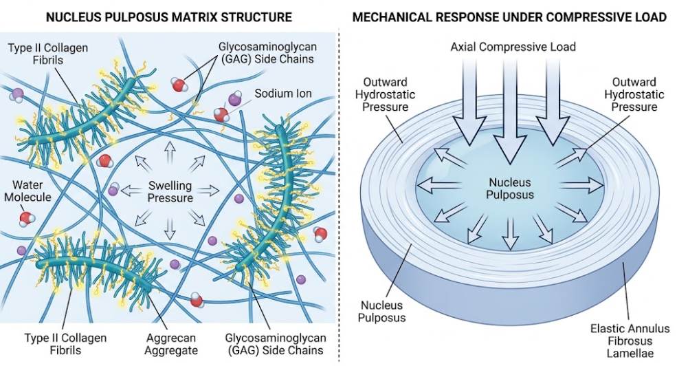
Figure 1. The Intervertebral Disc Shock-Absorbing Mechanism. Double view of the intervertebral disc with the microscopic structure of the nucleus pulposus causing internal swelling pressure (left), and its biomechanical ability to convert vertical compressive loads into outward hydrostatic pressure (right).
As in the Figure 1, the physiological nucleus pulposus is dependent on a dense aggregate of aggrecan molecules trapped in an elastic network of type II collagen fibrils. The high concentration of negatively charged glycosaminoglycans draws mobile cations and water into the matrix, generating a high swelling pressure that maintains disc height, and converts compressive spinal loads into distributed hydrostatic pressure(Ząbek et al., 2025).
In degenerative disc disease, a self-amplifying catabolic cascade disturbs the critical macromolecular balance between aggrecan and type II collagen. Degeneration begins with changes in the microenvironment that cause resident nucleus pulposus cells to switch from an anabolic to a catabolic, pro-inflammatory phenotype(Pattappa et al., 2012).
Overproduction of matrix degrading enzymes, mainly matrix metalloproteinases and ADAMTS aggrecanases, is a major molecular event. Pro-inflammatory cytokines, interleukin-1β and tumor necrosis factor-α, secreted by senescent disc cells and infiltrating immune cells, strongly up-regulate MMP-1, MMP-3, MMP-13, ADAMTS-4 and ADAMTS-5(Kong & Park, 2025; Huaizhen Liang et al., 2022).
The major aggrecanases responsible for initiating the proteoglycan degradation are ADAMTS-4 and ADAMTS-5, which cleave the aggrecan core protein within the interglobular domain between the G1 and G2 globular domains, especially at Glu373-Ala374. Cleavage releases the glycosaminoglycan-rich region from the hyaluronic acid-bound G1 domain. Such cleaved aggrecan fragments are no longer structurally attached, are no longer physically restrained by the collagen meshwork, and gradually diffuse out of the nucleus pulposus(Kibble et al., 2022; H. Liang et al., 2022).
Rapid depletion of chondroitin sulfate and keratan sulfate glycosaminoglycans occurs with loss of aggrecan fragments Reduced glycosaminoglycan content leads to enormous reduction of fixed charge density with subsequent loss of hyperosmotic gradient and water retention capacity(Hwang, Chen, Jing, Hoffman, & Setton, 2014; Mohd Isa et al., 2022). This dehydration appears as the dark disc sign on T2-weighted magnetic resonance imaging. Without adequate swelling pressure, the nucleus pulposus cannot maintain tissue turgor and disc height against physiological loads(Pattappa et al., 2012).
At the same time enzymatic degradation of type II collagen is greatly augmented. Matrix metalloproteinase-13 cleaves intact type II collagen fibers, resulting in denaturation and structural fragmentation. Type II collagen degradation induces a defective repair response by resident cells, which generate type I collagen. Type I collagen makes up thick, stiff fibers and lacks the isotropic elasticity of type II collagen. This phenotypic switch from a flexible cartilaginous matrix to rigid fibrotic matrix changes tissue biomechanics dramatically(Le Maitre, Pockert, Buttle, Freemont, & Hoyland, 2007).
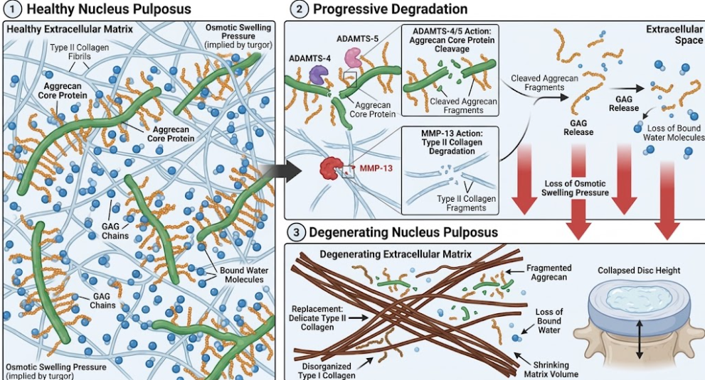
Figure 2. Pathophysiology of Degenerative Intervertebral Disc. Transition from healthy to degenerated state of the nucleus pulposus. ADAMTS and MMP-13 enzymatically degrade aggrecan and type II collagen, resulting in loss of osmotic pressure, disorganized collagen formation, and decreased disc height.
As illustrated in Figure 2, the catabolic cascade in degenerative disc disease involves cleavage of aggrecan core proteins by ADAMTS-4 and 5, leading to loss of glycosaminoglycans, decreased swelling pressure and tissue dehydration. Concurrently, MMP-13 degrades type II collagen leading to a shift in the synthesis toward fibrotic type I collagen, collapse of the matrix and loss of disc height(Pattappa et al., 2012).
This breakdown of the extracellular matrix in the nucleus pulposus initiates a biomechanical and biological feedback loop that drives the progression of spinal degeneration. The loss of aggrecan and the subsequent reduction in osmotic swelling pressure change how mechanical forces are transmitted through the spinal motion segment(Pattappa et al., 2012). In a normal disc, a compressive load creates a hydrostatic pressure that is shared equally between the endplates and the annulus fibrosus. In the degenerated disc, with loss of swelling pressure, compressive forces are transmitted directly and unevenly through solid matrix components(Hwang et al., 2014).
The loss of internal pressurization causes the annulus fibrosus to be axially loaded in excess. Under abnormal vertical compression, concentric lamellae of the annulus fibrosus will bulge, delaminate and microfissure. As radial tears develop, the dehydrated fibrotic nucleus pulposus tissue may herniate outward, causing direct mechanical compression of spinal nerves and the release of inflammatory mediators that trigger radicular pain(Hurri & Karppinen, 2004).
Moreover, the loss of aggrecan alters the immunological and neurovascular status of the disc. Healthy discs have a high fixed charge density and physical density of intact aggrecan that acts as a natural barrier to blood vessel and nerve ingrowth, thus maintaining the disc as an avascular and non-innervated organ. However, the loss of this protective barrier is due to proteolytic degradation of aggrecan(Ząbek et al., 2025). Neurotrophic factors like vascular endothelial growth factor and nerve growth factor and cytokines promote pathological neovascularization and nerve ingrowth into the nucleus pulposus. The primary origin of the chronic discogenic lower back pain is the sensitized sensory nerve fibers penetrating the degenerated disc by local acidic pH and inflammatory cytokines(Pattappa et al., 2012).
Understanding the macromolecular balance of aggrecan and type II collagen illustrates the drawbacks of current clinical interventions. Traditional conservative management and surgical procedures manage symptoms or remove damaged tissue but do not reverse matrix destruction. Advanced disease-modifying strategies should aim to restore matrix anabolism in conjunction with inhibition of catabolic degradation(P. J. Roughley, 2004). New tissue engineering strategies such as synthetic glycosaminoglycan-modified hydrogels, biomimetic aggrecan analogs, and injectable stem cell therapies seek to restore osmotic swelling pressure and enhance endogenous type II collagen production. However, these biological therapies need to be used early, before structural collapse has permanently destroyed the microenvironment(Huaizhen Liang et al., 2022; Ząbek et al., 2025).
The human intervertebral disc relies on a finely balanced extracellular matrix within the nucleus pulposus for its mechanical integrity and shock-absorbing function. The structural synergy of the large aggregating proteoglycan aggrecan with a flexible meshwork of type II collagen fibrils forms a pre-stressed hydrogel capable of producing high osmotic swelling pressure and converting axial spinal compression into distributed hydrostatic stress. In degenerative disc disease, the balance is disturbed by inflammatory signaling and mechanical overload, leading to upregulation of ADAMTS and MMP enzymes that cleave aggrecan and denature type II collagen. Loss of glycosaminoglycans leads to rapid tissue dehydration, loss of disc height, fibrotic transition to type I collagen, and catastrophic biomechanical failure. The next frontier of regenerative therapies for spinal degeneration, considering the limits of nutrient diffusion, involves targeting the molecular pathways of aggrecan proteolysis and restoring the capacity of matrix swelling.
Aspberg, A. (2012). The different roles of aggrecan interaction domains. J Histochem Cytochem, 60(12), 987-996. doi:10.1369/0022155412464376
Bezci, S. E., Nandy, A., & O’Connell, G. D. (2015). Effect of Hydration on Healthy Intervertebral Disc Mechanical Stiffness. J Biomech Eng, 137(10), 101007. doi:10.1115/1.4031416
Empere, M., Wang, X., Prein, C., Aspberg, A., Moser, M., Oohashi, T., . . . Alberton, P. (2023). Aggrecan governs intervertebral discs development by providing critical mechanical cues of the extracellular matrix. Front Bioeng Biotechnol, 11, 1128587. doi:10.3389/fbioe.2023.1128587
Gao, Y., Liu, S., Huang, J., Guo, W., Chen, J., Zhang, L., . . . Guo, Q. (2014). The ECM-cell interaction of cartilage extracellular matrix on chondrocytes. Biomed Res Int, 2014, 648459. doi:10.1155/2014/648459
Grant, M. P., Epure, L. M., Bokhari, R., Roughley, P., Antoniou, J., & Mwale, F. (2016). Human cartilaginous endplate degeneration is induced by calcium and the extracellular calcium-sensing receptor in the intervertebral disc. Eur Cell Mater, 32, 137-151. doi:10.22203/ecm.v032a09
Hurri, H., & Karppinen, J. (2004). Discogenic pain. Pain, 112(3), 225-228. doi:10.1016/j.pain.2004.08.016
Hwang, P. Y., Chen, J., Jing, L., Hoffman, B. D., & Setton, L. A. (2014). The role of extracellular matrix elasticity and composition in regulating the nucleus pulposus cell phenotype in the intervertebral disc: a narrative review. J Biomech Eng, 136(2), 021010. doi:10.1115/1.4026360
Kibble, M. J., Domingos, M., Hoyland, J. A., & Richardson, S. M. (2022). Importance of Matrix Cues on Intervertebral Disc Development, Degeneration, and Regeneration. Int J Mol Sci, 23(13). doi:10.3390/ijms23136915
Kong, C. G., & Park, J. B. (2025). Apoptotic Pathway in Intervertebral Disc Degeneration: From Molecular Pathways to Clinical Interventions. Diagnostics (Basel), 15(12). doi:10.3390/diagnostics15121510
Le Maitre, C. L., Pockert, A., Buttle, D. J., Freemont, A. J., & Hoyland, J. A. (2007). Matrix synthesis and degradation in human intervertebral disc degeneration. Biochem Soc Trans, 35(Pt 4), 652-655. doi:10.1042/bst0350652
Liang, H., Luo, R., Li, G., Zhang, W., Song, Y., & Yang, C. (2022). The Proteolysis of ECM in Intervertebral Disc Degeneration. Int J Mol Sci, 23(3). doi:10.3390/ijms23031715
Liang, H., Luo, R., Li, G., Zhang, W., Song, Y., & Yang, C. (2022). The Proteolysis of ECM in Intervertebral Disc Degeneration. Int J Mol Sci, 23(3), 1715.
Mohd Isa, I. L., Mokhtar, S. A., Abbah, S. A., Fauzi, M. B., Devitt, A., & Pandit, A. (2022). Intervertebral Disc Degeneration: Biomaterials and Tissue Engineering Strategies toward Precision Medicine. Adv Healthc Mater, 11(13), e2102530. doi:10.1002/adhm.202102530
Newell, N., Little, J. P., Christou, A., Adams, M. A., Adam, C. J., & Masouros, S. D. (2017). Biomechanics of the human intervertebral disc: A review of testing techniques and results. J Mech Behav Biomed Mater, 69, 420-434. doi:10.1016/j.jmbbm.2017.01.037
Ohshima, H., Tsuji, H., Hirano, N., Ishihara, H., Katoh, Y., & Yamada, H. (1989). Water diffusion pathway, swelling pressure, and biomechanical properties of the intervertebral disc during compression load. Spine (Phila Pa 1976), 14(11), 1234-1244. doi:10.1097/00007632-198911000-00017
Pattappa, G., Li, Z., Peroglio, M., Wismer, N., Alini, M., & Grad, S. (2012). Diversity of intervertebral disc cells: phenotype and function. J Anat, 221(6), 480-496. doi:10.1111/j.1469-7580.2012.01521.x
Roughley, P., Martens, D., Rantakokko, J., Alini, M., Mwale, F., & Antoniou, J. (2006). The involvement of aggrecan polymorphism in degeneration of human intervertebral disc and articular cartilage. Eur Cell Mater, 11, 1-7; discussion 7.
Roughley, P. J. (2004). Biology of intervertebral disc aging and degeneration: involvement of the extracellular matrix. Spine (Phila Pa 1976), 29(23), 2691-2699. doi:10.1097/01.brs.0000146101.53784.b1
Sakai, D., & Grad, S. (2015). Advancing the cellular and molecular therapy for intervertebral disc disease. Adv Drug Deliv Rev, 84, 159-171. doi:10.1016/j.addr.2014.06.009
Tam, V., Chen, P., Yee, A., Solis, N., Klein, T., Kudelko, M., . . . Chan, D. (2020). DIPPER, a spatiotemporal proteomics atlas of human intervertebral discs for exploring ageing and degeneration dynamics. Elife, 9. doi:10.7554/eLife.64940
Wu, Y., Cisewski, S. E., Sachs, B. L., Pellegrini, V. D., Jr., Kern, M. J., Slate, E. H., & Yao, H. (2015). The region-dependent biomechanical and biochemical properties of bovine cartilaginous endplate. J Biomech, 48(12), 3185-3191. doi:10.1016/j.jbiomech.2015.07.005
Ząbek, Z., Wyczałkowska-Tomasik, A., Poboży, K., Adamus, J. P., Turek, G., Ząbek, M., & Pączek, L. (2025). Understanding the Microenvironment of Intervertebral Disc Degeneration: A Comprehensive Review of Pathophysiological Insights and Therapeutic Implications. Int J Mol Sci, 26(20). doi:10.3390/ijms26209938
Zhang, Y., Lenart, B. A., Lee, J. K., Chen, D., Shi, P., Ren, J., . . . An, H. S. (2014). Histological features of endplates of the mammalian spine: from mice to men. Spine (Phila Pa 1976), 39(5), E312-317. doi:10.1097/brs.0000000000000174