Our Services
Others
- DFPP
- Shockwave
- IV Drip


“This review of the literature describes the role of dysregulation of embryonic neural progenitors in the mechanisms of atypical neurogenesis, cortical expansion and disorganization of minicolumns in early pathogenesis of Autism Spectrum Disorder.”
Traditionally, Autism Spectrum Disorder (ASD) has been viewed mainly in terms of postnatal behavioral and cognitive deficits. However, accumulating evidence, particularly from stem cell models, brain cortical organoids and longitudinal neuroimaging, has clearly changed the paradigm and identified ASD as a prenatal neurodevelopmental disorder. This pathology is founded on fundamental deviations in the earliest stages of brain formation. This review of the literature aims to provide a comprehensive synthesis of early brain development abnormalities in ASD with the key concepts of (1) atypical neuronal proliferation during embryogenesis, (2) altered cortical thickness and surface area expansion and (3) subsequent pathology of cortical minicolumns, all of which are interconnected. This review reveals the molecular, cellular, and macrostructural mechanisms that underlie the chain of events through which embryonic dysregulation results in the complex neuroarchitectural landscape of the autistic brain.
The development of the human cerebral cortex is an extraordinarily complex and prolonged process, dependent on a precisely orchestrated sequence of progenitor cell proliferation, neuronal specification, migration and connectivity.(Kriegstein & Alvarez-Buylla, 2009) This is a highly sensitive developmental window. If this window is disturbed by genetic perturbations or environmental insults during these early stages, the neuroarchitectural landscape can be fundamentally altered.(Guarnieri, de Chevigny, Falace, & Cardoso, 2018) These deviations are now more and more considered as the major causes of malformations of cortical development (MCDs) and a range of neurodevelopmental conditions, notably Autism Spectrum Disorder (ASD).(Leventer et al., 1999)
ASD has historically been defined by behavioral manifestations—such as impairments in social-communicative skills and repetitive behaviors—that are usually clinically detectable between 12 and 24 months of age.(E. Courchesne et al., 2024) Thus, much of the early clinical research focused on postnatal brain pathology and behavioral interventions.(Piven, Elison, & Zylka, 2017) However, in recent years there has been a major paradigm shift. Growing neuroimaging, genomic, and cellular evidence converges to support the notion that the pathophysiology of ASD begins prenatally, during embryogenesis and mid-fetal development.(Eric Courchesne, Gazestani, & Lewis, 2020) As shown in Figure 1, We currently understand ASD as a progressive disorder that begins early in the formation of the brain and not as a sudden postnatal onset, with early prenatal disruptions cascading to later developmental milestones.
Early brain overgrowth is one of the most consistent macroscopic findings in early ASD development.(Leventer et al., 1999) In longitudinal neuroimaging studies of high-risk infants a specific developmental trajectory has emerged: an abnormal hyper-expansion of the cortical surface area during the first year of life followed by brain volume overgrowth in the second year, coincident with the emergence of autistic social deficits.(Piven et al., 2017) The cellular mechanisms underlying this macroscopic overgrowth have historically been difficult to understand because the developing human fetal brain is inaccessible.(E. Courchesne et al., 2024) Fortunately, recent advances in the use of patient-derived induced pluripotent stem cells (iPSCs) and brain cortical organoids (BCOs) have made it possible to model early human neurodevelopment in vitro. These models have shown that ASD-derived embryonic organoids are significantly larger (39-41%) and grow faster than neurotypical controls.(E. Courchesne et al., 2024) The overgrowth is primarily due to dysregulated proliferation of neuronal progenitor cells and a profound deficit in the balance of excitatory cortical neuron subtypes.(Jourdon et al., 2023)
Importantly, such embryonic disruptions do not solely increase brain volume and surface area. They trigger a ripple effect that changes the basic micro-circuitry of the brain.(Piven et al., 2017) The hyper-proliferation of progenitor cells and the subsequent defects in cellular delamination and neuronal migration, ultimately corrupt the formation and the spatial organization of the cortical minicolumns, the basic vertical functional units of the cerebral cortex.(Beopoulos, Géa, Fasano, & Iris, 2022)
This literature review aims to integrate recent findings on the early neurodevelopmental origins of ASD to provide a cohesive understanding of this complex developmental cascade. Specifically, it focuses on three related areas: (1) atypical neurogenesis and progenitor proliferation during embryogenesis, (2) macrostructural alterations in cortical surface area and thickness, and (3) the derived minicolumn pathology. This article follows the pathogenesis of ASD from the early division of the neural stem cells to the disorganized macro- and micro-architecture of the autistic neocortex, and highlights the role of early embryonic deviations in the establishment of the clinical phenotype of autism, opening new critical windows for early intervention.
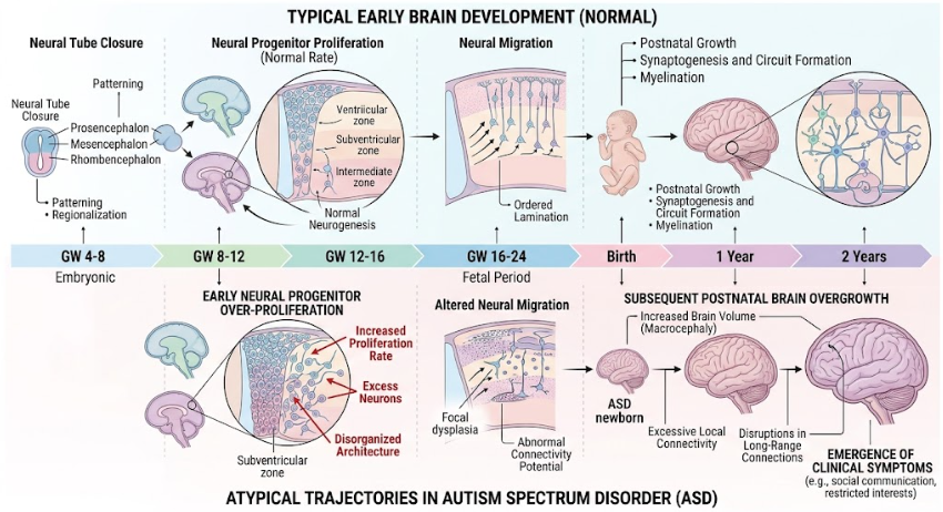
Figure 1. Comparative timeline of typical early brain development (top) and atypical trajectories in Autism Spectrum Disorder (bottom), showing major deviations in cell proliferation, migration and post-natal growth.
The mammalian cerebral cortex is built upon a long and tightly regulated neurogenic program that begins during embryogenesis (approximately at day 42 in humans) and continues through midgestation.(Stiles & Jernigan, 2010) neural progenitor cells, including apical radial glial cells (aRGCs), first undergo symmetric divisions to expand the progenitor pool, and then switch to asymmetric divisions to produce postmitotic neurons or intermediate progenitor cells (IPCs).(Sokpor, Brand-Saberi, Nguyen, & Tuoc, 2022; Subramanian, Bershteyn, Paredes, & Kriegstein, 2017) These IPCs and basal radial glial cells (bRGCs) migrate to the subventricular zone (SVZ) and function as neuron amplifying cells, driving the evolutionary expansion of the human neocortex.(Guarnieri et al., 2018; Sokpor et al., 2022) Cell Delamination, the detachment of differentiating cells from the apical junctional belt of the VZ, tightly regulates the balance between progenitor pool maintenance and neuronal differentiation.(Camargo Ortega et al., 2019)
Recent studies with iPSC- and patient-derived brain cortical organoids (BCOs) provided direct evidence that atypical neurogenesis is a major driver of ASD.(Urresti et al., 2021) Studies have shown that in the embryonic stage, toddlers with severe ASD have been found to have significantly larger BCOs (39% to 41%) and an accelerated growth rate nearly three times faster than controls. This early brain overgrowth is strongly linked to increased neurogenesis and abnormal regulation of cell cycle networks.(E. Courchesne et al., 2024)
This cellular makeup of these organoids confirms a profound imbalance of neuronal lineages. Idiopathic macrocephalic ASD exhibits an overproduction of excitatory neurons destined for the dorsal cortical plate (EN-DCP) with a relative loss of early generated preplate excitatory neurons (EN-PP) and inhibitory lineages. This imbalance suggests that in macrocephalic ASD, radial glial cells fail to exit the cell cycle appropriately and re-enter the cell cycle to expand the surface of the cortical plate. In contrast, normocephalic ASD models show the opposite trend: an early exit from the cell cycle, resulting in an overproduction of preplate neurons and a subsequent depletion of the cortical plate progenitor pool. As shown in Figure 2. (Jourdon et al., 2023)
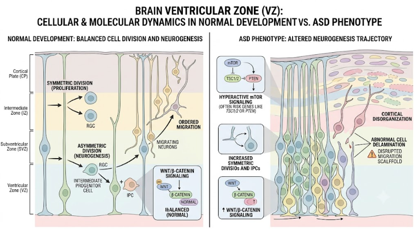
Figure 2. Cellular and molecular dynamics in brain ventricular zone. Normal balanced cell division and migration (left) and the ASD-associated trajectory of increased proliferation and disrupted cortical architecture (right).
The molecular mechanisms behind this aberrant proliferation converge on several of key genetic and signaling pathways.
β-catenin is a bifunctional protein, which takes part in cell adhesion and transcriptional activation.(Nelson & Nusse, 2004) β-catenin-mediated signaling is critical in the VZ to decide the fate of cortical precursors to either proliferate or differentiate. Overexpression or hyperactivation of β-catenin leads to an expansion of the neural precursor population and subsequent enlargement of the cortical surface area, while focal elimination of β-catenin results in forced premature neuronal differentiation. (Woodhead, Mutch, Olson, & Chenn, 2006) In a high-confidence ASD risk gene, Chd8-deficient mice, dysregulation of Wnt/β-catenin signaling transiently expands intermediate progenitors to cause an enlarged brain and autistic-like phenotypes. (Durak et al., 2016)
The mammalian target of rapamycin (mTOR) pathway is another major hub for regulation of cell growth, proliferation and protein synthesis.(Subramanian, Calcagnotto, & Paredes, 2019) Pathway hyperactivation caused by mutations in MTOR and upstream regulators of MTOR (eg, PTEN) has been directly linked to megalencephaly and focal cortical dysplasia.(Iffland & Crino, 2017) For example, PTEN mutations cause excess Wnt/β-catenin signaling, macrocephaly, and excess cortical neurons at birth, resulting in an imbalance in excitatory and inhibitory synaptic connections.(Vogt, Cho, Lee, Sohal, & Rubenstein, 2015)
The NDEL1 gene, which is essential for neuronal proliferation, neurite outgrowth and cell positioning, has also been implicated. In ASD BCO models Ndel1 activity and expression are significantly reduced and negatively correlated with organoid size and growth rate.(E. Courchesne et al., 2024) Ndel1 controls mitotic spindle function and its dysregulation is a direct driver of accelerated neurogenesis in severe ASD subtypes.(Feng & Walsh, 2004)
Neuronal migration and differentiation require delamination, the physical detachment of cells from the VZ. This process is regulated by epigenetic remodelers such as the BAF complex (e.g. BAF155, BAF170) and polarity related factors (e.g. LLGL1, CDC42).(Narayanan et al., 2018) Abnormal delamination due to mutation of these factors may lead to either depletion of the progenitor pool (microcephaly) or non-physiological expansion of the progenitor pool (megalencephaly and macrocephaly) emphasizing delamination as a critical checkpoint that may be compromised in certain neurodevelopmental profiles.(Jayaraman et al., 2016)
The hyper-proliferation of neural progenitors in embryogenesis sets the stage for macroscopic anatomic changes seen postnatally. Longitudinal neuroimaging studies of high-risk infant siblings have identified a specific temporal sequence: abnormal brain growth trajectory precedes the defining behavioral features of autism.(Piven et al., 2017) Most important of all, this enlargement of the brain is not, as a rule, present at birth. Instead, the brain exhibits a specific pattern of hyper-expansion of cortical surface area, mainly between 6 and 12 months of age. It is the primary force driving the brain volume overgrowth observed in the second year of life (12 to 24 months), the period when autistic social deficits appear and solidify.(Hazlett et al., 2017; Piven et al., 2017)
To better understand this overgrowth, it is necessary to distinguish between cortical surface area and cortical thickness, which are controlled by different genetic architectures and developmental mechanisms.(Chen et al., 2015) Rakic proposed “radial unit hypothesis” which states that the surface area of the cerebral cortex is determined by the number of radial columns produced by progenitor cells in the VZ and SVZ and the thickness of the cortex is determined by the number of cells in each column ,as shown in Figure 3.(Rakic, 1995) Thus, the hyper-expansion of cortical surface area in ASD is a direct macroscopic manifestation of the embryonic over proliferation of intermediate radial glia and progenitor cells.(Packer, 2016)
Early volume increases are driven by surface area but cortical thickness also follows an atypical trajectory. In typical development, there is an early increase in thickness followed by gradual thinning over the course of adolescence, due to synaptic pruning and myelination. In ASD, we see early increases in cortical thickness in toddlers (ages 2-4), followed by an accelerated rate of decline and thinning from adolescence into late middle age, suggestive of a neurodegenerative-like trajectory later in life.(Nunes et al., 2020) Moreover, this anomalous expansion is not uniform, and regional hyper-expansion is particularly prominent in the visual cortex (e.g., occipital gyrus, cuneus), which may explain early sensory-motor and visual orienting deficits in infants with ASD.(Hazlett et al., 2017)
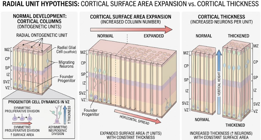
Figure 3. Cellular Dynamics of the Radial Unit Hypothesis. A visual contrast of two dimensions of development: horizontal expansion of surface area (due to radial column multiplication by symmetric progenitor division) versus vertical cortical thickening (due to increased neuronal output per column by asymmetric neurogenic division).
The structural and functional consequences of the altered neurogenesis and cortical expansion are finally expressed at the level of micro-circuitry, as cortical minicolumns.(Hazlett et al., 2017) The basic functional processing unit of the mature cerebral cortex is the minicolumn, a vertical chain of around 80 to 100 neurons running through the cortical layers II to VI with their dendrites and myelinated axons.(McKavanagh, Buckley, & Chance, 2015) In a “inside-out” manner (from the deepest layer VI to layer II), postmitotic excitatory pyramidal neurons migrate radially along radial glial fibers, establishing columns prenatally.(Beopoulos et al., 2022; Guarnieri et al., 2018; Subramanian et al., 2019) The spacing and architecture of these minicolumns permit the appropriate integration of thalamic input with corticocortical processing to discriminate signal from noise.(McKavanagh et al., 2015)
Autopsy studies of the autistic brain have shown profound abnormalities in minicolumnar architecture. Previous research has demonstrated that the minicolumns in the brains of people with ASD are smaller.(O’Reilly, Lewis, & Elsabbagh, 2017) However, subsequent large-scale analyses have shown that minicolumns in the ASD cortex are actually focally disorganized, with increased spacing between minicolumns and decreased cellular density within a given cortical region , as shown in Figure 4.(Donovan & Basson, 2017) This spacing irregularity affects all cortical regions by roughly 6-10%, but is most severe in higher-order association areas.(Donovan & Basson, 2017; McKavanagh et al., 2015)
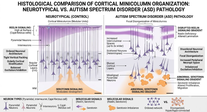
Figure 4. Histological comparison of neurotypical development (left) with orderly tightly packed cortical minicolumns, and ASD (right) with focal disorganization of cortical minicolumns, wider intercolumnar spaces and disrupted molecular signaling (Reelin and Serotonin).
The precise spacing and alignment of minicolumns are highly dependent on the glial scaffold and positioning cues during fetal development. One of the major orchestrators of this process is the Cajal-Retzius (CR) cell that is transiently present in the marginal zone (layer I) of the developing cortex.(Stiles & Jernigan, 2010) CR cells produce an extracellular glycoprotein, Reelin, which is essential for proper “inside-out” lamination of the cortex and is a critical regulator of neuronal migration and dendritic arborization.(Hirota & Nakajima, 2017)
In ASD, the misplacement and dysfunction of CR cells anatomically result in disorganized lamination and altered minicolumn distribution.(Donovan & Basson, 2017) CR cells physiology and Reelin secretion are strongly modulated by Serotonin (5-HT) thus providing a deeper mechanistic understanding. During the critical window of development. (Gestational Week 9 to 20) before the fetus is able to produce its own 5-HT, it depends on placental and maternal sources.(T. Vitalis & Parnavelas, 2003; Tania Vitalis & Verney, 2017) Examples of maternal inflammation insults include maternal inflammation. This can activate increased placental conversion of L-tryptophan to 5-HT, flooding the fetal brain with excess serotonin. This exogenous 5-HT is an abnormal axon guidance cue, which perturbs the delicate balance of CR cell signaling, modifies Reelin gradients, and consequently alters the radial migration of pyramidal neurons.(Beopoulos et al., 2022) The result is a very disorganized, widely spaced minicolumnar architecture which does not prune excess synapses correctly in the postnatal period, resulting in a catastrophic loss of signal-to-noise discrimination and the clinical manifestations of autism.(Donovan & Basson, 2017; Lieberman, McGuirt, Tang, & Sulzer, 2019)
Transcriptomic, neuroimaging, and stem-cell models converge to demonstrate that Autism Spectrum Disorder (ASD) is a progressive neurodevelopmental disorder with prenatal origins. The etiology is based on dysregulation of the embryonic neural progenitor.(E. Courchesne et al., 2024; Jourdon et al., 2023; Piven et al., 2017) In particular, the reduced expression and activity of the NDEL1 enzyme, which is essential for the regulation of the cell cycle, leads to a faster neural proliferation and, as a consequence, to brain cortical organoids (BCOs) that are significantly enlarged and develop more rapidly. In the macro scale, the enlarged progenitor pool leads to an increase in the number of radial columns, which directly contributes to the hyper-expansion of the cortical surface area observed in the first year of high-risk ASD infants.(Hazlett et al., 2017; Piven et al., 2017) Thus, this early expansion of surface area determines the later overgrowth of brain volume and abnormal trajectories of cortical thickness (early overgrowth followed by accelerated thinning) that characterize the autistic brain.
Early neurogenic disruption is pivotal in determining clinical severity and microstructural pathology. Macrocephalic ASD is marked by exuberant overproduction of cortical plate excitatory neurons and delayed cell-cycle exit, while normocephalic ASD is characterized by premature cell-cycle exit and preplate neuron overproduction.(Jourdon et al., 2023) This simple imbalance in the cell population eventually affects the spatial organization of cortical minicolumns, creating a broader disorganized micro-circuitry.(McKavanagh et al., 2015) These structural defects impair synaptic pruning and signal-to-noise discrimination and bias the brain’s functional networks toward a pattern of local overconnectivity and long-range underconnectivity, fitting nicely with the core social and sensory deficits of ASD.(O’Reilly et al., 2017)
These prenatal findings are still hard to translate into postnatal behavioral interventions, but 3D organoid models provide unprecedented platforms for early biomarker discovery and pharmacological therapy testing.(E. Courchesne et al., 2024; Urresti et al., 2021) For instance, rational early interventions are supported by proof-of-concept therapies targeting specific molecular dysregulations (e.g. RhoA inhibitors to rescue neuronal migration defects).(Urresti et al., 2021) Future research should focus on multi-modal approaches linking embryonic in vitro pathology with longitudinal neuroimaging and clinical phenotyping and should consider complex in utero environmental factors such as altered placental serotonin supply to fully understand the pathogenesis of ASD.
The pathogenesis of ASD is complicated and is closely related to the development of the brain in the embryonic and early postnatal periods. Recent multidisciplinary literature suggests the etiology of a cascading developmental framework: Genetic liabilities and environmental factors (e.g. altered 5-HT signaling) interfere with tightly controlled Wnt/β-catenin and mTOR pathways leading to an over-proliferation of intermediate neural progenitor cells and defective cell delamination. This anomaly in the embryo is seen macroscopically as an expansion of the cortical surface area in the first year of the infant’s life before the generalized overgrowth of brain volume. This early dysregulation affects the radial migration and positioning of the Cajal-Retzius cell-guided neurons at the microscopic level, leading to a focal disorganization of cortical minicolumns. Understanding these deeply interconnected mechanisms, from the initial division of a neural stem cell to the vast, disorganized architecture of the autistic neocortex, opens the path to identifying biomarkers and critical windows for targeted, rational early interventions before the behavioral hallmarks of ASD are fully consolidated.
Beopoulos, A., Géa, M., Fasano, A., & Iris, F. (2022). Autism spectrum disorders pathogenesis: Toward a comprehensive model based on neuroanatomic and neurodevelopment considerations. Front Neurosci, 16, 988735. doi:10.3389/fnins.2022.988735
Camargo Ortega, G., Falk, S., Johansson, P. A., Peyre, E., Broix, L., Sahu, S. K., . . . Götz, M. (2019). The centrosome protein AKNA regulates neurogenesis via microtubule organization. Nature, 567(7746), 113-117. doi:10.1038/s41586-019-0962-4
Chen, C. H., Peng, Q., Schork, A. J., Lo, M. T., Fan, C. C., Wang, Y., . . . Dale, A. M. (2015). Large-scale genomics unveil polygenic architecture of human cortical surface area. Nat Commun, 6, 7549. doi:10.1038/ncomms8549
Courchesne, E., Gazestani, V. H., & Lewis, N. E. (2020). Prenatal Origins of ASD: The When, What, and How of ASD Development. Trends in Neurosciences, 43(5), 326-342. doi:10.1016/j.tins.2020.03.005
Courchesne, E., Taluja, V., Nazari, S., Aamodt, C. M., Pierce, K., Duan, K., . . . Muotri, A. R. (2024). Embryonic origin of two ASD subtypes of social symptom severity: the larger the brain cortical organoid size, the more severe the social symptoms. Mol Autism, 15(1), 22. doi:10.1186/s13229-024-00602-8
Donovan, A. P., & Basson, M. A. (2017). The neuroanatomy of autism – a developmental perspective. J Anat, 230(1), 4-15. doi:10.1111/joa.12542
Durak, O., Gao, F., Kaeser-Woo, Y. J., Rueda, R., Martorell, A. J., Nott, A., . . . Tsai, L. H. (2016). Chd8 mediates cortical neurogenesis via transcriptional regulation of cell cycle and Wnt signaling. Nat Neurosci, 19(11), 1477-1488. doi:10.1038/nn.4400
Feng, Y., & Walsh, C. A. (2004). Mitotic spindle regulation by Nde1 controls cerebral cortical size. Neuron, 44(2), 279-293. doi:10.1016/j.neuron.2004.09.023
Guarnieri, F. C., de Chevigny, A., Falace, A., & Cardoso, C. (2018). Disorders of neurogenesis and cortical development. Dialogues Clin Neurosci, 20(4), 255-266. doi:10.31887/DCNS.2018.20.4/ccardoso
Hazlett, H. C., Gu, H., Munsell, B. C., Kim, S. H., Styner, M., Wolff, J. J., . . . Piven, J. (2017). Early brain development in infants at high risk for autism spectrum disorder. Nature, 542(7641), 348-351. doi:10.1038/nature21369
Hirota, Y., & Nakajima, K. (2017). Control of Neuronal Migration and Aggregation by Reelin Signaling in the Developing Cerebral Cortex. Frontiers in Cell and Developmental Biology, Volume 5 – 2017. doi:10.3389/fcell.2017.00040
Iffland, P. H., 2nd, & Crino, P. B. (2017). Focal Cortical Dysplasia: Gene Mutations, Cell Signaling, and Therapeutic Implications. Annu Rev Pathol, 12, 547-571. doi:10.1146/annurev-pathol-052016-100138
Jayaraman, D., Kodani, A., Gonzalez, D. M., Mancias, J. D., Mochida, G. H., Vagnoni, C., . . . Walsh, C. A. (2016). Microcephaly Proteins Wdr62 and Aspm Define a Mother Centriole Complex Regulating Centriole Biogenesis, Apical Complex, and Cell Fate. Neuron, 92(4), 813-828. doi:10.1016/j.neuron.2016.09.056
Jourdon, A., Wu, F., Mariani, J., Capauto, D., Norton, S., Tomasini, L., . . . Vaccarino, F. M. (2023). Modeling idiopathic autism in forebrain organoids reveals an imbalance of excitatory cortical neuron subtypes during early neurogenesis. Nat Neurosci, 26(9), 1505-1515. doi:10.1038/s41593-023-01399-0
Kriegstein, A., & Alvarez-Buylla, A. (2009). The glial nature of embryonic and adult neural stem cells. Annu Rev Neurosci, 32, 149-184. doi:10.1146/annurev.neuro.051508.135600
Leventer, R. J., Phelan, E. M., Coleman, L. T., Kean, M. J., Jackson, G. D., & Harvey, A. S. (1999). Clinical and imaging features of cortical malformations in childhood. Neurology, 53(4), 715-722. doi:10.1212/wnl.53.4.715
Lieberman, O. J., McGuirt, A. F., Tang, G., & Sulzer, D. (2019). Roles for neuronal and glial autophagy in synaptic pruning during development. Neurobiol Dis, 122, 49-63. doi:10.1016/j.nbd.2018.04.017
McKavanagh, R., Buckley, E., & Chance, S. A. (2015). Wider minicolumns in autism: a neural basis for altered processing? Brain, 138(Pt 7), 2034-2045. doi:10.1093/brain/awv110
Narayanan, R., Pham, L., Kerimoglu, C., Watanabe, T., Castro Hernandez, R., Sokpor, G., . . . Tuoc, T. (2018). Chromatin Remodeling BAF155 Subunit Regulates the Genesis of Basal Progenitors in Developing Cortex. iScience, 4, 109-126. doi:10.1016/j.isci.2018.05.014
Nelson, W. J., & Nusse, R. (2004). Convergence of Wnt, beta-catenin, and cadherin pathways. Science, 303(5663), 1483-1487. doi:10.1126/science.1094291
Nunes, A. S., Vakorin, V. A., Kozhemiako, N., Peatfield, N., Ribary, U., & Doesburg, S. M. (2020). Atypical age-related changes in cortical thickness in autism spectrum disorder. Scientific Reports, 10(1), 11067. doi:10.1038/s41598-020-67507-3
O’Reilly, C., Lewis, J. D., & Elsabbagh, M. (2017). Is functional brain connectivity atypical in autism? A systematic review of EEG and MEG studies. PLoS One, 12(5), e0175870. doi:10.1371/journal.pone.0175870
Packer, A. (2016). Neocortical neurogenesis and the etiology of autism spectrum disorder. Neurosci Biobehav Rev, 64, 185-195. doi:10.1016/j.neubiorev.2016.03.002
Piven, J., Elison, J. T., & Zylka, M. J. (2017). Toward a conceptual framework for early brain and behavior development in autism. Mol Psychiatry, 22(10), 1385-1394. doi:10.1038/mp.2017.131
Rakic, P. (1995). Radial versus tangential migration of neuronal clones in the developing cerebral cortex. Proceedings of the National Academy of Sciences, 92(25), 11323-11327. doi:doi:10.1073/pnas.92.25.11323
Sokpor, G., Brand-Saberi, B., Nguyen, H. P., & Tuoc, T. (2022). Regulation of Cell Delamination During Cortical Neurodevelopment and Implication for Brain Disorders. Front Neurosci, 16, 824802. doi:10.3389/fnins.2022.824802
Stiles, J., & Jernigan, T. L. (2010). The basics of brain development. Neuropsychol Rev, 20(4), 327-348. doi:10.1007/s11065-010-9148-4
Subramanian, L., Bershteyn, M., Paredes, M. F., & Kriegstein, A. R. (2017). Dynamic behaviour of human neuroepithelial cells in the developing forebrain. Nat Commun, 8, 14167. doi:10.1038/ncomms14167
Subramanian, L., Calcagnotto, M. E., & Paredes, M. F. (2019). Cortical Malformations: Lessons in Human Brain Development. Front Cell Neurosci, 13, 576. doi:10.3389/fncel.2019.00576
Urresti, J., Zhang, P., Moran-Losada, P., Yu, N. K., Negraes, P. D., Trujillo, C. A., . . . Iakoucheva, L. M. (2021). Cortical organoids model early brain development disrupted by 16p11.2 copy number variants in autism. Mol Psychiatry, 26(12), 7560-7580. doi:10.1038/s41380-021-01243-6
Vitalis, T., & Parnavelas, J. G. (2003). The role of serotonin in early cortical development. Dev Neurosci, 25(2-4), 245-256. doi:10.1159/000072272
Vitalis, T., & Verney, C. (2017). Sculpting Cerebral Cortex with Serotonin in Rodent and Primate. In K. Fatima-Shad (Ed.), Serotonin – A Chemical Messenger Between All Types of Living Cells. London: IntechOpen.
Vogt, D., Cho, K. K. A., Lee, A. T., Sohal, V. S., & Rubenstein, J. L. R. (2015). The parvalbumin/somatostatin ratio is increased in Pten mutant mice and by human PTEN ASD alleles. Cell Rep, 11(6), 944-956. doi:10.1016/j.celrep.2015.04.019
Woodhead, G. J., Mutch, C. A., Olson, E. C., & Chenn, A. (2006). Cell-autonomous beta-catenin signaling regulates cortical precursor proliferation. J Neurosci, 26(48), 12620-12630. doi:10.1523/jneurosci.3180-06.2006