Our Services
Others
- DFPP
- Shockwave
- IV Drip


“This extensive literature review presents the cellular, biophysical and molecular mechanisms of arteriogenesis that result in coronary collateral vessel growth to rescue ischemic myocardium in cardiovascular diseases.”
Coronary artery disease (CAD) is still the leading cause of morbidity and mortality worldwide. When epicardial coronary blood flow is severely compromised by atherosclerotic stenosis or acute arterial occlusion the heart relies on a natural compensatory by-pass network, the coronary collateral circulation. Unlike hypoxia-driven angiogenesis, which results in the formation of fragile capillary sprouts, arteriogenesis is defined as the outward remodeling and maturation of pre-existing, dormant arteriolar anastomoses into high-capacity, muscular conductive arteries capable of restoring significant blood flow to ischemic myocardium. The current literature review offers a systematic analysis of the biophysical, cellular and molecular orchestrations that regulate collateral artery expansion. We describe the transformation of kinetic blood flow into fluid shear stress by hydrodynamic pressure gradients, which activate mechanosensitive endothelial ion channels and primary intracellular cascades. The role of monocyte recruitment, matrix metalloproteinase-mediated degradation of the extracellular matrix, phenotypic switching of smooth muscle cells and rebuilding of the arterial coats is discussed. This review also evaluates the clinical collateral assessment modalities, comparing qualitative angiographic grading with invasive pressure derived collateral flow index measurements. Finally, we address therapeutic arteriogenesis, highlighting the shift from pro-inflammatory cytokine therapies to non-inflammatory mechanical shear-stress triggers such as exercise training and enhanced external counterpulsation, and emerging microRNA biomarker panels for non-invasive diagnostic stratification.
Keywords: Coronary Collateral Circulation, Arteriogenesis, Fluid Shear Stress, Myocardial Ischemia, Endothelial Mechanotransduction, Monocyte Recruitment, Cardiovascular Diseases
Cardiovascular diseases (CVDs), particularly coronary artery disease (CAD), are the leading cause of mortality and long-term disability worldwide. The clinical evolution of coronary artery disease is characterized by chronic lipid accumulation in the subendothelium, vascular inflammation and progressive narrowing of the lumen. As described in our previous reviews on the dynamics of plaque rupture in cardiovascular Atherosclerosis and Coronary Artery Plaque Rupture Dynamics in Cardiovascular Diseases, sudden structural disruption of a vulnerable atheromatous plaque leads to a rapid thrombotic cascade that can abruptly occlude blood flow to downstream tissue, resulting in acute myocardial infarction or sudden cardiac death(Degen, Millenaar, & Schirmer, 2014; Seiler, 2003).
Severe stenosis or chronic occlusion of a major epicardial coronary artery results in a severe perfusion mismatch in the downstream myocardium. The existence and degree of an alternative vascular system, the so-called coronary collateral circulation, represents the ultimate factor in determining myocardial survival, preservation of left ventricular function and, ultimately, the longevity of the patient under these critical conditions(Meier et al., 2013). The coronary collateral circulation is a specialized interarterial network of pre-existing anastomotic bridges that link divergent coronary vascular beds or neighboring segments of the same epicardial vessel(Hakimzadeh & Piek, 2013; Helfant, Vokonas, & Gorlin, 1971).
To understand how the cardiovascular system compensates arterial occlusion a fundamental distinction has to be made between the different types of vascular growth. Vascular growth was formerly referred to as angiogenesis in general. Modern vascular biology, however, recognizes three different physiological processes: vasculogenesis, angiogenesis, and arteriogenesis(Degen et al., 2014). Vasculogenesis is the formation of primitive blood vessels from embryonic angioblasts or progenitor cells. Angiogenesis is the sprouting and intussusceptive branching of new capillary-like networks from pre-existing microscopic vessels, primarily induced by severe tissue hypoxia and hypoxia-inducible factor signals. Angiogenesis increases the microvascular density in ischemic areas. The microscopic capillaries lack smooth muscle coats and elastic laminae. Capillaries are therefore hydraulically very resistant and cannot deliver bulk blood flow to salvage a large myocardial territory(Bavry, Kumbhani, Rassi, Bhatt, & Askari, 2006; O’Donoghue et al., 2008).
Arteriogenesis is the active outward growth, circumferential widening, and maturation of pre-existing, microscopic arteriolar channels into large, muscular conduit arteries. Arteriogenesis, largely stimulated by physical hydrodynamics and fluid shear stress rather than parenchymal hypoxia, converts low-capacity, 30- to 50-micrometer-diameter anastomotic vessels into robust, muscular collateral arteries that can increase their caliber up to twentyfold(Nickolay, Nichols, Ingle, & Hoye, 2020). This structural expansion can, on the basis of physical principles of vascular resistance, increase the blood flow capacity up to 25 times, providing a natural biological bypass, preserving contractility and limiting infarction in jeopardize myocardial zones(Koerselman, van der Graaf, de Jaegere, & Grobbee, 2003; Pipp et al., 2004).
The initiation of arteriogenesis is essentially a biophysical process that is driven by fluid mechanics. Under basal physiological conditions, existing inter-arterial collateral anastomoses are structurally dormant and exhibit minimal blood flow. This quiescent state is maintained because neighboring epicardial coronary arteries have similar perfusion pressures, so that there is little pressure gradient across the connecting anastomotic bridges(Seiler, 2010).
However, if atherosclerotic plaque deposition leads to severe luminal narrowing or total occlusion of a recipient coronary artery, the hemodynamic balance is immediately lost. Distal arteriolar vasodilation in an attempt to maintain tissue perfusion results in a precipitous fall in perfusion pressure in the vascular bed distal to the occlusion. By contrast, the perfusion pressure in the unobstructed donor artery is at systemic arterial levels. The resulting difference leads to a large transmural pressure gradient (ΔP = Pdonor – Precipient) across the pre-existing collateral channel(Hakimzadeh & Piek, 2013).
Driven by this elevated pressure gradient, blood flow through the high-resistance anastomotic arterioles increases dramatically. Fluid flow across a vessel wall exerts a frictional force per unit surface area directly onto the apical membrane of endothelial cells, defined as fluid shear stress (τ). Mathematically, fluid shear stress is directly proportional to blood viscosity (μ) and flow velocity (Q), and inversely proportional to the third power of the internal vessel radius (r3):
The sudden increase in blood velocity results in extraordinary localized increases in tangential fluid shear stress due to the microscopic radii of pre-existing collateral channels. This physical force is the major biological trigger that activates quiescent endothelial cells, transducing mechanical energy into intracellular signaling cascades that initiate structural arterial remodeling(Koerselman et al., 2003; Meier et al., 2013; Nickolay et al., 2020).
The physical laws governing flow dynamics change with both outward remodeling of the collateral vessel and expansion of the luminal cross-sectional area. The Hagen-Poiseuille equation predicts that the vascular resistance (R) is inversely proportional to the fourth power of the vessel radius (R ∝ 1/r4). Therefore, even a small change in the internal diameter results in an exponential change in vascular resistance(Van Royen, Piek, Schaper, Bode, & Buschmann, 2001). As the collateral channel turns into a large conductive artery, blood flow to the distal ischemic bed increases and distal post-stenotic pressure rises gradually. This re-establishment of distal pressure gradually erodes the driving pressure gradient across the collateral network. Eventually fluid shear stress drops below the level needed to stimulate further vascular growth and arteriogenesis reaches a natural physiological plateau(Choo, 2015).
Experimental swine and rodent occlusion models showed this self-limiting feedback loop by surgically creating arteriovenous shunts distal to an arterial occlusion. Investigators kept pressure gradients and shear stress high and artificial across the collateral network by continuously shunting blood into the low-pressure venous system(Pipp et al., 2004). This sustained physical stimulus pushed collateral expansion well above normal physiologic limits, restoring downstream perfusion to nearly twice that of baseline healthy controls. These results demonstrated that fluid shear stress is the predominant rate-limiting driver of collateral expansion(Eitenmüller et al., 2006; Schaper, 2009).
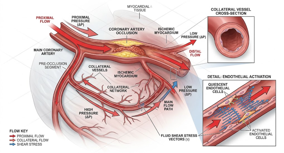
Figure 1. Hemodynamic Mechanisms of Coronary Collateral Growth and Endothelial Activation. Diagrammatic representation of collateral blood flow by pressure gradient (ΔP) following coronary artery occlusion. The corresponding vectors of fluid shear stress (τ) induce endothelial cell activation for arteriogenesis to reperfuse the ischemic myocardium. Insets show cross-section of collateral vessel and detailed endothelial activation mediated by shear stress.
As shown in Figure 1, the sharp decline in distal perfusion pressure leads to an enhanced pressure gradient across the anastomotic bridge, directing high fluid shear stress vectors along the luminal endothelial membrane that initiate the early stages of collateral remodeling(Hakimzadeh & Piek, 2013).
Conversion of fluid shear stress into active structural vessel growth depends on an elaborate biological apparatus that can perform mechanotransduction, the process by which physical forces are transduced into chemical signals. The vascular endothelium is the main mechanosensor, using a wide range of surface molecules, structural complexes and membrane microdomains(Constantinescu, Micheu, Liehn, & Udrişte, 2026; Naylor, O’Driscoll, Fitzsimons, Arnolda, & Green, 2006).
Mechanosensors, including transient receptor potential vanilloid 4 (TRPV4) ion channels in endothelial plasma membranes, are important in this process. Increased fluid shear stress causes deformation of the endothelial membrane, leading to conformational changes in TRPV4 channels, which results in a rapid influx of extracellular calcium (Ca2+) ions(C. Troidl et al., 2009). This localized calcium transient activates calmodulin and the downstream endothelial nitric oxide synthase (eNOS), resulting in a strong release of nitric oxide (NO). Nitric oxide is an acute, immediate vasodilator that reduces mechanical wall tension and modulates the downstream smooth muscle cell responses(K. Troidl et al., 2010).
Biophysical signals are transmitted simultaneously through the endothelial glycocalyx, a carbohydrate-rich pericellular layer coating the luminal surface, and structural cell–cell adhesion complexes, including the VE-cadherin–platelet endothelial cell adhesion molecule 1 (PECAM-1)–vascular endothelial growth factor receptor 2 (VEGFR2) mechanosensory complex. Transduction via such complexes activates integrins (αvβ3 and α5β1) at the basal focal adhesion sites(Cronin, Dawson, & DeMali, 2024). Binding of integrins results in autophosphorylation of the focal adhesion kinase (FAK) and recruitment of the Src kinase, which in turn phosphorylates the Paxillin and Cas. The integrin-FAK-Src signaling axis controls the reorganization of the cytoskeletal actin filaments and allows the endothelial cells to align in parallel with the direction of fluid flow(Ballermann, Dardik, Eng, & Liu, 1998).
Downstream of focal adhesion assembly, increased shear stress activates the protein kinase C (PKC) pathway and the mitogen-activated protein kinase (MAPK) cascade, which causes rapid and transient activation of extracellular signal-regulated kinases 1 and 2 (ERK1/2). Activation of ERK1/2 promotes endothelial cell survival, gene transcription and metabolic adaptation(Cronin et al., 2024). The Nrf2 master transcriptional regulator and heme oxygenase-1 (HO-1) antioxidant pathway is upregulated to maintain cellular resilience during turbulent mechanical stress, along with shear stress signaling through AMP-activated protein kinase (AMPK) and glycogen synthase kinase-3β (GSK-3β)(Hong et al., 2024).
Activation of mechanoreceptors induces a pro-inflammatory, synthetic phenotype in endothelial cells. They upregulate nuclear factor kappa B (NF-κB) and activator protein-1 (AP-1) transcription factors resulting in the robust surface expression of cell adhesion molecules including vascular cell adhesion molecule-1 (VCAM-1) and intercellular adhesion molecule-1 (ICAM-1). Activated endothelial cells produce and secrete potent chemoattractant cytokines, principally monocyte chemoattractant protein-1 (MCP-1/CCL2), transforming growth factor-beta (TGF-β) and basic fibroblast growth factor (bFGF)(Constantinescu et al., 2026; Naylor et al., 2006).
MCP-1 secretion creates a local chemotactic gradient that recruits circulating CC-chemokine receptor 2 (CCR2)-positive monocytes from the blood stream. Monocytes tether to up-regulated VCAM-1 and ICAM-1 receptors on collateral endothelium and roll, firmly adhere and transmigrate across the endothelial junctional barrier into the subendothelial matrix and adventitia. The most crucial cellular checkpoint in collateral vessel growth is the infiltration of circulating monocytes. Once in the perivascular space, monocytes differentiate into active tissue macrophages(Meier et al., 2013).
Perivascular macrophages drive extensive extracellular matrix remodeling. They secrete matrix metalloproteinases (MMPs) such as interstitial collagenases (MMP-1), gelatinases (MMP-2 and MMP-9) and membrane type metalloproteinases (MMP-14). These collagenolytic, zinc-dependent endopeptidases enzymatically digest the thick collagenous extracellular matrix and the internal elastic lamina surrounding the thin collateral vessel. This controlled matrix degradation removes the rigid mechanical constraint on the arteriole, creating physical space for vessel expansion(Nickolay et al., 2020).
Activated macrophages and endothelial cells release mitogenic and chemotactic growth factors such as platelet-derived growth factor (PDGF), tumor necrosis factor-alpha (TNF-α) and vascular endothelial growth factor-A (VEGF-A). PDGF and TGF-β act on resting vascular smooth muscle cells (VSMCs) in the tunica media. In response, VSMCs switch phenotype from a contractile, non-dividing to a synthetic, proliferative phenotype. Proliferating smooth muscle cells migrate outward along nitric oxide gradients and produce novel elastin and structural collagen rich extracellular scaffolding(Meier et al., 2013; Nickolay et al., 2020).
As the vessel diameter increases, a process known as “vascular pruning” takes place. First, the collateral plexus is recruited through multiple parallel anastomotic channels. But as some channels enlarge faster flow physics dictates that larger vessels have less hydraulic resistance and pull the vast majority of blood flow. The smaller collateral channels in the area are exposed to lower shear stress and regress, leaving a few large-calibre muscular collateral arteries that are highly efficient(Choo, 2015; Nickolay et al., 2020).
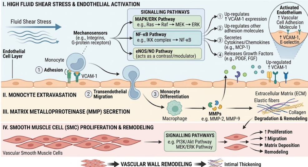
Figure 2. Arteriogenesis: Cellular and Molecular Cascade. Cellular diagram of high fluid shear stress on endothelial mechanoreceptors (TRPV4, Integrins, FAK/Src) leading to VCAM-1 expression, monocyte adhesion and transmigration into the adventitia. Macrophages are shown releasing MMP-1/9 to degrade the basement membrane while secreting PDGF and TGF-β to promote smooth muscle cell proliferation and outward vessel expansion.
As shown in Figure 2, fluid shear stress activates endothelial cells to upregulate adhesion molecules and chemoattractant that recruit circulating monocytes, which initiate matrix degradation and coat proliferation of smooth muscle cells needed for outward arterial remodeling(Yang et al., 2008).
This is important as accurate and objective diagnostic tools are needed to assess the clinical functional significance of coronary collateral circulation. Traditionally, collateral assessment was mainly based on visual evaluation during routine contrast coronary angiography(Choo, 2015). The Rentrop grading scale is the most widely recognized visual scoring system and was reported by Rentrop and colleagues. This angiographic scale classifies collateral filling into four discrete grades:
In everyday clinical practice the Rentrop scale is still often used because of its simplicity, but has major physiological and technical limitations. The spatial resolution limit for coronary angiography is about 100 µm Thus, the microscopic collateral connections, on the order of 30 to 90 micrometers, are completely invisible in standard angiograms despite their collective ability to carry functionally important blood flow. Moreover, standard resting angiography only visualizes spontaneously active collaterals; it does not measure “recruitable” collaterals that open only during acute arterial occlusion or maximal physical exertion(Hakimzadeh & Piek, 2013).
To overcome these visual limitations, Seiler, Pijls and colleagues pioneered quantitative pressure- and velocity-derived invasive measurements establishing the Collateral Flow Index (CFI) as physiological gold standard(Hakimzadeh & Piek, 2013). Collateral flow index (CFIp) derived from the pressure during temporary balloon occlusion during percutaneous coronary intervention is expressed as a fraction of flow through the same vessel when totally patent:
where P occl is the mean coronary wedge pressure distal to the inflated balloon measured using a sensor-tipped pressure guidewire, Pao is the mean aortic pressure measured simultaneously through the guiding catheter, and CVP is the central venous pressure representing back-pressure(Meier et al., 2013; Seiler, 2003).
A CFIp threshold of ≥ 0.25 has been shown in large clinical validation studies to indicate a functionally sufficient collateral circulation that can prevent electrocardiographic ST-segment elevation and myocardial ischemia during acute vessel occlusion. Patients with high collateral flow index (≥ 0.25) have significantly smaller infarct size, lower peak serum cardiac enzymes, preserved left ventricular ejection fraction, lower ventricular aneurysm formation and almost 35% reduction in long-term cardiovascular mortality compared with patients with low collateral capacity(Hakimzadeh & Piek, 2013; Meier et al., 2013).
The development of therapeutic interventions to actively stimulate arteriogenesis, so-called “biological revascularization,” is a major goal in cardiovascular medicine because of the tremendous prognostic benefit of a well-developed collateral circulation. Treatment methods can be generally classified into two groups: delivery of chemical/pharmacological agents and augmentation of mechanical shear-stress(Degen et al., 2014).
Initial experimental attempts were focused mostly on systemic or intracoronary administration of recombinant proangiogenic proteins and growth factors. Important clinical trials such as the VIVA (Vascular Endothelial Growth Factor in Ischemia for Vascular Angiogenesis) trial using recombinant human VEGF, and the FIRST and AGENT trials using recombinant basic fibroblast growth factor (FGF-2) and adenoviral FGF-4, respectively, sought to enhance coronary perfusion. However, this landmark phase II and III double-blind randomized clinical trials failed to show statistically significant improvement in primary endpoints such as exercise treadmill duration, regional myocardial perfusion, or angina frequency versus placebo(Buschmann, Heil, Jost, & Schaper, 2003).
The clinical failure of protein and gene therapy trials was caused by a fundamental mechanistic misunderstanding: administration of angiogenic growth factors mainly induced sprouting of primitive, high resistance capillary beds (angiogenesis) rather than muscular maturation of conductive arterioles (arteriogenesis) (Seiler, 2003). Furthermore, in dysvascular patients with underlying endothelial dysfunction, diabetes and hypercholesterolemia, recombinant growth factors did not elicit normal cellular responses because of impaired intracellular signal transduction(Meier et al., 2013).
Then, therapeutic approaches were developed targeting the inflammatory core of arteriogenesis by administration of cytokines capable of mobilizing and activating circulating monocytes. Granulocyte colony stimulating factor (G-CSF) and granulocyte macrophage colony stimulating factor (GM-CSF) have been tested in clinical trials by Seiler et al., 2003 (Seiler, 2003). Invasive measurements of collateral flow index and monocyte counts were significantly increased after subcutaneous administration of G-CSF and GM-CSF in patients with stable coronary disease, but safety concerns were raised. A trial of subcutaneous GM-CSF therapy was stopped early because 2 patients developed acute coronary syndromes(Meier et al., 2013). Inflammatory pathways overlap between arteriogenesis and atherogenesis, and systemic therapies with pro-inflammatory cytokines risk destabilizing vulnerable atherosclerotic plaques, driving cap degradation and triggering acute luminal thrombosis.
To mitigate the risks of cytokine-induced plaque rupture, research has been directed at the mechanical and physiologic augmentation of endogenous fluid shear stress without inducing systemic inflammation. Increased collateral growth can be achieved in the most effective natural way through regular physical activity training(Degen et al., 2014). Physical exercise increases heart rate, stroke volume and myocardial oxygen demand. Therefore, a coronary blood flow velocity increases approximately 5 times. This transient pulse increases the shear stress of fluid on the coronary endothelium. In addition, exercise -induced bradycardia prolongs the duration of cardiac diastole, the phase in which the majority of coronary perfusion occurs, thereby prolonging the temporal window of increased shear stress(Duncker & Bache, 2008).
Landmark studies, such as the EXCITE (Impact of Intensive Exercise Training on Coronary Collateral Circulation in Patients with Stable Coronary Artery Disease) trial performed by Möbius-Winkler and colleagues, have evaluated stable angina patients undergoing four weeks of high-intensity exercise training by randomized controlled clinical trials. The high-intensity exercise group showed significant improvements in invasive collateral flow index (CFIp) as well as improvements in peak oxygen consumption (VO2peak) and ischemic thresholds without regression of primary atherosclerotic lesions(Wu, Lin, Hsu, Lai, & Sheu, 2022).
Enhanced External Counterpulsation (EECP) is another very effective non-invasive mechanical strategy. In EECP therapy, pneumatic cuffs are placed on the patient’s lower extremities and inflate sequentially during cardiac diastole to hydraulic pressures of 300 mmHg, rapidly deflating just prior to systole. This simultaneous inflation dramatically increases diastolic aortic pressure and venous return, improving coronary perfusion pressure and increasing fluid shear stress over collateral beds. Randomized controlled trials show significant improvement in CFIp, reduction in angina frequency and improvement in myocardial perfusion following 30 hours of high-pressure EECP in patients with end-stage non-revascularizable coronary artery disease(Nickolay et al., 2020).
Similarly, pharmacological reduction of heart rate by ivabradine, a selective inhibitor of the sinoatrial node If pacemaker current, prolongs diastolic perfusion time. In clinical studies, Gloekler and colleagues confirmed that long-term ivabradine therapy significantly increased collateral flow index in patients with stable coronary artery disease, and demonstrated that the reduction of target heart rate provides an effective pro-arteriogenic mechanical stimulus, without inducing pro-inflammatory side effects(Degen et al., 2014).
Further, besides therapeutic interventions, advanced diagnostic research has identified non-invasive expression profiles of circulating microRNA (miRNA) as predictive biomarkers of collateral capacity. Constantinescu et al recently reported comprehensive profiling studies demonstrating distinct transcoronary microRNA gradients in patients with well-developed collateral circulation after myocardial infarction. Higher levels of pro-arteriogenic and mechanosensitive miRNAs, including miR-210, miR-143, miR-379, miR-146a, and miR-155, are strongly associated with high collateral flow index measurements and strong outward arterial remodeling. Conversely, high expression levels of anti-arteriogenic transcripts such as miR-329, miR-494 and miR-495 correlate with low monocyte activation and poor collateral capacity. The multiplex biomarker panels that combine the circulating microRNAs with serological cytokines (MCP-1, TGF-β, Adropin, sCD93/MMR-2) hold great promise for non-invasively stratifying a patient’s innate capacity for collateral development following infarction(Constantinescu et al., 2026).
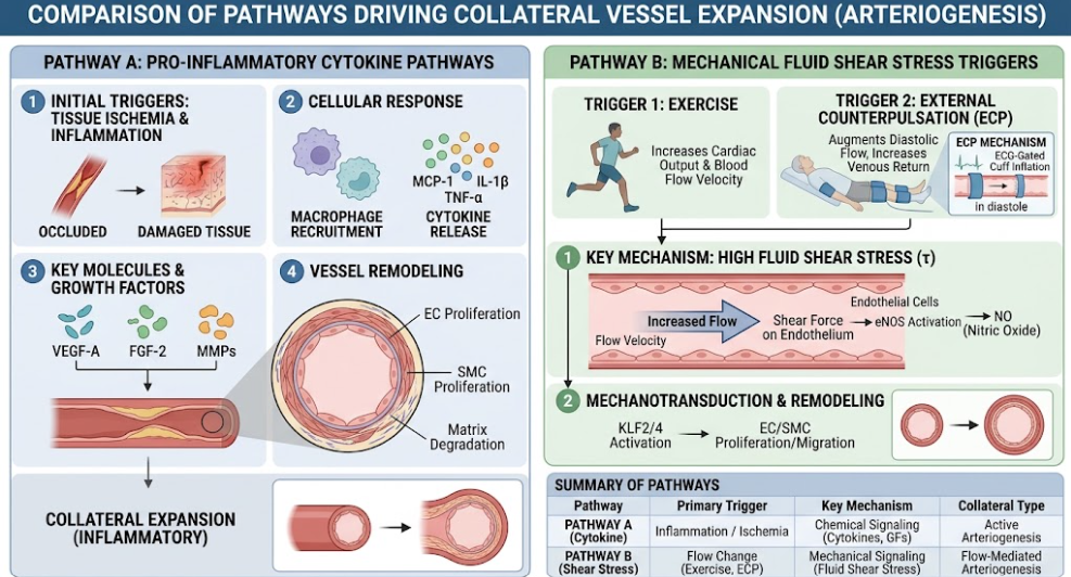
Figure 3. Therapeutic Approaches to Enhance Coronary Arteriogenesis. A conceptual comparative infographic comparing chemical cytokine delivery (VEGF, GM-CSF) versus mechanical shear-stress augmentation modalities (Physical Exercise, Enhanced External Counterpulsation, Ivabradine). The diagram illustrates how mechanical strategies can safely increase diastolic fluid shear stress and eNOS activation without systemic inflammation or plaque rupture.
Mechanical modalities such as exercise training and external counterpulsation increase physiologic shear stress and endothelial nitric oxide pathways without activating systemic inflammatory cascades that destabilize atherosclerotic plaques (see Figure 3)(Yang et al., 2008).
The study of coronary collateral circulation and arteriogenesis emphasizes a fascinating physiological adaptation protecting myocardial tissue in the setting of chronic ischemia or acute coronary events. Translating basic vascular biology into safe and effective clinical therapies, however, presents a number of difficult challenges(Degen et al., 2014).
One of the major challenges in therapeutic arteriogenesis is the physiological paradox of inflammation. As illustrated, arteriogenesis is dependent on local perivascular inflammation, monocyte infiltration, and matrix metalloproteinase activity to degrade the restricting adventitia and to allow outward expansion of the artery. However, systemic administration of pro-inflammatory cytokines may promote acceleration of atherogenesis and deterioration of the fibrous caps of vulnerable plaques in other areas of the coronary tree(Hakimzadeh & Piek, 2013). Matrix metalloproteinase degradation of interstitial collagen within a thin-cap fibroatheroma dramatically increases plaque structural stress and predisposing the lesion to catastrophic structural rupture, as we have already addressed in our previous reviews on coronary plaque dynamics in cardiovascular Atherosclerosis and Coronary Artery Plaque Rupture Dynamics in Cardiovascular Diseases. Therefore, future pharmacological agents should be designed to target localized endothelial mechanotransduction or monocyte CCR2 signaling in high-shear collateral channels without generalized systemic inflammatory activation(Degen et al., 2014; Hakimzadeh & Piek, 2013).
In addition, co-morbidities of pathological states often compromise the endogenous capacity for collateral expansion of the patient. Patients with advanced type 2 diabetes mellitus, metabolic syndrome and intense oxidative stress have severely blunted arteriogenic responses. Hyperglycemia and dyslipidemia induce severe endothelial dysfunction, leading to uncoupling of eNOS, impairments in monocyte chemotactic responsiveness to MCP-1, and downregulation of hypoxia-inducible factor 1-alpha (HIF-1α) expression. Thus, pro-arteriogenic therapies that work in young, healthy animal models often fail in heterogeneous human clinical cohorts with widespread metabolic disease(Chilian et al., 2012).
Another interesting biomechanical aspect is the possibility of competitive flow and restenosis after revascularization(Constantinescu et al., 2026). In a chronically occluded recipient artery with a well-developed collateral network, successful percutaneous coronary intervention (PCI) and stenting would suddenly restore antegrade blood flow. The rapid rise in antegrade pressure lowers the pressure gradient across the collateral pathway, causing the fluid shear stress within the collateral vessel to collapse(Chilian et al., 2012). This process of the collateral artery shrinking back and lying dormant is called collateral regression. However, when a well-developed collateral network is present along with a newly stented native vessel, the ensuing competitive blood flow may change the local wall shear stress patterns in the stented segment, which has been correlated with an increased risk for in-stent restenosis in some clinical intravascular ultrasound studies(Constantinescu et al., 2026).
Looking ahead, precision cardiovascular medicine needs to integrate non-invasive molecular diagnostic profiling with targeted mechanical therapies. By generating comprehensive biomarker panels that measure mechanosensitive microRNAs (such as miR-210, miR-143, miR-379) together with serological growth factors, clinicians will be able to identify early “poor collateral formers” during acute myocardial infarction(Constantinescu et al., 2026). Patients with documented suppressed collateral capacity can be triaged into intensive mechanical shear-stress therapies, including structured high-intensity exercise rehabilitation and bespoke external counterpulsation protocols, or selected for emerging targeted microRNA therapeutics that silence anti-arteriogenic signaling cascades without compromising plaque stability(Wu et al., 2022).
The coronary collateral circulation is an important physiological defense mechanism which may alleviate myocardial ischemia, preserve left ventricular function and significantly reduce the cardiovascular death rate in patients with severe coronary artery disease. Arteriogenesis is the outward structural expansion and maturation of pre-existing arteriolar anastomoses into high-capacity muscular conductive arteries The fundamental initiator and maintainer of arteriogenesis are the hydrodynamic pressure gradients and fluid shear stress. This mechanical force is translated into active vessel growth through a precisely orchestrated biological cascade, including endothelial mechanoreceptors, integrin-FAK activation, nitric oxide synthesis, monocyte extravasation, matrix metalloproteinase activity mediating extracellular matrix remodeling, and reconstruction of the smooth muscle cell coat.
Early clinical trials with chemical revascularization using pro-angiogenic growth factors or pro-inflammatory cytokines failed because of misguided capillary sprouting or unacceptable risks of plaque destabilization. Conversely, mechanical approaches that augment the natural fluid shear stress, namely physical exercise training, enhanced external counterpulsation, and heart rate reduction, have emerged as highly effective, safe, and clinically validated pro-arteriogenic therapies. With the development of advanced imaging, invasive pressure-derived collateral flow index assessments, and circulating microRNA biomarker panels, the merger of biomechanical insight and molecular diagnostics will enable modern cardiology to maximize biological revascularization, ultimately transforming patient care and reducing the global burden of ischemic heart disease.
Ballermann, B. J., Dardik, A., Eng, E., & Liu, A. (1998). Shear stress and the endothelium. Kidney Int Suppl, 67, S100-108. doi:10.1046/j.1523-1755.1998.06720.x
Bavry, A. A., Kumbhani, D. J., Rassi, A. N., Bhatt, D. L., & Askari, A. T. (2006). Benefit of early invasive therapy in acute coronary syndromes: a meta-analysis of contemporary randomized clinical trials. J Am Coll Cardiol, 48(7), 1319-1325. doi:10.1016/j.jacc.2006.06.050
Buschmann, I., Heil, M., Jost, M., & Schaper, W. (2003). Influence of inflammatory cytokines on arteriogenesis. Microcirculation, 10(3-4), 371-379. doi:10.1038/sj.mn.7800199
Chilian, W. M., Penn, M. S., Pung, Y. F., Dong, F., Mayorga, M., Ohanyan, V., . . . Yin, L. (2012). Coronary collateral growth–back to the future. J Mol Cell Cardiol, 52(4), 905-911. doi:10.1016/j.yjmcc.2011.12.006
Choo, G. H. (2015). Collateral Circulation in Chronic Total Occlusions – an interventional perspective. Curr Cardiol Rev, 11(4), 277-284. doi:10.2174/1573403×11666150909112548
Constantinescu, A., Micheu, M. M., Liehn, E. A., & Udrişte, A. S. (2026). Predictive Biomarkers for Coronary Collateral Circulation Development After Myocardial Infarction. Int J Mol Sci, 27(8). doi:10.3390/ijms27083671
Cronin, N. M., Dawson, L. W., & DeMali, K. A. (2024). Shear stress-stimulated AMPK couples endothelial cell mechanics, metabolism and vasodilation. J Cell Sci, 137(24). doi:10.1242/jcs.262232
Degen, A., Millenaar, D., & Schirmer, S. H. (2014). Therapeutic approaches in the stimulation of the coronary collateral circulation. Curr Cardiol Rev, 10(1), 65-72. doi:10.2174/1573403×113099990027
Duncker, D. J., & Bache, R. J. (2008). Regulation of coronary blood flow during exercise. Physiol Rev, 88(3), 1009-1086. doi:10.1152/physrev.00045.2006
Eitenmüller, I., Volger, O., Kluge, A., Troidl, K., Barancik, M., Cai, W. J., . . . Schaper, W. (2006). The range of adaptation by collateral vessels after femoral artery occlusion. Circ Res, 99(6), 656-662. doi:10.1161/01.RES.0000242560.77512.dd
Hakimzadeh, N., & Piek, J. J. (2013). The coronary collateral circulation revisited. Neth Heart J, 21(3), 144-145. doi:10.1007/s12471-013-0376-0
Helfant, R. H., Vokonas, P. S., & Gorlin, R. (1971). Functional importance of the human coronary collateral circulation. N Engl J Med, 284(23), 1277-1281. doi:10.1056/nejm197106102842301
Hong, S. G., Ashby, J. W., Kennelly, J. P., Wu, M., Steel, M., Chattopadhyay, E., . . . Mack, J. J. (2024). Mechanosensitive membrane domains regulate calcium entry in arterial endothelial cells to protect against inflammation. J Clin Invest, 134(13). doi:10.1172/jci175057
Koerselman, J., van der Graaf, Y., de Jaegere, P. P., & Grobbee, D. E. (2003). Coronary collaterals: an important and underexposed aspect of coronary artery disease. Circulation, 107(19), 2507-2511. doi:10.1161/01.Cir.0000065118.99409.5f
Meier, P., Schirmer, S. H., Lansky, A. J., Timmis, A., Pitt, B., & Seiler, C. (2013). The collateral circulation of the heart. BMC Med, 11, 143. doi:10.1186/1741-7015-11-143
Naylor, L. H., O’Driscoll, G., Fitzsimons, M., Arnolda, L. F., & Green, D. J. (2006). Effects of training resumption on conduit arterial diameter in elite rowers. Med Sci Sports Exerc, 38(1), 86-92. doi:10.1249/01.mss.0000181220.03855.1c
Nickolay, T., Nichols, S., Ingle, L., & Hoye, A. (2020). Exercise Training as a Mediator for Enhancing Coronary Collateral Circulation: A Review of the Evidence. Curr Cardiol Rev, 16(3), 212-220. doi:10.2174/1573403×15666190819144336
O’Donoghue, M., Boden, W. E., Braunwald, E., Cannon, C. P., Clayton, T. C., de Winter, R. J., . . . Sabatine, M. S. (2008). Early invasive vs conservative treatment strategies in women and men with unstable angina and non-ST-segment elevation myocardial infarction: a meta-analysis. Jama, 300(1), 71-80. doi:10.1001/jama.300.1.71
Pipp, F., Boehm, S., Cai, W. J., Adili, F., Ziegler, B., Karanovic, G., . . . Schmitz-Rixen, T. (2004). Elevated fluid shear stress enhances postocclusive collateral artery growth and gene expression in the pig hind limb. Arterioscler Thromb Vasc Biol, 24(9), 1664-1668. doi:10.1161/01.ATV.0000138028.14390.e4
Schaper, W. (2009). Collateral circulation: past and present. Basic Res Cardiol, 104(1), 5-21. doi:10.1007/s00395-008-0760-x
Seiler, C. (2003). The human coronary collateral circulation. Heart, 89(11), 1352-1357. doi:10.1136/heart.89.11.1352
Seiler, C. (2010). The human coronary collateral circulation. Eur J Clin Invest, 40(5), 465-476. doi:10.1111/j.1365-2362.2010.02282.x
Troidl, C., Troidl, K., Schierling, W., Cai, W. J., Nef, H., Möllmann, H., . . . Schaper, W. (2009). Trpv4 induces collateral vessel growth during regeneration of the arterial circulation. J Cell Mol Med, 13(8b), 2613-2621. doi:10.1111/j.1582-4934.2008.00579.x
Troidl, K., Tribulova, S., Cai, W. J., Rüding, I., Apfelbeck, H., Schierling, W., . . . Schaper, W. (2010). Effects of endogenous nitric oxide and of DETA NONOate in arteriogenesis. J Cardiovasc Pharmacol, 55(2), 153-160. doi:10.1097/FJC.0b013e3181c9556f
Van Royen, N., Piek, J. J., Schaper, W., Bode, C., & Buschmann, I. (2001). Arteriogenesis: mechanisms and modulation of collateral artery development. J Nucl Cardiol, 8(6), 687-693. doi:10.1067/mnc.2001.118924
Wu, W. T., Lin, T. H., Hsu, P. C., Lai, W. T., & Sheu, S. H. (2022). Prediction for Coronary Collateral Development. Acta Cardiol Sin, 38(1), 27-28. doi:10.6515/acs.202201_38(1).20211208a
Yang, H. T., Prior, B. M., Lloyd, P. G., Taylor, J. C., Li, Z., Laughlin, M. H., & Terjung, R. L. (2008). Training-induced vascular adaptations to ischemic muscle. J Physiol Pharmacol, 59 Suppl 7(Suppl 7), 57-70.