Our Services
Others
- DFPP
- Shockwave
- IV Drip


“This literature review provides a comprehensive mechanistic synthesis of how the strict nutrient diffusion limits of the human intervertebral disc—the largest avascular structure in the body drive metabolic stress, cellular fate transitions, and extracellular matrix breakdown in Degenerative Disc Disease.”
The human intervertebral disc (IVD) is the largest avascular organ in the human body, creating an extraordinary biological paradigm where resident cells operate at the extreme threshold of metabolic survival. Lacking a direct microvascular network within the central nucleus pulposus (NP) and inner annulus fibrosus (AF), the IVD relies almost exclusively on passive molecular diffusion from capillary beds terminating at the cartilaginous endplates (CEPs) and outer AF margins. These structural constraints establish severe physiological gradients: oxygen tension, glucose concentration, and extracellular pH diminish precipitously toward the disc center, compelling resident cells to depend heavily on anaerobic glycolysis for adenosine triphosphate (ATP) production. This narrative literature review synthesizes current evidence on the physiological and pathological nutrient transport mechanisms within the IVD and explores how biological diffusion limits drive the onset and progression of Degenerative Disc Disease (DDD). Pathologic changes such as calcification of the CEP, sclerosis of the subchondral bone, dehydration of the matrix and continuous mechanical compression reduce the diffusivity of solutes and increase the resistance to transport. This microenvironmental deprivation triggers a detrimental biochemical cascade characterized by severe intracellular acidosis, reactive oxygen species (ROS) accumulation, expression of catabolic enzymes (MMPs and ADAMTS), cellular senescence, and various forms of regulated cell death (apoptosis, pyroptosis, necroptosis, and ferroptosis). Understanding diffusion-driven degenerative mechanisms is of utmost importance for early biomarker identification and regenerative therapy development that can survive the hostile metabolic environment of the degenerating disc.
Keywords: Intervertebral Disc Degeneration, Avascular Organ, Passive Diffusion, Cartilage Endplate, Metabolic Stress, Cellular Senescence, Extracellular Matrix Degradation
Degenerative Disc Disease (DDD) is one of the most important causes of chronic low back pain and spinal disability worldwide, with enormous socioeconomic costs, and reduction of workforce productivity(Gradisnik, Kocivnik, Maver, & Velnar, 2025). Anatomically, the intervertebral disc (IVD) is a complex, highly specialized fibrocartilaginous structure situated between adjacent vertebral bodies(Yurube, Takeoka, Kanda, Kuroda, & Kakutani, 2023). Spinal motion is allowed, axial compressive loads are transmitted, and the biomechanical alignment is retained through the motion segments(Qiao, Zhang, & Zhao, 2025). It serves as a dynamic mechanical shock absorber.
The IVD consists of three distinct anatomical compartments that function together:
The most fundamental physiological hallmark of the mature human IVD being its position as the largest avascular organ of the human body. Transient vascular channels in the endplates are early embryonic and infant precursors that regress during early childhood and disappear by adolescence(Dowdell et al., 2017; Murphy, Lufkin, & Kraus, 2023). Consequently, the mature NP and inner AF possess no direct microvascular supply, leaving central disc cells up to 7 to 8 millimeters away from the nearest functional blood vessel(Nishida et al., 1999).
This avascular architecture obligates the IVD to rely almost entirely on passive molecular diffusion across matrix gradients to receive vital nutritional solutes—such as glucose and oxygen—and to evacuate toxic metabolic byproducts like lactic acid. This delicate diffusive equilibrium is absolutely essential for cellular survival and extracellular matrix (ECM) homeostasis, and any physical or structural impediment to nutrient transport results in severe metabolic stress. This literature review delves into the inherent biological limitations of nutrient diffusion in the IVD, the pathophysiological processes leading to impaired solute transport and the long term nutritional deprivation that drives the cellular and matrix destruction that is characteristic of Degenerative Disc Disease(Urban, Smith, & Fairbank, 2004).
Because the interior compartments of the IVD lack direct vascularization, solute supply and waste elimination depend on two peripheral microvascular routes:
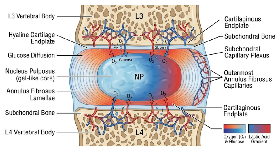
Figure 1. Mechanism of nutrient exchange in lumbar inter-vertebral disc. This schematic diagram shows how nutrients (oxygen and glucose) diffuse from the subchondral capillary networks and the outer annulus fibrosus, through the cartilaginous endplates into the core of the nucleus pulposus.You see the concentration gradients here: nutrients decline toward the avascular center (red > blue), and the lactic acid gradient (blue > red).
Solutes are transported in the dense ECM of the IVD by two physical mechanisms: passive molecular diffusion and convective fluid flow. Passive diffusion is based on Fick’s laws and driven by the concentration gradients across the tissue(Hickman, Rathan-Kumar, & Peck, 2022). It is the major transport pathway for uncharged low molecular weight solutes such as glucose (~ 180 Da), dissolved oxygen (~ 32 Da) and lactate (~ 89 Da)(De Geer, 2018). Convective fluid transport, driven by hydraulic pressure gradients generated during cyclic biomechanical loading, plays a secondary role for small solutes but is critical for transporting larger macromolecules, including growth factors, cytokines, and enzymes(De Geer, 2018).
The central nucleus pulposus, as seen in Figure 1, is mainly dependent on passive diffusion through the microvasculature of the cartilage endplate, resulting in significant solute gradients from the periphery to the core that depend on distance. Transport efficiency of small solutes is governed by effective diffusion coefficient (Deff) and partition coefficient (K) that are strongly modulated by tissue hydration, fixed charge density and structural anisotropy(Jackson, Yuan, Huang, Brown, & Gu, 2012).
Experimental measurements in human AF tissue indicate that the diffusivity of nutrients is highly anisotropic, i.e. the rates of diffusion depend on the spatial orientation. In particular, the radial diffusivity of solute is significantly smaller than in the axial or circumferential directions(Jackson et al., 2012). This directional diffusion is related to the highly organized lamellar structure of the AF in which the parallel collagen fiber bundles and interlamellar microtubes provide preferential transport pathways along the axial and circumferential planes while radial transport across the concentric lamellae is highly resistant(Urban, Holm, Maroudas, & Nachemson, 1982). Hence the radial diffusion from the peripheral AF is fundamentally inefficient in satisfying the metabolic demands of the central NP when the CEP route is compromised during disease(Nachemson, Lewin, Maroudas, & Freeman, 1970).
The IVD’s unique avascular anatomy leads to an extreme microenvironment with severe nutrient deprivation, chronic hypoxia, hyperosmolality and acidic pH. Resident disc cells are under severe metabolic constraints, and they need to adapt their bioenergetics to survive on the edge of biological viability(Nishida et al., 1999).
Glucose is the main substrate for energy production in the cells of the intervertebral disc. In normal adults, the physiological concentration of glucose in the outer periphery of the AF and CEP interface is around 5.0 mM, and in the central NP it is 0.5 to 1.0 mM(Zhang et al., 2022). Oxygen levels are extremely low in the central NP, so disc cells generate almost all of their adenosine triphosphate (ATP) through anaerobic glycolysis. Glycolysis is the process in which each molecule of glucose is converted into two molecules of lactic acid and produces only 2 ATP molecules instead of 36 ATP molecules produced through oxidative phosphorylation(Urban et al., 2004).
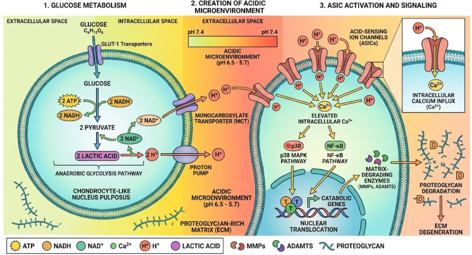
Figure 2. Metabolic acidosis and ASIC activation in nucleus pulposus cells. Diagram of a chondrocyte-like nucleus pulposus cell in a matrix rich in proteoglycans. Anaerobic glycolysis leads to the production of ATP and lactic acid, which results in an acidic extracellular microenvironment (pH 6.5-5.7). This acidity triggers Acid-Sensing Ion Channels (ASICs) that cause an intracellular calcium influx and downstream catabolic signaling, resulting in matrix degeneration.
As shown in the Figure 2, nucleus pulposus cells rely on anaerobic glycolysis to convert glucose to ATP and lactic acid. This process is highly sensitive to the drop of extracellular pH and exhaustion of glucose. Healthy NP has a relatively low cell density (~4,000 cells/mm3), compared to the AF (~9,000 cells/mm3), an evolutionary adaptation to limit total metabolic demand over long diffusion distances(Johnson, Stephan, & Roberts, 2008).
Survival of cells in this low-glucose environment is determined by strict biological thresholds:
Oxygen partial pressure declines sharply from approximately 10% to 13% at the disc margins to 1% to 2% (approx. 1.0 to 3.6 kPa) in the center of the NP. A unique molecular machinery is possessed by the resident NP cells to survive in this hypoxic niche, with it being mainly driven by the constitutive expression of Hypoxia Inducible Factor 1-alpha (HIF-1α)(Karchevskaya, Poluektov, & Korolishin, 2023; Oichi, Taniguchi, Oshima, Tanaka, & Saito, 2020). Unlike peripheral cells that degrade HIF-1α under normoxia conditions, NP cells constantly stabilize HIF-1α, which transactivates critical glycolytic enzymes, glucose transporters and extracellular matrix genes including aggrecan(Luo et al., 2021).
Oxygen is not the major determinant of disc cell survival but is an important regulatory factor in extracellular matrix biosynthesis. Matrix production, i.e. synthesis of sulfated glycosaminoglycans (sGAG) and protein complexes, occurs most rapidly at an optimal oxygen concentration of 5%. At oxygen tensions <5% and in extreme hypoxia (<1%) matrix synthesis rates are reduced significantly compromising tissue repair potential(Grunhagen, Wilde, Soukane, Shirazi-Adl, & Urban, 2006).
A direct consequence of intense anaerobic glycolysis in an avascular tissue is the generation and retention of lactic acid. Protons and lactate ions exported into the narrow intercellular spaces cannot be rapidly cleared due to long diffusion distances. A healthy young IVD maintains the extracellular pH at a slightly acidic pH of 7.0 to 7.2. However, early degeneration causes an impairment in the clearance of metabolic waste, resulting in the accumulation of lactic acid and a decrease of local extracellular pH to 6.5 and 5.7 in the moderately and severely degenerated discs respectively(Horner & Urban, 2001; Murphy et al., 2023).
The extracellular acidification has devastating catabolic and cytotoxic consequences:
Degenerative Disc Disease begins and progresses when the delicate balance between the supply of nutrients and the demand from cells is upset. Convergence of structural, mechanical and systemic factors impairs matrix permeability and creates physical barriers that choke off solute diffusion(Chan, Sze, Samartzis, Leung, & Chan, 2011).
Because the CEP is the primary gateway for solute transport into the NP, morphological alterations in the CEP represent a critical initiating trigger for disc degeneration. With advancing age and pathological stress, the hyaline matrix of the CEP undergoes progressive calcification, mineralization, and endochondral-like ossification(Nishida et al., 1999).
This pathological remodeling consists of:
The kinetics of solute transport is highly dependent on mechanical loading. During diurnal loading cycles of daytime weight bearing, IVD can lose up to 20% of its fluid volume that is typically replenished during nighttime rest. However, abnormal, excessive or sustained static compressive loading results in extrusion of interstitial water from the ECM and matrix compaction(De Geer, 2018; Jackson et al., 2012; Murphy et al., 2023).
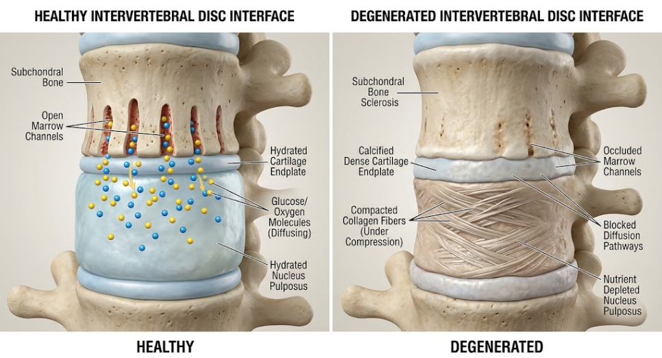
Figure 3. Healthy vs. Degenerated Intervertebral Disc Interface. A side-by-side illustration comparing a healthy intervertebral disc with open marrow channels and active nutrient diffusion (left) to a degenerated disc exhibiting subchondral bone sclerosis, calcified endplates, compacted collagen fibers, and blocked diffusion pathways leading to nutrient depletion (right).
As in the figure 3, structural calcification of the cartilage endplates and compression-induced matrix compaction severely restrict solute diffusion into the central nucleus pulposus. Compaction reduces the effective pore size of the extracellular matrix. Because solute diffusivity is directly proportional to tissue water content and pore dimensions, compression-induced fluid exudation significantly lowers the diffusion coefficients of both glucose and oxygen. Further, radial diffusion across concentric AF lamellae is intrinsically slower due to the structural anisotropy and static compression further compromises periannular solute delivery, making the central and posterior regions of the disc highly prone to nutrient deprivation(Jackson et al., 2012).
Systemic conditions that impair microvascular blood flow to the vertebrae increase diffusion distances in the disc:
Combination of theories of mechano-electrochemical transport with three-dimensional finite element models has provided key spatial insights into how patterns of degeneration are dictated by specific pathway impairments:
When solute diffusion is below critical physiological thresholds, resident disc cells experience a series of catastrophic fate transitions. Starving, acidotic cells shift from anabolic extracellular matrix maintenance to active catabolism, inflammatory signaling, and programmed cell death(Urban, 2002).
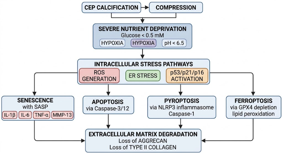
Figure 4. Pathways of Nucleus Pulposus Degeneration. The flowchart illustrates the molecular phenomena of nucleus pulposus cells under severe microenvironmental stress (low glucose, hypoxia, acidic pH). It shows how mechanical and nutritional stimuli induce intracellular stress, different modes of cell death and the subsequent loss of aggrecan and type II collagen.
In response to severe nutrient stress disc cells downregulate a number of key anabolic transcription factors, most notably SOX9, resulting in a dramatic reduction in synthesis of aggrecan and type II collagen. Concomitantly, cells upregulate the expression of catabolic proteases such as Matrix Metalloproteinases (MMP-1, MMP-3, MMP-13), and A Disintegrin and Metalloproteinase with Thrombospondin Motifs (ADAMTS-4, ADAMTS-5)(Wu, Kim, & Jang, 2020).
These catabolic enzymes cleave the core protein of aggrecan, releasing hydrophilic glycosaminoglycan side-chains into the tissue. The fixed charge density and the osmotic swelling pressure of the NP decrease as the GAG content decreases. The NP loses its hydration and hydrostatic pressurization resulting in a reduction of disc height and redistribution of axial compressive loads directly onto the fibrotic annulus fibrosus. When exposed to altered mechanical strain, the cells remaining switch from the synthesis of type II to type I collagen, and the gel-like NP becomes disorganized, fibrotic scar tissue(Shnayder et al., 2023).
Resident disc cells undergo stress-induced premature senescence (SIPS) as a consequence of chronic metabolic stress, DNA damage and ROS accumulation. Senescent cells are characterized by an irreversible cell-cycle arrest that is mediated by upregulation of the cyclin-dependent kinase inhibitors, p16INK4a and p21CIP1/WAF1 via the p53-p21-Rb pathway (Wang, Cai, Shi, Wang, & Wu, 2016). Senescent cells are metabolically active but non- dividing cells that adopt a Senescence Associated Secretory Phenotype (SASP).
The Senescence Associated Secretory Phenotype (SASP) secretome is characterized by an increased secretion of a variety of factors including:
Moreover, SASP factors induce senescence of the adjacent healthy cells by autocrine and paracrine signaling, which further promotes the degradation of the extracellular matrix in local area and generates a self-feedback inflammatory microenvironment(Gradisnik et al., 2025).
As in the figure 4, Chronic nutrient deprivation initiates multiple cell death pathways and secretion of catabolic enzymes that maintain a cycle of tissue degeneration. Regulated cell death involves four types and is induced by severe glucose withdrawal and acidic microenvironmental stress, which exceed cellular repair mechanisms:
Early mild nutrient deprivation triggers autophagy, an intracellular self-cannibalization and recycling pathway, in disc cells to clear damaged organelles and produce transient ATP. PI3K/Akt/mTOR pathway is a regulator of autophagic flux. Mild starvation induces autophagy to protect NP cells from apoptosis. However, when degeneration advances to the stage of complete nutrient deprivation, autophagic capacity is depleted or dysregulated and cannot block cell death(Yurube et al., 2023).
Healthy adult discs have a dense matrix structure and endogenous anti-angiogenic/anti-neurogenic factors (e.g., Aggrecan fixed charge, Semaphorin-3A) that inhibit blood vessel and nerve ingrowth(Karchevskaya et al., 2023). But nutrient starvation causes loss of aggrecan and secretion of SASP factors such as VEGF, NGF and BDNF which results in vascular and neural fibers growing in from peripheral tissues through annular fissures and calcified endplates. Vascular ingrowth exposes the immune-privileged NP matrix to the systemic circulation and induces autoimmune responses and macrophage infiltration. Local IL-1β, TNF-α and acidic pH sensitize non-myelinated nociceptive nerve fibers that follow these vascular channels deep into the degenerated disc generating chronic discogenic low back pain(Murphy et al., 2023; Veronesi et al., 2026).
The synthesis of literature presented in this review establishes that the strictly avascular architecture of the human intervertebral disc represents a double-edged evolutionary trade-off. While avascularity and high swelling pressure enable the IVD to withstand massive, dynamic mechanical loads across a lifespan, it leaves resident cells operating at the absolute margin of bioenergetic viability(Urban et al., 2004).
The primary initiating event in poor-nutrition-related DDD is a reduction in solute transport below critical biological thresholds. Computational simulations and experimental transport studies show that cellular repair mechanisms catastrophically fail at glucose concentrations below 0.5 mM or extracellular pH below 6.5. Loss of functional cells leads to an irreversible reduction in proteoglycan production, dehydration of the matrix, loss of disc height and mechanical disequilibrium, which propagates injury to adjacent spinal structures(Chan et al., 2011).
The design of successful biological and cell-based therapies for DDD requires a thorough understanding of the strict biological limits of nutrient diffusion:
Despite the great progress in understanding the IVD transport biology, there are still a few limitations with current research:
The intervertebral disc exists at the ultimate boundary of biological transport constraint. As the largest avascular organ in the human body, its structural survival is intrinsically bound to passive molecular diffusion across matrix gradients. Pathological calcification of the cartilage endplates, mechanical compaction under sustained compressive loading, and systemic microvascular compromise disrupt solute diffusivity, starving resident cells of glucose and oxygen while trapping acidic metabolic byproducts. This microenvironment failure triggers a destructive molecular cascade that involves catabolic protease dominance, stress-induced cellular senescence, inflammatory amplification by SASP and multiple regulated cell death pathways. Future therapeutic breakthroughs in Degenerative Disc Disease will depend on strategies that restore matrix permeability and overcome nutrient diffusion limits, creating a viable biological niche for true tissue regeneration.
Chan, W. C., Sze, K. L., Samartzis, D., Leung, V. Y., & Chan, D. (2011). Structure and biology of the intervertebral disk in health and disease. Orthop Clin North Am, 42(4), 447-464, vii. doi:10.1016/j.ocl.2011.07.012
De Geer, C. M. (2018). Intervertebral Disk Nutrients and Transport Mechanisms in Relation to Disk Degeneration: A Narrative Literature Review. J Chiropr Med, 17(2), 97-105. doi:10.1016/j.jcm.2017.11.006
Dowdell, J., Erwin, M., Choma, T., Vaccaro, A., Iatridis, J., & Cho, S. K. (2017). Intervertebral Disk Degeneration and Repair. Neurosurgery, 80(3s), S46-s54. doi:10.1093/neuros/nyw078
Gradisnik, L., Kocivnik, N., Maver, U., & Velnar, T. (2025). Degenerative Disease of Intervertebral Disc: A Narrative Review of Pathogenesis, Clinical Implications and Therapies. Bioengineering (Basel), 13(1). doi:10.3390/bioengineering13010040
Grunhagen, T., Wilde, G., Soukane, D. M., Shirazi-Adl, S. A., & Urban, J. P. (2006). Nutrient supply and intervertebral disc metabolism. J Bone Joint Surg Am, 88 Suppl 2, 30-35. doi:10.2106/jbjs.E.01290
Hickman, T. T., Rathan-Kumar, S., & Peck, S. H. (2022). Development, Pathogenesis, and Regeneration of the Intervertebral Disc: Current and Future Insights Spanning Traditional to Omics Methods. Front Cell Dev Biol, 10, 841831. doi:10.3389/fcell.2022.841831
Horner, H. A., & Urban, J. P. (2001). 2001 Volvo Award Winner in Basic Science Studies: Effect of nutrient supply on the viability of cells from the nucleus pulposus of the intervertebral disc. Spine (Phila Pa 1976), 26(23), 2543-2549. doi:10.1097/00007632-200112010-00006
Jackson, A. R., Yuan, T. Y., Huang, C. Y., Brown, M. D., & Gu, W. Y. (2012). Nutrient transport in human annulus fibrosus is affected by compressive strain and anisotropy. Ann Biomed Eng, 40(12), 2551-2558. doi:10.1007/s10439-012-0606-4
Johnson, W. E., Stephan, S., & Roberts, S. (2008). The influence of serum, glucose and oxygen on intervertebral disc cell growth in vitro: implications for degenerative disc disease. Arthritis Res Ther, 10(2), R46. doi:10.1186/ar2405
Karchevskaya, A. E., Poluektov, Y. M., & Korolishin, V. A. (2023). Understanding Intervertebral Disc Degeneration: Background Factors and the Role of Initial Injury. Biomedicines, 11(10), 2714.
Kos, N., Gradisnik, L., & Velnar, T. (2019). A Brief Review of the Degenerative Intervertebral Disc Disease. Med Arch, 73(6), 421-424. doi:10.5455/medarh.2019.73.421-424
Li, Z., Zhang, K., Li, X., Pan, H., Li, S., Chen, F., . . . Liu, H. (2018). Wnt5a suppresses inflammation-driven intervertebral disc degeneration via a TNF-α/NF-κB-Wnt5a negative-feedback loop. Osteoarthritis Cartilage, 26(7), 966-977. doi:10.1016/j.joca.2018.04.002
Luo, L., Jian, X., Sun, H., Qin, J., Wang, Y., Zhang, J., . . . Zhou, Y. (2021). Cartilage endplate stem cells inhibit intervertebral disc degeneration by releasing exosomes to nucleus pulposus cells to activate Akt/autophagy. Stem Cells, 39(4), 467-481. doi:10.1002/stem.3322
Maldonado, E. N., Sheldon, K. L., DeHart, D. N., Patnaik, J., Manevich, Y., Townsend, D. M., . . . Lemasters, J. J. (2013). Voltage-dependent anion channels modulate mitochondrial metabolism in cancer cells: regulation by free tubulin and erastin. J Biol Chem, 288(17), 11920-11929. doi:10.1074/jbc.M112.433847
Murphy, K., Lufkin, T., & Kraus, P. (2023). Development and Degeneration of the Intervertebral Disc-Insights from Across Species. Vet Sci, 10(9). doi:10.3390/vetsci10090540
Nachemson, A., Lewin, T., Maroudas, A., & Freeman, M. A. (1970). In vitro diffusion of dye through the end-plates and the annulus fibrosus of human lumbar inter-vertebral discs. Acta Orthop Scand, 41(6), 589-607. doi:10.3109/17453677008991550
Naqvi, S. M., & Buckley, C. T. (2015). Extracellular matrix production by nucleus pulposus and bone marrow stem cells in response to altered oxygen and glucose microenvironments. J Anat, 227(6), 757-766. doi:10.1111/joa.12305
Nishida, K., Kang, J. D., Gilbertson, L. G., Moon, S. H., Suh, J. K., Vogt, M. T., . . . Evans, C. H. (1999). Modulation of the biologic activity of the rabbit intervertebral disc by gene therapy: an in vivo study of adenovirus-mediated transfer of the human transforming growth factor beta 1 encoding gene. Spine (Phila Pa 1976), 24(23), 2419-2425. doi:10.1097/00007632-199912010-00002
Ohnishi, T., Novais, E. J., & Risbud, M. V. (2020). Alterations in ECM signature underscore multiple sub-phenotypes of intervertebral disc degeneration. Matrix Biol Plus, 6-7, 100036. doi:10.1016/j.mbplus.2020.100036
Oichi, T., Taniguchi, Y., Oshima, Y., Tanaka, S., & Saito, T. (2020). Pathomechanism of intervertebral disc degeneration. JOR Spine, 3(1), e1076. doi:10.1002/jsp2.1076
Qiao, H., Zhang, K., & Zhao, J. (2025). Mechanobiology of intervertebral disc degeneration: From pathological mechanisms to therapeutic approaches. Mechanobiol Med, 3(4), 100162. doi:10.1016/j.mbm.2025.100162
Roughley, P. J. (2004). Biology of intervertebral disc aging and degeneration: involvement of the extracellular matrix. Spine (Phila Pa 1976), 29(23), 2691-2699. doi:10.1097/01.brs.0000146101.53784.b1
Shirazi-Adl, A., Taheri, M., & Urban, J. P. (2010). Analysis of cell viability in intervertebral disc: Effect of endplate permeability on cell population. J Biomech, 43(7), 1330-1336. doi:10.1016/j.jbiomech.2010.01.023
Shnayder, N. A., Ashhotov, A. V., Trefilova, V. V., Nurgaliev, Z. A., Novitsky, M. A., Vaiman, E. E., . . . Nasyrova, R. F. (2023). Cytokine Imbalance as a Biomarker of Intervertebral Disk Degeneration. Int J Mol Sci, 24(3). doi:10.3390/ijms24032360
Urban, J. P. (2002). The role of the physicochemical environment in determining disc cell behaviour. Biochem Soc Trans, 30(Pt 6), 858-864. doi:10.1042/bst0300858
Urban, J. P., Holm, S., Maroudas, A., & Nachemson, A. (1982). Nutrition of the intervertebral disc: effect of fluid flow on solute transport. Clin Orthop Relat Res(170), 296-302.
Urban, J. P., & Roberts, S. (2003). Degeneration of the intervertebral disc. Arthritis Res Ther, 5(3), 120-130. doi:10.1186/ar629
Urban, J. P., Smith, S., & Fairbank, J. C. (2004). Nutrition of the intervertebral disc. Spine (Phila Pa 1976), 29(23), 2700-2709. doi:10.1097/01.brs.0000146499.97948.52
Veronesi, F., Salamanna, F., Tedesco, G., Ruffilli, A., Rosa, F., Faldini, C., & Giavaresi, G. (2026). Mapping the degenerating intervertebral disc: a systematic review of histological evidence. Front Med (Lausanne), 13, 1753988. doi:10.3389/fmed.2026.1753988
Von Forell, G. A., Nelson, T. G., Samartzis, D., & Bowden, A. E. (2014). Changes in vertebral strain energy correlate with increased presence of Schmorl’s nodes in multi-level lumbar disk degeneration. J Biomech Eng, 136(6), 061002. doi:10.1115/1.4027301
Wang, F., Cai, F., Shi, R., Wang, X. H., & Wu, X. T. (2016). Aging and age related stresses: a senescence mechanism of intervertebral disc degeneration. Osteoarthritis Cartilage, 24(3), 398-408. doi:10.1016/j.joca.2015.09.019
Wu, P. H., Kim, H. S., & Jang, I. T. (2020). Intervertebral Disc Diseases PART 2: A Review of the Current Diagnostic and Treatment Strategies for Intervertebral Disc Disease. Int J Mol Sci, 21(6). doi:10.3390/ijms21062135
Yurube, T., Takeoka, Y., Kanda, Y., Kuroda, R., & Kakutani, K. (2023). Intervertebral disc cell fate during aging and degeneration: apoptosis, senescence, and autophagy. N Am Spine Soc J, 14, 100210. doi:10.1016/j.xnsj.2023.100210
Zhang, J., Zhang, W., Sun, T., Wang, J., Li, Y., Liu, J., & Li, Z. (2022). The Influence of Intervertebral Disc Microenvironment on the Biological Behavior of Engrafted Mesenchymal Stem Cells. Stem Cells Int, 2022, 8671482. doi:10.1155/2022/8671482
Zhu, Q., Gao, X., Levene, H. B., Brown, M. D., & Gu, W. (2016). Influences of Nutrition Supply and Pathways on the Degenerative Patterns in Human Intervertebral Disc. Spine (Phila Pa 1976), 41(7), 568-576. doi:10.1097/brs.0000000000001292