Our Services
Others
- DFPP
- Shockwave
- IV Drip


“This review of the literature compiles scientific evidence that misfolded toxic protein aggregates spread in a self-propagating prion-like fashion along axonal tracts through interconnected neural networks to promote progressive neurodegeneration.”
Neurodegenerative diseases such as Alzheimer disease, Parkinson disease, amyotrophic lateral sclerosis, and frontotemporal lobar degeneration are characterized pathologically by the selective accumulation of misfolded protein aggregates in vulnerable regions of the central nervous system. Such phenomena are historically considered to be cell autonomous, where the affected neurons are clustered in a localized fashion independently from one another, but compelling scientific evidence now establishes a paradigm of non-cell autonomous network driven phenomena. In this novel paradigm, the pathological proteins tau, α-synuclein, TAR DNA binding protein 43 and amyloid-β act as prionoids or self-propagating propagons. These toxic conformers recruit the native monomeric counterparts by template assisted misfolding and seeded aggregation. Once initiated, pathological seeds can breach plasma membrane barriers through specialized release and uptake mechanisms, propagate long anatomical distances within axonal tracts by anterograde and retrograde molecular transport machinery, and transsynaptically transmit to second order target neurons. This literature review offers an in-depth synthesis of the biophysical, cellular and neuroanatomical mechanisms involved in the axonal propagation of pathogenic protein aggregates. We describe the kinetics of nucleation dependent polymerization, liquid phase separation, pathways of vesicle mediated and direct intercellular transfer, axonal transport dynamics by kinesin and dynein motors and protein specific neuropathological spreading patterns across major neurological conditions. Finally, we discuss the clinical, diagnostic and disease modifying therapeutic implications of targeting the extracellular and trans synaptic phases of neuropathological progression.
Keywords: Prion like propagation, Axonal transport, Seeded aggregation, Trans synaptic spreading, TDP-43, Tauopathy, α-synuclein, Neurodegenerative disorders
Neurodegenerative diseases are a growing worldwide health problem characterized by progressive death of specific groups of neurons and neurological function . The key commonality of these clinically heterogeneous diseases is the accumulation of characteristic proteinaceous aggregates in the brain and spinal cord(Brettschneider, Del Tredici, Lee, & Trojanowski, 2015). For decades classical neuropathological paradigms have been based on a purely cell autonomous hypothesis. Disease progression was traditionally thought to be the result of independent aggregation events proceeding in parallel, synchronized across isolated cells sharing intrinsic metabolic or genetic vulnerabilities(Brundin, Melki, & Kopito, 2010). However, clinical observations of stereotypical topographically predictable disease progression, together with landmark pathological findings, have revolutionized this consensus to a cell nonautonomous network based model of neuropathology(Brettschneider et al., 2015; McAlary, Plotkin, Yerbury, & Cashman, 2019).
The dominant paradigm is that neurodegenerative disease progression is mediated via transfer of pathological protein seeds between cells and their self-propagation via interconnected anatomical pathways. We refer to the phenomenon as prion-like propagation. In classical prion diseases, the cellular prion protein is converted into a β-sheet rich scrapie isoform which is infectious, replicates by template directed conversion and transmits between organisms (Brundin et al., 2010). Alternatively, the pathological proteins associated with common neurodegenerative diseases such as tau, α-synuclein, TDP-43 and fused in sarcoma share fundamental molecular mechanisms of templated misfolding, cell to cell transfer and structural strain diversity but generally lack inter host infectivity. These non-infectious entities are called prionoids or propagons to emphasize their intra host spread and to distinguish them from true infectious prions(Brettschneider et al., 2015).
To unravel disease trajectory, it is critical to understand how these toxic aggregates travel the long anatomical distances of axonal tracts. Axons of projection neurons and motor nerve fibers extend over large spatial areas and therefore are uniquely dependent on the integrity of the cytoskeleton and intracellular transport machinery(Koshy, Kincaid, & Bartz, 2022). Pathological seeds travel anterogradely or retrogradely through these axonal conduits between anatomically linked brain regions, causing localized molecular misfolding to become widespread network failure(Pongrácová, Buratti, & Romano, 2024).
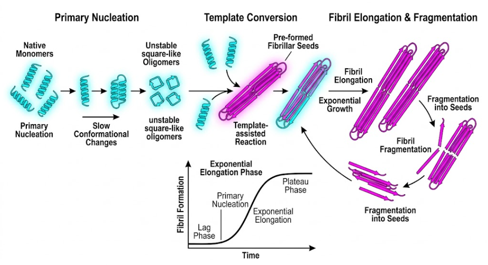
Figure 1 The Biophysical Cascade of Misfolded Proteins. The figure illustrates the conversion of native helical proteins into β-sheet amyloid fibrils. The primary nucleation, template-assisted conversion and fibril elongation are the main steps, which are directly correlated with the different phases of the sigmoidal kinetic growth curve.
Primary nucleation of native monomeric proteins involves a rate limiting conformational transition to form stable aggregation nuclei; addition of pre formed pathogenic seeds to the system abolishes the nucleation lag phase, resulting in rapid exponential elongation and fibril fragmentation into multiple active seeds((Brundin et al., 2010) , as shown in Figure 1.
Conversion of soluble functional proteins into insoluble pathogenic aggregates is a nucleation-dependent polymerization process with a characteristic sigmoidal kinetic profile. Native functional proteins are in thermodynamically stable conformations at physiological conditions(McAlary et al., 2019). Primary nucleation, the initial formation of a stable, self-propagating oligomeric assembly, is thermodynamically unfavorable and is the slow, rate-limiting lag phase of disease initiation. In this phase, native monomers undergo transient conformational shifts that expose hydrophobic amino acid side chains, which are normally buried within the folded structure of the protein(Brundin et al., 2010).
After a stable oligomeric nucleus or seed is formed, the reaction proceeds with an exponential elongation stage. This step involves rapid incorporation of soluble monomeric proteins into the growing aggregate tips in the aberrant, β-sheet rich template conformation(Brundin et al., 2010). Moreover, as large amyloid fibrils accumulate, fibril fragmentation occurs due to mechanical shear forces or cellular chaperones, resulting in a plethora of smaller, highly reactive seed fragments. These newly formed seeds provide further surfaces for elongation and initiate a self-propagating cascade of aggregation. For exogenous pre formed aggregates or patient derived seeds entering a naive recipient cell the primary nucleation lag phase is completely circumvented, dramatically accelerating the conversion of endogenous native monomers(Brundin et al., 2010).
There are two main mechanistic models explaining the process of pathogenic seeds corrupting native protein populations in the neuronal cytoplasm:
Recent research advances have pointed to liquid liquid phase separation as a major contributor to pathological aggregation especially of RNA binding proteins such as TDP-43 and FUS (Pongrácová et al., 2024). TDP-43 has a flexible intrinsically disordered low complexity C terminal domain enriched in glycine, glutamine and asparagine residues, which confer strong self-assembly properties. In cellular stress conditions, TDP-43 changes its normal location in the nucleus into cytoplasm and aggregates in liquid like biomolecular condensates like stress granules(François-Moutal et al., 2019; Kumar et al., 2023).
Physiological stress granules are readily disassembled upon stress resolution, but disease-associated genetic alterations, persistent oxidative stress or abnormal post-translational modifications such as hyperphosphorylation, ubiquitination and C-terminal cleavage to 25-kDa and 35-kDa fragments increase condensate viscosity. This changes the biomolecular state by an irreversible transition from liquid to solid phase that leads to the assembly of toxic oligomers and insoluble fibrillar inclusions(Chen et al., 2010; Pongrácová et al., 2024).
The presence of structural strains, which are different conformational variants of the same protein polymer encoding different pathological phenotypes is one of the hallmarks taken from classical prion biology. High-resolution structural studies reveal distinct tertiary folds, filament dimensions, and biochemical stabilities for tau, α-synuclein, and TDP-43 aggregates derived from different clinical disease subtypes(Carta & Aguzzi, 2022). In cell or animal models, this unique brain derived aggregate strain faithfully self-propagates its exact molecular conformation, inducing subtype specific inclusion morphologies, anatomical spreading velocities and cell type tropisms(Laferrière et al., 2019). This strain diversity provides a strong molecular basis for the fact that a single protein such as TDP-43 can manifest as ALS, frontotemporal dementia or limbic predominant age related TDP-43 encephalopathy (Mompeán et al., 2014).
Cytosolic or nuclear aggregates must transit a multi-step extracellular phase to propagate across interconnected neural networks. This procedure has strict cellular requirements: recruitment of soluble monomers to the aggregate and fragmentation, continuous synthesis of native monomeric substrate in recipient cells, active or passive secretion of seeds to the extracellular space, and binding, internalization and cytoplasmic escape within the recipient neuron(Brundin et al., 2010).
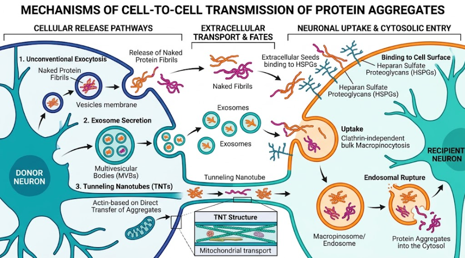
Figure 2. Nerve Cell Transmission. This figure shows the transfer of protein aggregates from a donor to a recipient neuron by exocytosis, exosomes and tunneling nanotubes.
Pathogenic aggregate seeds are released from donor cells through unconventional exocytosis, exosomal secretion and tunneling nanotubes, and subsequently taken up by recipient neurons through heparan sulfate proteoglycan mediated macropinocytosis or receptor operated endocytosis, as illustrated in Figure 2(Ren et al., 2009).
Pathogenic protein aggregates lack the classical signal peptides that are required for classical secretion pathways from the ER to the Golgi(Colin et al., 2020). but their release into the interstitial fluid is mediated by specialized active and passive non-classical mechanisms:
Once in the extracellular interstitial space seeds interact with specific recipient cell surface receptors to induce internalization:
Astrocytes and microglia are actively modulating prion-like propagation kinetics. Microglia take up extracellular aggregates via scavenger receptors, clearing some and thus reducing the seed concentration, but an excessive build-up of aggregates can trigger inflammasome activation and secretion of pro-inflammatory cytokines (Braak & Del Tredici, 2013). Exosomes released by activated microglia have also been shown to contain seeds that promote the spread of tau and TDP-43 to neighboring neurons. Astrocytes also internalize neuronal aggregates but can become metabolically exhausted and release processed seeds back into the neuropil (Braak & Del Tredici, 2013; Goedert et al., 2017).
Once inside the neuronal cytoplasm, protein seeds use the endogenous cytoskeletal transport machinery of the neuron to move the long anatomical distances along axonal tracts (Koshy et al., 2022). Axonal transport depends on polarized microtubule tracks, where:
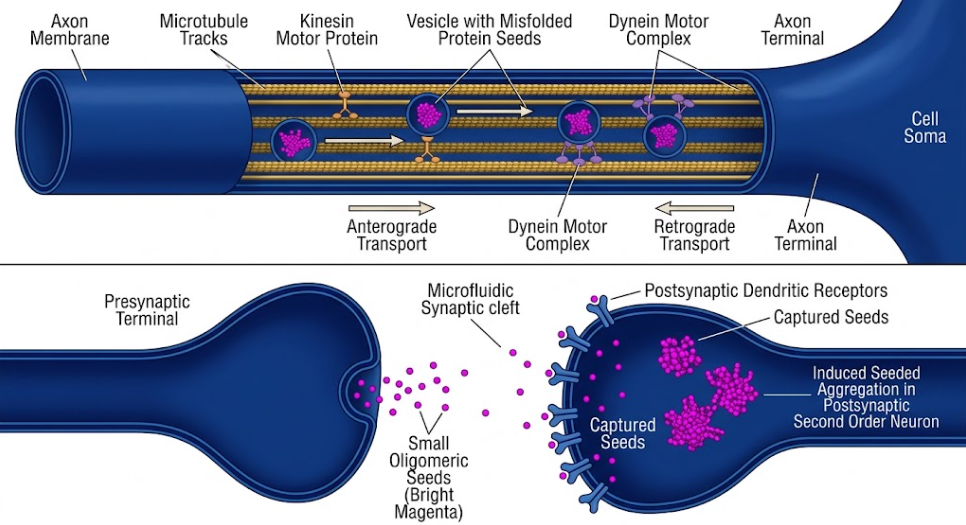
Figure 3. Axonal transport and transsynaptic propagation of misfolded proteins. Schematic of the bidirectional transport of misfolded protein seeds along axonal microtubules and their trans-synaptic propagation to postsynaptic neurons.
Internalized aggregate seeds are actively transported bidirectionally along microtubule tracks by kinesin motors (anterograde) and dynein motors (retrograde). Figure 3 shows the resulting trans-synaptic secretion of aggregate seeds into the synaptic cleft and uptake by postsynaptic second order neurons. In vitro models using microfluidic chambers that physically separate cell bodies from axon terminals show that tau, α-synuclein, TDP-43 and prion seeds are taken up in either the somatodendritic or axonal terminal compartments and transported bidirectionally over long distances (Freundt et al., 2012).
Axonal transport is running in different velocity regimes:
Live cell imaging and radiolabeled tracking studies indicate that the propagation velocity of prion-like aggregates of about 1 to 5 millimeters per day is compatible with the rate of slow component b transport. This rate is similar to observed clinical propagation speeds of pathology along central white matter tracts in human neurodegenerative conditions (Brettschneider et al., 2015; Kaufman & Diamond, 2013). A motor driven transport of discrete vesicular cargoes has been developed, and a complementary domino like membrane propagation model has been established. In this model, membrane-anchored seeds induce a sequential conformational conversion of native proteins continuously along the axonal plasma membrane, propagating a wave of misfolding down the nerve fiber (Koshy et al., 2022).
Activity-dependent exocytosis releases aggregate seeds into the synaptic cleft when reaching axon terminals. Increased firing of the synapses and depolarization of the neurons improve the release of tau and α-synuclein into the extra-cellular space, which in turn speeds up the trans synaptic transfer toward the post-synaptic neurons. This connectivity dependent transfer accounts for why pathology spreads along functional neuronal networks rather than simply diffusing into contiguous anatomical space (Brettschneider et al., 2015).
Pathogenic aggregates accumulate in nerve fibers and disrupt transport mechanics actively. TDP-43 and prion aggregates abnormally activate Casein Kinase 2, which phosphorylates kinesin light chains, causing premature dissociation of kinesin motors from microtubule tracks. This impairment hampers the fast axonal transport of vital cellular cargos such as mitochondria, neurofilaments and neurotrophic factor receptors, leading to axonal dying back pathology and synaptic dysfunction before the death of the somatic cell (Colin et al., 2020; Zamponi et al., 2017).
Aggregated α-synuclein inclusions are a hallmark of Parkinson disease, dementia with Lewy bodies and multiple system atrophy. Neuropathological staging has demonstrated that Lewy pathology begins at specific induction sites, particularly the anterior olfactory bulb and the dorsal motor nucleus of the vagus nerve in the brainstem. Pathology then spreads in a stereotyped fashion rostrally along anatomical fiber tracts through the pons, midbrain substantia nigra, basal forebrain and ultimately the neocortex (Jellinger, 2009).
The gut-brain hypothesis proposes that α-synuclein misfolding is initiated by environmental triggers in postganglionic enteric neurons of the gastrointestinal tract. These seeds migrate retrogradely in vagal nerve fibers to the dorsal motor nucleus of the central nervous system. Clinical evidence for cell-to-cell transfer is provided by post-mortem analyzes of Parkinson disease patients treated with embryonic neural grafts. Years after the surgery, a proportion of the grafted dopamine neurons developed classical α-synuclein positive Lewy bodies, showing host to graft transmission of pathology in the human brain (Braak et al., 2003; Freed et al., 2001).
Tau is a soluble microtubule associated protein that stabilizes the neuronal cytoskeleton. In Alzheimer disease and primary tauopathies, hyperphosphorylated tau detaches from microtubules and aggregates into paired helical filaments and neurofibrillary tangles (Brettschneider et al., 2015; Goedert et al., 2017). Tau pathology progresses in a strict hierarchical manner that can be charted in discrete stages:
This model of connectivity is supported by transgenic mouse models where human mutant tau is expressed only in the entorhinal cortex, and human tau pathology spreads over time trans synaptically to dentate gyrus granule cells and hippocampal pyramidal neurons receiving direct synaptic projections from the entorhinal cortex. Importantly, anatomical dissemination is halted by surgical or traumatic transection of axonal tracts (Goedert et al., 2017).
TAR DNA binding protein 43 (TDP-43) is a nuclear protein that binds nucleic acids and regulates RNA splicing, transport and stability. In the vast majority of ALS cases and in about half of frontotemporal dementia cases, TDP-43 is translocated to the cytoplasm and forms hyperphosphorylated, ubiquitinated inclusions (Brettschneider et al., 2015; Pongrácová et al., 2024).
The neuropathological stage models describe the progression of TDP-43 in ALS as a four stage corticofugal spreading:
Experimental studies in rodents show that stereotaxic injection of patient derived frontotemporal dementia or ALS brain extracts into the forebrain or motor tracts induces widespread, time dependent phosphorylated TDP-43 pathology along connected white matter tracts and ALS like motor deficits (Brettschneider et al., 2015).
In brains affected by neurodegeneration, pathological proteins often interact. Cross seeding is a process in which an aggregate seed from one protein recruits and templates the misfolding of a heterologous protein species:
Network-based recognition of axonal propagation has revolutionized clinical diagnostic paradigms. Diffusion tensor imaging white matter tractography and whole-brain functional connectivity MRI show that regional patterns of cortical atrophy and axonal degeneration map onto functional connectivity networks rather than spatial proximity (Brettschneider et al., 2015; Colin et al., 2020). Advanced tau and α-synuclein positron emission tomography tracers allow physicians to map the longitudinal evolution of aggregate seeds along axonal tracts in living patients, allowing real time biomarker validation of neuropathological staging schemes (Okamura et al., 2014).
The knowledge of seed-dependent polymerization kinetics has allowed the design of ultrasensitive diagnostic tools such as Real Time Quaking Induced Conversion and Seeding Amplification Assays. These assays (e.g. TDP-43 SAA and α-synuclein SAA) amplify minute amounts of pathological seeds that are present in biospecimens e.g. cerebrospinal fluid, blood plasma or nasal olfactory mucosa biopsies by incubation with recombinant monomeric substrates under intermittent shaking. The assays have high diagnostic sensitivity and specificity for detection of pre symptomatic proteinopathy, opening the door to early therapeutic intervention before irreversible neurodegeneration (Fontana et al., 2024).
An extracellular phase for interneuronal seed transit provides a reachable therapeutic window for disease modifying interventions
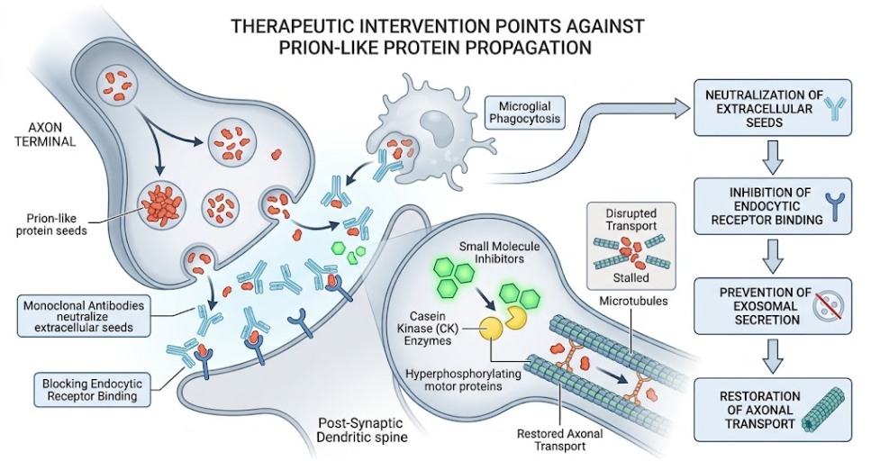
Figure 4. Targeting Prion-Like Protein Propagation for Therapeutic Intervention. Schematic representation of therapeutic targets and strategies to interfere with the propagation and toxicity of prion-like proteins in neurons.
Therapeutic approaches (Figure 4) target several nodes in the propagation cascade such as small molecule inhibition of intracellular aggregation, antibody-mediated neutralization of extracellular seeds and inhibition of exosomal packaging (Pozzi et al., 2019).
The paradigm shift from cell autonomous pathology to prion-like axonal propagation has altered our view of neurodegenerative disorders. Pathogenic protein aggregates are self-propagating propagons that exploit the axonal transport system of neurons and trans synaptic connectivity to sequentially disseminate through neural networks. By deepening our molecular understanding of template assisted conversion, endosomal escape mechanisms, molecular motor transport, and strain diversity, we can reveal critical vulnerabilities in the spreading cascade. A promising approach to stop the progression of devastating neurodegenerative diseases is to target the extracellular phase of this process with combination therapies pairing immunotherapies with kinase inhibitors restoring transport and proteostasis enhancers.
Braak, H., & Del Tredici, K. (2013). Evolutional aspects of Alzheimer’s disease pathogenesis. J Alzheimers Dis, 33 Suppl 1, S155-161. doi:10.3233/jad-2012-129029
Braak, H., Del Tredici, K., Rüb, U., de Vos, R. A., Jansen Steur, E. N., & Braak, E. (2003). Staging of brain pathology related to sporadic Parkinson’s disease. Neurobiol Aging, 24(2), 197-211. doi:10.1016/s0197-4580(02)00065-9
Brettschneider, J., Del Tredici, K., Lee, V. M., & Trojanowski, J. Q. (2015). Spreading of pathology in neurodegenerative diseases: a focus on human studies. Nat Rev Neurosci, 16(2), 109-120. doi:10.1038/nrn3887
Brundin, P., Melki, R., & Kopito, R. (2010). Prion-like transmission of protein aggregates in neurodegenerative diseases. Nat Rev Mol Cell Biol, 11(4), 301-307. doi:10.1038/nrm2873
Carta, M., & Aguzzi, A. (2022). Molecular foundations of prion strain diversity. Curr Opin Neurobiol, 72, 22-31. doi:10.1016/j.conb.2021.07.010
Chen, A. K., Lin, R. Y., Hsieh, E. Z., Tu, P. H., Chen, R. P., Liao, T. Y., . . . Huang, J. J. (2010). Induction of amyloid fibrils by the C-terminal fragments of TDP-43 in amyotrophic lateral sclerosis. J Am Chem Soc, 132(4), 1186-1187. doi:10.1021/ja9066207
Colin, M., Dujardin, S., Schraen-Maschke, S., Meno-Tetang, G., Duyckaerts, C., Courade, J.-P., & Buée, L. (2020). From the prion-like propagation hypothesis to therapeutic strategies of anti-tau immunotherapy. Acta Neuropathologica, 139(1), 3-25. doi:10.1007/s00401-019-02087-9
Fontana, E., Bongianni, M., Benussi, A., Bronzato, E., Scialo, C., Sacchetto, L., . . . Zanusso, G. (2024). Detection of TDP-43 seeding activity in the olfactory mucosa from patients with frontotemporal dementia. Alzheimers Dement, 20(2), 1156-1165. doi:10.1002/alz.13541
François-Moutal, L., Perez-Miller, S., Scott, D. D., Miranda, V. G., Mollasalehi, N., & Khanna, M. (2019). Structural Insights Into TDP-43 and Effects of Post-translational Modifications. Front Mol Neurosci, 12, 301. doi:10.3389/fnmol.2019.00301
Freed, C. R., Greene, P. E., Breeze, R. E., Tsai, W. Y., DuMouchel, W., Kao, R., . . . Fahn, S. (2001). Transplantation of embryonic dopamine neurons for severe Parkinson’s disease. N Engl J Med, 344(10), 710-719. doi:10.1056/nejm200103083441002
Freundt, E. C., Maynard, N., Clancy, E. K., Roy, S., Bousset, L., Sourigues, Y., . . . Brahic, M. (2012). Neuron-to-neuron transmission of α-synuclein fibrils through axonal transport. Ann Neurol, 72(4), 517-524. doi:10.1002/ana.23747
Goedert, M., Masuda-Suzukake, M., & Falcon, B. (2017). Like prions: the propagation of aggregated tau and α-synuclein in neurodegeneration. Brain, 140(2), 266-278. doi:10.1093/brain/aww230
Jellinger, K. A. (2009). Formation and development of Lewy pathology: a critical update. J Neurol, 256 Suppl 3, 270-279. doi:10.1007/s00415-009-5243-y
Kaufman, S. K., & Diamond, M. I. (2013). Prion-like propagation of protein aggregation and related therapeutic strategies. Neurotherapeutics, 10(3), 371-382. doi:10.1007/s13311-013-0196-3
Kfoury, N., Holmes, B. B., Jiang, H., Holtzman, D. M., & Diamond, M. I. (2012). Trans-cellular propagation of Tau aggregation by fibrillar species. J Biol Chem, 287(23), 19440-19451. doi:10.1074/jbc.M112.346072
Koshy, S. M., Kincaid, A. E., & Bartz, J. C. (2022). Transport of Prions in the Peripheral Nervous System: Pathways, Cell Types, and Mechanisms. Viruses, 14(3), 630.
Kumar, S. T., Nazarov, S., Porta, S., Maharjan, N., Cendrowska, U., Kabani, M., . . . Lashuel, H. A. (2023). Seeding the aggregation of TDP-43 requires post-fibrillization proteolytic cleavage. Nat Neurosci, 26(6), 983-996. doi:10.1038/s41593-023-01341-4
Laferrière, F., Maniecka, Z., Pérez-Berlanga, M., Hruska-Plochan, M., Gilhespy, L., Hock, E. M., . . . Polymenidou, M. (2019). TDP-43 extracted from frontotemporal lobar degeneration subject brains displays distinct aggregate assemblies and neurotoxic effects reflecting disease progression rates. Nat Neurosci, 22(1), 65-77. doi:10.1038/s41593-018-0294-y
McAlary, L., Plotkin, S. S., Yerbury, J. J., & Cashman, N. R. (2019). Prion-Like Propagation of Protein Misfolding and Aggregation in Amyotrophic Lateral Sclerosis. Frontiers in Molecular Neuroscience, Volume 12 – 2019. doi:10.3389/fnmol.2019.00262
Mompeán, M., Buratti, E., Guarnaccia, C., Brito, R. M., Chakrabartty, A., Baralle, F. E., & Laurents, D. V. (2014). “Structural characterization of the minimal segment of TDP-43 competent for aggregation”. Arch Biochem Biophys, 545, 53-62. doi:10.1016/j.abb.2014.01.007
Okamura, N., Furumoto, S., Fodero-Tavoletti, M. T., Mulligan, R. S., Harada, R., Yates, P., . . . Villemagne, V. L. (2014). Non-invasive assessment of Alzheimer’s disease neurofibrillary pathology using 18F-THK5105 PET. Brain, 137(Pt 6), 1762-1771. doi:10.1093/brain/awu064
Pongrácová, E., Buratti, E., & Romano, M. (2024). Prion-like Spreading of Disease in TDP-43 Proteinopathies. Brain Sci, 14(11). doi:10.3390/brainsci14111132
Pozzi, S., Thammisetty, S. S., Codron, P., Rahimian, R., Plourde, K. V., Soucy, G., . . . Julien, J. P. (2019). Virus-mediated delivery of antibody targeting TAR DNA-binding protein-43 mitigates associated neuropathology. J Clin Invest, 129(4), 1581-1595. doi:10.1172/jci123931
Ren, P. H., Lauckner, J. E., Kachirskaia, I., Heuser, J. E., Melki, R., & Kopito, R. R. (2009). Cytoplasmic penetration and persistent infection of mammalian cells by polyglutamine aggregates. Nat Cell Biol, 11(2), 219-225. doi:10.1038/ncb1830
Wang, L. J., Huang, H. Y., Huang, M. P., Liou, W., Chang, Y. T., Wu, C. C., . . . Chang, Y. S. (2014). The microtubule-associated protein EB1 links AIM2 inflammasomes with autophagy-dependent secretion. J Biol Chem, 289(42), 29322-29333. doi:10.1074/jbc.M114.559153
Zamponi, E., Buratti, F., Cataldi, G., Caicedo, H. H., Song, Y., Jungbauer, L. M., . . . Pigino, G. F. (2017). Prion protein inhibits fast axonal transport through a mechanism involving casein kinase 2. PLoS One, 12(12), e0188340. doi:10.1371/journal.pone.0188340