Our Services
Others
- DFPP
- Shockwave
- IV Drip


“This review explores how the loss of E-cadherin acts as a molecular trigger that uncouples intestinal desmosomes, breaking the physical barrier of the crypt wall and fueling chronic inflammation in Crohn’s disease.”
The epithelial monolayer lining the mammalian gastrointestinal tract represents an extraordinary mechanical and physiological boundary, tasked with absorbing essential nutrients while resisting substantial luminal shear stress and excluding trillions of luminal microbes. While traditional paradigms in Crohn’s disease have centered heavily on immune dysregulation, emerging biophysical and biochemical evidence identifies the structural breakdown of intercellular junctions as an inciting pathophysiological driver. Intercellular cohesion within the crypts of Lieberkühn depends on the cooperative mechanical coupling between adherens junctions and subjacent desmosomes. Epithelial cadherin (E-cadherin) forms the core adhesive backbone of the adherens junction, binding neighbor cells through calcium-dependent homophilic trans-dimerization and transmitting mechanical loads to the cortical actomyosin cytoskeleton via catenin cofactors. Desmosomes in simple intestinal epithelia are composed of desmoglein-2 and desmocollin-2 alone, and they are linked to the cytokeratin network, conferring structural resilience and high tensile strength. This tensile bridge is dismantled under chronic mucosal inflammation by pathological shedding and proteolytic cleavage of E-cadherin by cell surface metalloproteinases. Loss of membranous E-cadherin destabilizes cytoskeletal anchorage, releases intracellular catenin signaling cascades, and produces a catastrophic mechanical domino effect that uncouples neighboring desmosomal spot welds. Herein, we integrate the current literature on the molecular, ultrastructural and biomechanical cleavage of E-cadherin, collapse of crypt structural architecture and subsequent junctional uncoupling that results in breakdown of the mucosal barrier observed in Crohn’s disease.
The mammalian intestinal epithelium is a single cell layer that forms the major physical and immunological interface between the internal host environment and the external contents of the gut lumen.(Lessey, Robinson, Chaudhary, & Daniel, 2022) This thin cellular monolayer is rapidly renewed every 3 to 5 days, fueled by vigorously cycling stem cells located deep within the invaginations called the crypts of Lieberkühn.(Rees, Tandun, Yau, Zachos, & Steiner, 2020) Specialized junctional complexes along the lateral membranes of adjacent intestinal epithelial cells (IECs) fuse these cells into a coherent mechanical continuum to preserve barrier integrity despite ongoing peristaltic motion, fluid flux, and tissue morphogenesis.(Lessey et al., 2022)
The traditional view of intestinal barrier maintenance has almost exclusively centered on the apical-most tight junctions (TJs) that establish epithelial cell polarity and form the first paracellular barrier to selective ion, water and macromolecular transport.(Landy et al., 2016) However, the paracellular cleft is regulated by a tripartite junctional architecture called the “terminal bar” or apical junctional complex, consisting of tight junctions, adherens junctions (AJs), and desmosomes (DMs). Adherens junctions, directly subjacent to tight junctions, initiate and stabilize cell-to-cell adhesion. Desmosomes are electron-dense “spot welds” further basolateral that resist external shear and tensile stress.(Raya-Sandino et al., 2021)
Based on our previous observations of Paneth cell defensin deficiency and ileal mucosal barrier breakdown, it was realized that chemical defense mechanisms, specifically secretion of α-defensins (HD5 and HD6), work in tandem with physical cellular barriers to protect the vulnerable stem cell niche at the crypt base.(Salzman, 2010) Our previous review concerned the failure of this chemical shield but recent molecular results suggest that chemical susceptibility is inherently linked to mechanical instability.(Ali, Tan, & Kaiko, 2020) In patients with Crohn’s disease (CD) – a chronic inflammatory bowel disease characterized by transmural inflammation, granulomas, and segmental ulceration – loss of mucosal mechanical cohesion is an early hallmark of pathology, rather than an incidental byproduct of tissue damage.(Schlegel, Boerner, & Waschke, 2021) The key structural failure is the uncoupling of adherens junctions and desmosomes, which is mainly due to the pathological cleavage and shedding of the transmembrane anchor protein, epithelial cadherin (E-cadherin).(Lessey et al., 2022)
To understand the mechanisms that underlie junctional failure, it is first necessary to dissect the complex structural hierarchy that connects these multiprotein complexes into an integrated mechanotransductive network.(Rübsam et al., 2018) In the simple monolayer of the intestinal epithelium, the mechanical continuity between adjacent enterocytes is determined by different but cross-talking cadherin superfamilies. (Raya-Sandino et al., 2021)
The adherens junction is built around classical epithelial cadherin (E-cadherin) a single-pass transmembrane glycoprotein whose extracellular domain is made of five tandem cadherin repeats (EC1–EC5).(Lessey et al., 2022) Adjacent E-cadherin molecules interact through trans-homophilic binding across the intercellular space and cis-dimerization laterally in the presence of extracellular calcium, forming a flexible zipper-like adhesion plane. In the intracellular region, the highly conserved cytoplasmic tail of E-cadherin interacts directly with members of the Armadillo family of proteins, especially β-catenin and p120-catenin (p120ctn). (McCrea & Gottardi, 2016) The distal carboxyl terminus of E-cadherin binds β-catenin, which in turn recruits α-catenin. α-catenin bridges the junctional complex to the cortical filamentous actin (F-actin) cytoskeleton.(Lessey et al., 2022) At the same time, p120ctn binds to the juxtamembrane domain (JMD) of E-cadherin and serves as a physical cap to block clathrin-dependent endocytosis and to control the steady-state degradation and turnover of E-cadherin at the basolateral membrane . In some cases γ-catenin (plakoglobin) may substitute for β-catenin and create an alternate bridge to α-catenin, promoting junctional stability.(Smalley-Freed et al., 2010)
Desmosomes, which provide hyper-adhesive mechanical resistance, are located immediately basal to the adherens junctions. Stratified squamous epithelia (such as the epidermis) express a variety of desmosomal cadherins (desmoglein 1-4 and desmocollin 1-3), whereas simple intestinal epithelia express only desmoglein-2 (Dsg2) and desmocollin-2 (Dsc2).(Raya-Sandino et al., 2021) The extracellular domains of Dsg2 and Dsc2 interact across the intercellular space to mediate calcium-dependent and calcium-independent adhesion. The cytoplasmic tails of Dsg2 and Dsc2 anchor inside the cell to an electron dense plaque formed by armadillo proteins (mainly plakoglobin and plakophilins, predominantly PKP2/PKP3) and the cytolinker desmoplakin (DP I/II). Desmoplakin is the linker of the entire desmosomal plaque to the intermediate filament network of cytokeratin 8 and 18 (K8/K18), which serves as an energy-absorbing and dissipating mechanical strain across the whole cell sheet.(Arsenovic et al., 2016) Adherens junctions and desmosomes build an ordered, interdependent network of junctions along the lateral membranes of intestinal epithelial cells, physically coupling cortical actin networks to resilient cytokeratin filaments (Figure 1.).(Schlegel et al., 2021)
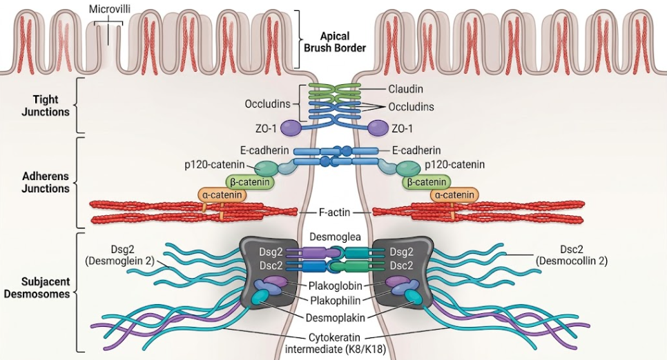 Figure 1. Schematic organization of the apical junctional complex and desmosome-keratin scaffold in normal intestinal crypt cells. Apical tight junctions (claudins, occludin, ZO-1) seal the intercellular space. E-cadherin homodimers are linked to subjacent adherens junctions via β-catenin and α-catenin to perijunctional F-actin bundles, providing force-sensitive mechanical resilience. Desmosomes (Dsg2 and Dsc2) form an intracellular plaque (plakoglobin, plakophilin, desmoplakin) basolaterally, which directly links to cytokeratin intermediate filaments (K8/K18) to form a cohesive tensile network throughout the crypt epithelium.
The adherens junction is not just a static adhesive tether, but an active, dynamic mechanosensitive engine.(Hatzfeld, Keil, & Magin, 2017) Continuous calibration of mechanical tension versus tissue-level deformations is required for the physical integrity of the crypt epithelium.(Price et al., 2018)
When mechanical tension is applied across the epithelial sheet, for example during peristalsis or crypt invagination, E-cadherin trans-dimers transmit these tensile loads intracellularly via the catenin complex.(Price et al., 2018) At low pulling forces, the α-catenin-actin filament association is a “catch bond” in which the bond lifetime is strongly increased with tension, up to a physiological limit. Simultaneously, mechanical tension unrolls α-catenin, exposing a cryptic binding site for the cytoskeletal protein vinculin.(Mège & Ishiyama, 2017) Vinculin recruitment strengthens the link between the cadherin-catenin complex and the actin cytoskeleton, thereby increasing the junctional stiffness in response to mechanical strain.(Ladoux, Nelson, Yan, & Mège, 2015)
The anchoring of E-cadherin to the actomyosin cytoskeleton is essential to maintain the unique crypt geometry.(Sumigray, Terwilliger, & Lechler, 2018) Morphometric analyzes of crypt morphogenesis show that progenitor cells undergo non-muscle myosin II-dependent apical constriction. This contractility relies on stable E-cadherin junctions for the transmission of circumferential forces without ripping the epithelium.(Sumigray et al., 2018) In addition, biophysical measurements using FRET-based tension sensors have demonstrated that E-cadherin is under constant actomyosin-generated mechanical tension.(Acharya et al., 2017) Intact E-cadherin provides this resting tension, which sets the density of epithelial cells, dictates the direction of cell division, and establishes a stiff mechanical barrier that protects the crypt base from excess luminal stress.(Van Patten, Parkash, & Jain, 2010)
In the inflamed mucosa of Crohn’s disease, the homeostatic mechanical equilibrium of the crypt is shattered. A central biochemical hallmark of this disruption is the accelerated shedding of the extracellular ectodomain of E-cadherin and the intracellular cleavage of its cytoplasmic scaffolding.(Lessey et al., 2022)
In chronic inflammation of the intestine, the mucosal microenvironment is filled with a variety of pro-inflammatory cytokines, especially Tumor Necrosis Factor-alpha (TNF- α) and Interferon-gamma (IFN-γ).(Borisova et al., 2020) Enterocytes exposed to TNF-α and IFN-γ quickly activate intracellular stress cascades, especially the p38 mitogen-activated protein kinase (p38MAPK) and Src kinase pathways. This inflammatory signaling induces the expression and membrane translocation of matrix metalloproteinases (MMPs) and “A Disintegrin and Metalloproteinase” family members, ADAM10 and ADAM17.(Schlegel et al., 2021) ADAM10, ADAM17 and related metalloproteinases cleave the extracellular domain of membrane E-cadherin. These proteases perform an endopeptidic cleavage in proximity to the juxtamembrane extracellular border, cleaving the intact ~120 kDa full-length E-cadherin molecule.(Zhao et al., 2019) This cleavage releases a soluble ~80 kDa extracellular fragment (sE-cad) into the crypt lumen and tissue interstitial space, leaving behind a membrane-bound ~38 kDa C-terminal fragment (E-cad/CTF1).(Zhao et al., 2019) The physical implication is immediate: the remaining stub of membrane, devoid of its extracellular repeats, cannot participate in homophilic trans-dimerization with opposite epithelial cells, immediately dismantling the mechanical bridge that maintains adjacent enterocytes together.(Schlegel et al., 2021)
Following ectodomain shedding, the truncated membrane-bound fragment is further processed by stepwise intramembrane proteolysis. The γ-secretase complex (presenilin-dependent) cleaves E-cad/CTF1 within the lipid bilayer, generating an intracellular soluble cytoplasmic fragment designated as E-cad/CTF2 (~33 kDa). Rather than being inertly degraded, E-cad/CTF2 translocates into the cell nucleus, aided in part by chaperone interactions with p120ctn. In the nucleus, cleaved E-cadherin fragments interact with transcription factors and modify canonical Wnt/β-catenin signaling.(Ferber et al., 2008) Nuclear E-cadherin directly competes with the transcription factor TCF-4 (TCF7L2) for binding to the Armadillo repeats of β-catenin, perturbing the canonical Wnt gene transcription programs required for intestinal stem cell maintenance and crypt regeneration.(Su, Chang, Lin, Liang, & Lee, 2015)
In the meantime, the genomics and proteomics of Crohn’s disease mucosal biopsies reveal a phenomenon called “cadherin switching”. In active inflammatory lesions, CDH1 gene expression (E-cadherin) is strongly down-regulated, often via epigenetic mechanisms or transcriptionally by Kaiso and Snail. Instead, P-cadherin (CDH3) is inappropriately up-regulated in enterocytes.(Lessey et al., 2022) P-cadherin is a classical cadherin that mediates much weaker homophilic adhesive bonds and lacks the strong mechanosensitive catch-bond properties of E-cadherin. Thus, cadherin switching does not restore tissue level tensile strength, leaving the crypt wall mechanically vulnerable.(Naydenov et al., 2022) As in Figure 2, Inflammatory activation of cell-surface metalloproteinases cleaves full-length E-cadherin, shedding its adhesive ectodomain and liberating cytoplasmic fragments that invade the nucleus to impair stem cell transcription.(Schlegel et al., 2021)
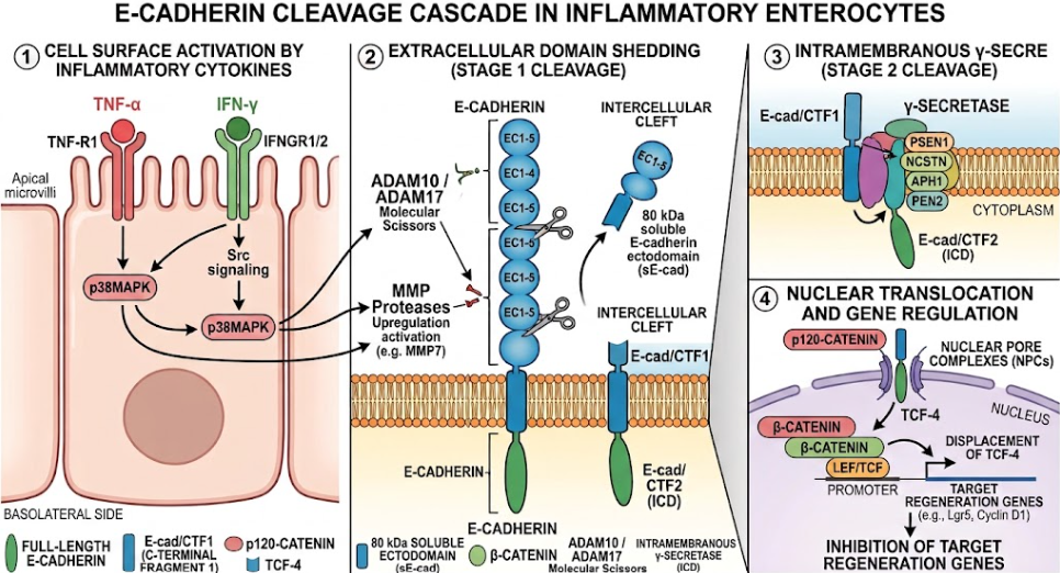
Figure 2. Molecular mechanism of E-cadherin shedding, intramembrane cleavage and nuclear translocation under inflammatory stress. Pro-inflammatory cytokines (TNF-α, IFN-γ) induce metalloproteinases (ADAM10, ADAM17, MMPs) to cleave full-length E-cadherin, shedding its ~80 kDa extracellular domain (sE-cad) and disrupting intercellular adhesion. γ-secretase then mediates intramembrane proteolysis releasing a ~33 kDa fragment of the cytoplasm (E-cad/CTF2) which translocates to the nucleus with p120-catenin, displaces TCF-4 from β-catenin and represses Wnt target genes that are essential for crypt stem cell regeneration.
Destruction of adherens junctions is not a discrete event but rather triggers a catastrophic structural uncoupling of subjacent desmosomes. Desmosomes provide the greatest static tensile strength in epithelial tissues but their assembly, structural maturation and maintenance are fundamentally dependent on the presence of intact adherens junctions.(Lessey et al., 2022)
Early biophysical studies and recent live-cell imaging show that adherens junctions are essential positional cues for desmosome nucleation. Under baseline conditions, E-cadherin-mediated cell-cell contact brings the opposing plasma membranes into close spatial proximity (~15-20 nm).(Raya-Sandino et al., 2021) This mechanical proximity allows desmosomal cadherins (Dsg2 and Dsc2) with shorter, intrinsically flexible extracellular domains to meet and form homophilic and heterophilic trans-interactions.(Tariq et al., 2015) The cleavage and shedding of E-cadherin from the membrane causes a broadening of the physical distance between neighboring cells, augmenting the intermembrane space (IMS). Biophysical measurements reveal that desmosomal cadherins are not competent to form cis-oligomers in the absence of pre-existing membrane alignment, thereby preventing de novo desmosome assembly.(Tariq et al., 2015)
In healthy tissues desmosomes go through a unique physiological maturation process termed “hyperadhesion” in which their intercellular cadherin domains are organized into a highly ordered, crystal-like array that is resistant to calcium chelation and extreme pulling forces.(Raya-Sandino et al., 2021) Transmission electron microscopy studies of biopsies from Crohn’s disease demonstrate a severe disruption in the normal regular sequence of tight junctions, adherens junctions and desmosomes. In inflamed Crohn tissues, the number of desmosomes, which are structurally asymmetric with small and irregular plaques and disrupted central midlines, is drastically reduced.(Schlegel et al., 2021)
Biochemical analysis of Crohn’s resection specimens shows a marked depletion of total intestinal protein levels of Dsg2, to ~63% of control levels, whereas enteroids derived from the crypt stem cell compartments of Crohn’s patients retain this defective desmosomal phenotype through multiple ex vivo passages.(Schlegel et al., 2021) Moreover, experiments using inducible epithelial-specific knock-downs of desmocollin-2 (Dsc2ERΔIEC) show that loss or uncoupling of desmosomal cadherins directly increases intermembrane space between enterocytes, reduces overall plaque dimensions and causes immediate barrier failure.(Raya-Sandino et al., 2021)
Extensive mediation of desmosomal uncoupling downstream of E-cadherin loss through cytoskeletal reorganization. In response to pro-inflammatory signaling cascades, cytokeratins 8 and 18 are rapidly hyperphosphorylated, especially by p38MAPK.(Hatzfeld et al., 2017) Keratin retraction is characterized by the detachment of phosphorylated cytokeratins from desmoplakin plaques and their collapse into dense perinuclear aggregates. At the same time, the phosphorylation status of desmoplakin at Ser2849 is altered, and dephosphorylation or loss of regulatory coordination locks desmoplakin into a dysfunctional, hyper-tensioned state that promotes junctional rupture under mechanical stress.(Price et al., 2018)
Using FRET-based tension biosensors, we show that disruption of adherens junctions results in pathological accumulation of mechanical strain on the remaining desmosomes. The desmosomal spot welds then snap sequentially under luminal shear stress, without the shock-absorbing capacity of the actin-adherens network.(Hatzfeld et al., 2017) These uncoupling changes the crypt epithelium from a robust, mechanically integrated sheet to a suspended, weakly connected population of fragile cells.(Hatzfeld et al., 2017)
6. Pathophysiological consequences in Crohn’s disease: From mechanical disruption to transmural inflammation
The uncoupling of adherens junctions and desmosomes in intestinal crypts initiates a multistep pathological cascade that ultimately ends up in the chronic debilitating inflammation that is characteristic of Crohn’s disease.(Lessey et al., 2022)
As newly generated daughter cells move upwards toward the villus or surface epithelium, enterocytes and stem cells are continuously displaced spatially within the geometrical constraints of the crypt base.(Sumigray et al., 2018) When lateral cell-cell pinning is abolished by E-cadherin shedding, enterocytes lose their anchorage-dependent survival cues. This loss induces a specialized form of detachment-induced cell death, termed anoikis, as well as accelerated epithelial extrusion and premature shedding into the crypt lumen.(Yulis, Quiros, Hilgarth, Parkos, & Nusrat, 2018)
This vulnerability is powerfully exemplified by in vivo animal models of conditional intestinal E-cadherin deletion (VilCre: Cdh1loxP/loxP) and intestinal p120ctn ablation: mice develop extensive mucosal erosions, crypt architecture is completely disintegrated, bloody diarrhea and fatal luminal hemorrhage occur within days. The structural failure of the crypt walls results in the classic histological hallmarks of Crohn’s disease specimens seen by pathologists: crypt abscesses, crypt architectural distortion, and branching.(Lessey et al., 2022)
Irreversible damage to epithelial barrier selectivity results from physical uncoupling of lateral junctions.(Borisova et al., 2020) Healthy apical junctions restrict paracellular permeation of solutes to small ions (<8 Å pore pathway) but loss of mechanical tension across adherens junctions disrupts tight junctional composition.(Rübsam et al., 2018) In enterocytes, the barrier-forming proteins claudin-1 and occludin are strongly down-regulated, and the pore-forming protein claudin-2 is aberrantly up-regulated.(Borisova et al., 2020) As shown in Figure 3, the structural breakdown of adherens junctions and desmosomes leads to the widening of the lateral intercellular cleft to > 100 Å (the non-selective “leak” pathway), allowing uncontrolled passage of macromolecules, bacterial flagellin, lipopolysaccharide (LPS), and even entire microbial organisms directly into the sterile lamina propria.(Lessey et al., 2022; Raya-Sandino et al., 2021)
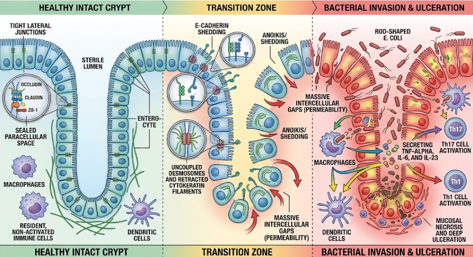 Figure 3. Pathological development of junctional uncoupling, crypt barrier breakdown and transmural immune activation in Crohn’s disease. (Left) Epithelial continuity is maintained by tight junctions, which prevent microbial entry. (Center) E-cadherin shedding widens the intercellular cleft, uncouples desmosomes and induces cytokeratin retraction by p38MAPK phosphorylation, initiating enterocyte anoikis and crypt distortion. (Right) The dilated paracellular leak pathway facilitates the translocation of bacteria into the lamina propria, resulting in Th1/Th17 immune activation and mucosal ulceration.
After the mechanical barrier is broken, translocated microbes are exposed to antigen presenting cells of the subepithelial space. Recognition of microbial components by pattern recognition receptors on lamina propria macrophages and dendritic cells leads to massive waves of interleukin-1beta (IL-1β), interleukin-6 (IL-6), interleukin-23 (IL-23) and TNF-α.(Schlegel et al., 2021) This surge of cytokines drives naive CD4+ T cells toward a pro-inflammatory T helper 17 (Th17) and T helper 1 (Th1) phenotype.(Schlegel et al., 2021)
Importantly, Th17-derived cytokines (including IL-17A and IL-22) as well as TNF-α act directly back on the epithelial monolayer to further promote ADAM10, ADAM17, and MMP transcription, and also to expedite p38MAPK-dependent keratin retraction. This establishes a vicious, self-sustaining feed-forward loop: mechanical failure enables microbial invasion, microbial invasion promotes immune activation, and immune activation accelerates enzymatic junctional destruction.(Lessey et al., 2022) Over time this non-resolving inflammation extends through all the layers of the intestinal wall creating the transmural lymphoid aggregates, fistulae and fibrotic strictures that characterize the end stage of Crohn’s disease.(Schlegel et al., 2021)
Modern biochemical, genetic and biomechanical studies show a force to rethink the pathogenesis of Crohn’s disease at a fundamental level.(Schlegel et al., 2021) For decades, the prevailing clinical hypothesis regarded epithelial barrier breakdown as a passive, late event of an overly aggressive mucosal immune system.(Schlegel et al., 2021) However, clinical observations show that unaffected first-degree relatives of patients with Crohn’s disease frequently have increased intestinal epithelial permeability in the absence of active clinical inflammation or systemic cytokine elevation.(Borisova et al., 2020) In addition, ex vivo crypt enteroids generated from patients with Crohn’s disease maintain inherent deficits in junctional stability (e.g., ongoing Dsg2 loss) after extensive passaging in sterile, cytokine-free media.(Schlegel et al., 2021)
These key findings make a compelling case that mechanical vulnerability, in particular an intrinsic fragility of adherens junction and desmosomal coupling, can be a primary predisposing defect that reduces the threshold for disease onset. Triggering the inflammatory cascade is the focal rupture of an epithelium with defective mechanical cohesion under environmental stress, metabolic exhaustion (e.g., epithelial ATP depletion), or minor microbial challenges.(Borisova et al., 2020)
The current therapeutic options for Crohn’s disease are mainly systemic anti-inflammatory agents, including corticosteroids, antimetabolites and biologic therapies (anti-TNF, anti-IL-12/23 and anti-integrin monoclonal antibodies). Although these therapies are effective to control the downstream immune fire, they do not achieve full histological and mechanical “mucosal healing” in a large proportion of patients.(Schlegel et al., 2021) Non-responders and patients with secondary loss of response still have underlying junctional defects which make them susceptible to relapse, creeping fat deposition and fibrostenotic stricture formation requiring surgical bowel resection.(Schlegel et al., 2021)
True durable mucosal restitution requires next generation therapeutic approaches that target stabilization of the crypt’s mechanical architecture. Several new molecular approaches show great promise in preclinical models:
Adherens junctions and desmosomes cooperate in exquisite and tightly orchestrated mechanical cooperation to maintain the structural integrity of the intestinal crypt epithelium. E-cadherin is the fundamental component of this architecture, forming the first intercellular bridge, transmitting forces to the actomyosin cytoskeleton, and providing the spatial scaffold for the assembly of desmosomes and their anchoring to intermediate filaments.
In Crohn’s disease, inflammatory metalloproteinases cleave and shed E-cadherin, disrupting this important mechanical bridge. This uncoupling of adherens junctions and desmosomes results in dilation of the paracellular cleft, detachment of enterocytes and crypt distortion, and enables uncontrolled invasion of luminal microbes into the lamina propria. By identifying junctional uncoupling as a major factor in barrier failure, gastroenterology can go beyond the paradigm of conventional immunosuppression, and move toward novel barrier-stabilizing drugs that aim to reinforce the physical-mechanical fortress of the gut.
Acharya, B. R., Wu, S. K., Lieu, Z. Z., Parton, R. G., Grill, S. W., Bershadsky, A. D., . . . Yap, A. S. (2017). Mammalian Diaphanous 1 Mediates a Pathway for E-cadherin to Stabilize Epithelial Barriers through Junctional Contractility. Cell Rep, 18(12), 2854-2867. doi:10.1016/j.celrep.2017.02.078
Ali, A., Tan, H., & Kaiko, G. E. (2020). Role of the Intestinal Epithelium and Its Interaction With the Microbiota in Food Allergy. Front Immunol, 11, 604054. doi:10.3389/fimmu.2020.604054
Arsenovic, P. T., Ramachandran, I., Bathula, K., Zhu, R., Narang, J. D., Noll, N. A., . . . Conway, D. E. (2016). Nesprin-2G, a Component of the Nuclear LINC Complex, Is Subject to Myosin-Dependent Tension. Biophys J, 110(1), 34-43. doi:10.1016/j.bpj.2015.11.014
Borisova, M. A., Achasova, K. M., Morozova, K. N., Andreyeva, E. N., Litvinova, E. A., Ogienko, A. A., . . . Kozhevnikova, E. N. (2020). Mucin-2 knockout is a model of intercellular junction defects, mitochondrial damage and ATP depletion in the intestinal epithelium. Sci Rep, 10(1), 21135. doi:10.1038/s41598-020-78141-4
Ferber, E. C., Kajita, M., Wadlow, A., Tobiansky, L., Niessen, C., Ariga, H., . . . Fujita, Y. (2008). A role for the cleaved cytoplasmic domain of E-cadherin in the nucleus. J Biol Chem, 283(19), 12691-12700. doi:10.1074/jbc.M708887200
Hatzfeld, M., Keil, R., & Magin, T. M. (2017). Desmosomes and Intermediate Filaments: Their Consequences for Tissue Mechanics. Cold Spring Harb Perspect Biol, 9(6). doi:10.1101/cshperspect.a029157
Ladoux, B., Nelson, W. J., Yan, J., & Mège, R. M. (2015). The mechanotransduction machinery at work at adherens junctions. Integr Biol (Camb), 7(10), 1109-1119. doi:10.1039/c5ib00070j
Landy, J., Ronde, E., English, N., Clark, S. K., Hart, A. L., Knight, S. C., . . . Al-Hassi, H. O. (2016). Tight junctions in inflammatory bowel diseases and inflammatory bowel disease associated colorectal cancer. World J Gastroenterol, 22(11), 3117-3126. doi:10.3748/wjg.v22.i11.3117
Lessey, L. R., Robinson, S. C., Chaudhary, R., & Daniel, J. M. (2022). Adherens junction proteins on the move-From the membrane to the nucleus in intestinal diseases. Front Cell Dev Biol, 10, 998373. doi:10.3389/fcell.2022.998373
McCrea, P. D., & Gottardi, C. J. (2016). Beyond β-catenin: prospects for a larger catenin network in the nucleus. Nat Rev Mol Cell Biol, 17(1), 55-64. doi:10.1038/nrm.2015.3
Mège, R. M., & Ishiyama, N. (2017). Integration of Cadherin Adhesion and Cytoskeleton at Adherens Junctions. Cold Spring Harb Perspect Biol, 9(5). doi:10.1101/cshperspect.a028738
Naydenov, N. G., Lechuga, S., Zalavadia, A., Mukherjee, P. K., Gordon, I. O., Skvasik, D., . . . Ivanov, A. I. (2022). P-Cadherin Regulates Intestinal Epithelial Cell Migration and Mucosal Repair, but Is Dispensable for Colitis Associated Colon Cancer. Cells, 11(9). doi:10.3390/cells11091467
Price, A. J., Cost, A. L., Ungewiß, H., Waschke, J., Dunn, A. R., & Grashoff, C. (2018). Mechanical loading of desmosomes depends on the magnitude and orientation of external stress. Nat Commun, 9(1), 5284. doi:10.1038/s41467-018-07523-0
Raya-Sandino, A., Luissint, A. C., Kusters, D. H. M., Narayanan, V., Flemming, S., Garcia-Hernandez, V., . . . Nusrat, A. (2021). Regulation of intestinal epithelial intercellular adhesion and barrier function by desmosomal cadherin desmocollin-2. Mol Biol Cell, 32(8), 753-768. doi:10.1091/mbc.E20-12-0775
Rees, W. D., Tandun, R., Yau, E., Zachos, N. C., & Steiner, T. S. (2020). Regenerative Intestinal Stem Cells Induced by Acute and Chronic Injury: The Saving Grace of the Epithelium? Front Cell Dev Biol, 8, 583919. doi:10.3389/fcell.2020.583919
Rübsam, M., Broussard, J. A., Wickström, S. A., Nekrasova, O., Green, K. J., & Niessen, C. M. (2018). Adherens Junctions and Desmosomes Coordinate Mechanics and Signaling to Orchestrate Tissue Morphogenesis and Function: An Evolutionary Perspective. Cold Spring Harb Perspect Biol, 10(11). doi:10.1101/cshperspect.a029207
Salzman, N. H. (2010). Paneth cell defensins and the regulation of the microbiome: détente at mucosal surfaces. Gut Microbes, 1(6), 401-406. doi:10.4161/gmic.1.6.14076
Schlegel, N., Boerner, K., & Waschke, J. (2021). Targeting desmosomal adhesion and signalling for intestinal barrier stabilization in inflammatory bowel diseases—Lessons from experimental models and patients. Acta Physiologica, 231(1), e13492. doi:https://doi.org/10.1111/apha.13492
Smalley-Freed, W. G., Efimov, A., Burnett, P. E., Short, S. P., Davis, M. A., Gumucio, D. L., . . . Reynolds, A. B. (2010). p120-catenin is essential for maintenance of barrier function and intestinal homeostasis in mice. J Clin Invest, 120(6), 1824-1835. doi:10.1172/jci41414
Su, Y. J., Chang, Y. W., Lin, W. H., Liang, C. L., & Lee, J. L. (2015). An aberrant nuclear localization of E-cadherin is a potent inhibitor of Wnt/β-catenin-elicited promotion of the cancer stem cell phenotype. Oncogenesis, 4(6), e157. doi:10.1038/oncsis.2015.17
Sumigray, K. D., Terwilliger, M., & Lechler, T. (2018). Morphogenesis and Compartmentalization of the Intestinal Crypt. Dev Cell, 45(2), 183-197.e185. doi:10.1016/j.devcel.2018.03.024
Tariq, H., Bella, J., Jowitt, T. A., Holmes, D. F., Rouhi, M., Nie, Z., . . . Tabernero, L. (2015). Cadherin flexibility provides a key difference between desmosomes and adherens junctions. Proc Natl Acad Sci U S A, 112(17), 5395-5400. doi:10.1073/pnas.1420508112
Van Patten, K., Parkash, V., & Jain, D. (2010). Cadherin expression in gastrointestinal tract endometriosis: possible role in deep tissue invasion and development of malignancy. Mod Pathol, 23(1), 38-44. doi:10.1038/modpathol.2009.127
Yulis, M., Quiros, M., Hilgarth, R., Parkos, C. A., & Nusrat, A. (2018). Intracellular Desmoglein-2 cleavage sensitizes epithelial cells to apoptosis in response to pro-inflammatory cytokines. Cell Death Dis, 9(3), 389. doi:10.1038/s41419-018-0380-9
Zhao, Y., Yu, T., Zhang, N., Chen, J., Zhang, P., Li, S., . . . Liu, F. (2019). Nuclear E-Cadherin Acetylation Promotes Colorectal Tumorigenesis via Enhancing β-Catenin Activity. Mol Cancer Res, 17(2), 655-665. doi:10.1158/1541-7786.Mcr-18-0637
By Dr.Nattida Kampuang