Our Services
Others
- DFPP
- Shockwave
- IV Drip


For patients navigating progressive ocular disease, standard-of-care treatments often reach a frustrating ceiling. When researching advanced alternatives, stem cell therapy in ophthalmology frequently emerges as an investigational path, with Thailand operating as a highly visible center for these interventions. But let’s cut through the noise. The global regenerative medicine market is aggressively saturated with conflicting data. This obscures the critical line between biologically plausible, peer-reviewed clinical applications and predatory commercial marketing.
Patients face the daunting task of deciphering cellular mechanisms, navigating unverified claims, and assessing true clinical risks. It isn’t easy.
By the end of this guide, you will understand the exact biological mechanisms of ocular stem cell therapies, the current clinical trial data, and the objective cost variables driving medical travel. This analysis dissects targeted applications like macular degeneration, details Thailand’s
This article is for educational purposes only and does not replace consultation with a qualified medical professional.
Stem cell therapy in ophthalmology utilizes cellular derivatives, specifically mesenchymal stromal cells (MSCs), to target senescent ocular tissues and mitigate progressive vision loss. Rather than functioning as a direct structural replacement a pervasive and highly inaccurate misconception these therapies operate through complex paracrine signaling. They secrete highly targeted growth factors that promote wound healing, inhibit excessive tissue fibrosis, and actively modify the localized immune environment within the eye.
Understanding this strict biological distinction is the absolute foundation of realistic patient expectations. When an unregulated clinic aggressively advertises a sudden vision restoration breakthrough, they drastically oversimplify the biology to sell hope to desperate patients. A broad scientific evaluation of recent clinical research indicates that the data paints a vastly different, far more nuanced picture of cellular mechanics.
Stem cells introduced into the ocular environment do not automatically “know” how to build a new retina from scratch. Instead, they operate as micro-pharmacological dispensaries. This localized biochemical interaction actively modifies the aggressive immune response, slowing down the apoptosis (programmed cell death) of existing, healthy retinal cells. A theoretically sound cellular mechanism does not automatically translate to functional human vision, rendering many commercial claims vastly premature.
While grasping the cellular mechanics is critical, these biological principles must be rigorously evaluated against their practical application in specific ocular pathologies.
The persistent myth in regenerative medicine is structural replacement the idea that injected cells simply morph into missing photoreceptors and instantly wire themselves into the optic nerve. That simply isn’t how this works. Umbilical Cord Mesenchymal Stromal Cells (UC-MSCs), an abundant source of active progenitor cells, function as biological factories within the ocular environment. Rather than building a new retina, they release a highly specific cocktail of proteins, exosomes, and cytokines into the surrounding tissue.
These complex secretomes contain vital components like Vascular Endothelial Growth Factor (VEGF), which stimulates local new vessel formation, alongside essential epidermal and basic fibroblast growth factors. When introduced into a deteriorating, oxygen-starved ocular environment, these molecules synthesize extracellular matrix proteins and actively reduce damaging oxidative stress via the SIRT1 pathway. For conditions like severe dry eye syndrome, researchers are exploring stem cell eye treatment precisely because of this profound anti-inflammatory capacity. Mesenchymal stem cells mitigate the hyperosmolarity of the tear film by rebuilding damaged lacrimal gland structures through extensive cytokine secretion.
Critically, this macrophage reprogramming shifts the environment from a hostile, pro-inflammatory state to a healing, anti-inflammatory one. We routinely see the ERK MAPK signaling pathway activated, suppressing the hostile immune responses that typically accelerate cellular degeneration.
| 📌 If you’d like the bigger picture of how MSCs regulate immune activity beyond the eye specifically, our article, Mesenchymal Stem Cell Therapy for Immune Modulation, covers the underlying mechanisms in more depth. |
However, we must explicitly address the massive gap between biological plausibility in a laboratory and proven functional clinical benefits in human subjects. While animal models demonstrate robust macrophage reprogramming, human eyes present a far more complex immunological challenge. The therapy essentially buys crucial time, altering the hostile environment so surviving native cells can function longer.
When we shift our focus from retinal layers to neural pathways, the biological hurdles multiply exponentially. Intervening in central nervous system (CNS) tissue repair is notoriously arduous because the optic nerve is stubbornly resistant to spontaneous regeneration. Once an axon is severed or severely damaged by elevated intraocular pressure, local glial cells rapidly form a dense
So, can stem cell therapy to regenerate the optic nerve actually overcome this intensely hostile environment? Mesenchymal stem cells and iPSCs have demonstrated the ability to promote axonal regeneration in experimental optic nerve damage models (Nature, 2021) providing the foundational science for neurotrophic interventions. Over 70% of experimental models show axonal sprouting proving biological plausibility is achievable under strictly controlled laboratory conditions.
These foundational cellular mechanisms provide the scientific rationale for utilizing induced pluripotent stem cells (iPSCs) to provide targeted neurotrophic support. The injected cells attempt to bathe the damaged optic nerve in brain-derived neurotrophic factor (BDNF) and nerve growth factor (NGF). To overcome the formidable glial scar, the cells must secrete matrix metalloproteinases (MMPs) to break down the scar tissue while coaxing surviving retinal ganglion cells to extend new axons.
Measuring success in these experimental models relies heavily on microscopic clinical endpoints like biomarker improvements. Translating that microscopic sprouting into meaningful human visual acuity changes remains a highly investigational frontier that requires extreme caution.
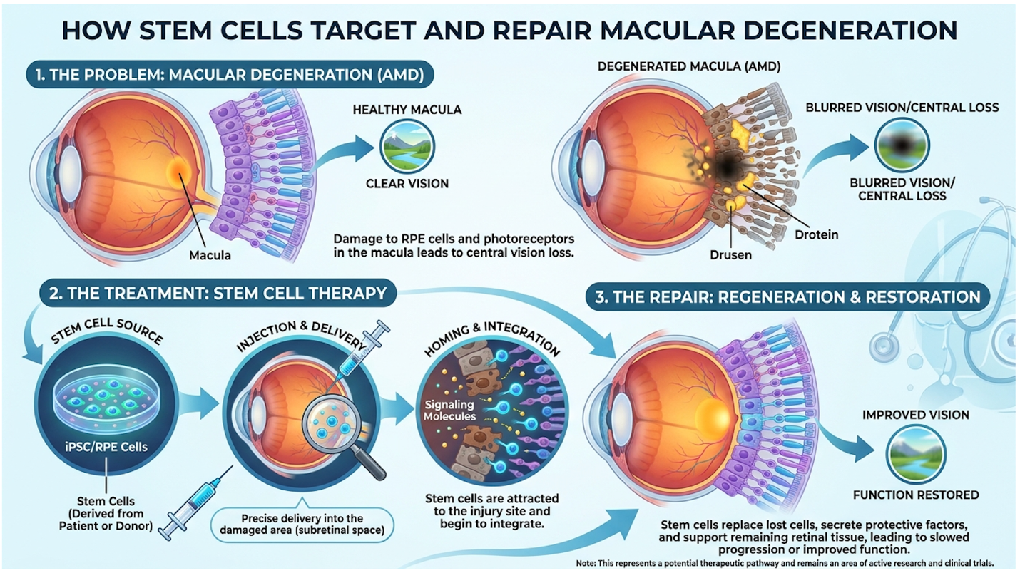
Specific ocular pathologies dictate the clinical reality of biological theory, requiring patients to examine targeted applications rather than broad regenerative promises. While the foundational premise of cellular regeneration is broad, current, peer-reviewed evidence heavily restricts viable clinical applications to distinct, well-researched conditions. The efficacy of stem cell interventions varies significantly depending on whether the target is the retinal pigmented epithelium, the optic nerve, or the intraocular pressure mechanisms associated with severe glaucoma.
Competitors and unregulated clinics frequently group all eye diseases together under one umbrella of regenerative potential. Lumping these distinct pathologies under a single regenerative banner is clinically irresponsible and highly misleading. A patient facing age-related macular degeneration (AMD) has a vastly different clinical trajectory and surgical protocol than a patient dealing with advanced optic nerve atrophy.
To make deeply informed decisions, you have to parse the evidence explicitly for your specific diagnosis. The exact location of the cellular damage heavily dictates the administration route whether subretinal injection, intravitreal injection, or intravenous infusion and drastically impacts the likelihood of biological uptake. We need to look closely at the data separating conventional
Efficacy data reveals a stark spectrum: AMD trials show documented structural promise, whereas ONA and glaucoma applications remain deeply experimental. As the clinical data evolves, patients frequently seek out specialized international institutions equipped to safely handle these highly advanced protocols.
Age-Related Macular Degeneration (AMD), a leading cause of severe, irreversible vision loss, currently represents the strongest area of clinical stem cell research. Conventional treatments generally rely on AREDS2 vitamin supplementation, which only slows the disease progression, or anti-VEGF injections for the wet form of the disease. Macular degeneration stem cells take a radically different approach by attempting to target the exhausted retinal pigmented epithelium (RPE) directly.
Standard anti-VEGF injections temporarily suppress leaky blood vessels, but they do absolutely nothing to replace dead RPE cells. Subretinal stem cell implantation targets this exact cellular deficit. Surgeons perform a highly precise pars plana vitrectomy, creating a microscopic blister under the retina to carefully deposit a suspension of cultivated RPE cells exactly where geographic atrophy is most severe. Phase I/IIa clinical trials have demonstrated the early safety and tolerability of human embryonic stem cell-derived retinal tissue for treating advanced macular degeneration (NIH, 2020) establishing a critical baseline for retinal regeneration protocols.
Phase I trials report up to 40% anatomical stabilization in AMD subjects a crucial step toward preventing complete photoreceptor death. However, functional integration and actual vision improvement remain highly variable. Some patients successfully stabilized their visual fields, while others continued to experience gradual decline despite a successful cellular graft. The therapy shows profound biological promise in modifying the disease environment, but it is not a verified method for returning vision to a pristine state.
While AMD research centers extensively on retinal layers, entirely distinct clinical approaches are required for neuropathies affecting the neural pathways. Stem cell treatment for optic nerve atrophy involves attempting to bridge the structural gap left by the irreversible death of retinal ganglion cells (RGCs). Rescuing these severely damaged cells is arduous because the central nervous system completely lacks the innate regenerative capacity found in peripheral nerves.
Glaucoma, often misunderstood merely as high eye pressure, is fundamentally an optic neuropathy. Approximately 30% of glaucoma standard-of-care patients experience progressive vision loss despite having heavily controlled intraocular pressure. Because of this, cellular neuroprotection is rapidly becoming a mandatory research frontier. Glaucoma stem cell therapy largely ignores mechanical intraocular pressure and focuses entirely on preserving the fragile neural pathway. The injected MSCs secrete neurotrophic factors specifically designed to shield the remaining, highly stressed RGCs from the apoptosis cascade.
However, the timeline for successful intervention is aggressively narrow. Once a retinal ganglion cell completely undergoes apoptosis, the corresponding axon degenerates all the way up to the brain’s lateral geniculate nucleus. Stem cells must be introduced during the critical “apoptotic window” the precise period when the cell is metabolically stressed but not yet dead. Clinical trials have not reliably demonstrated that science can rebuild a completely atrophied optic nerve once that critical intervention window officially closes.
Beyond pressure-induced and atrophic damage, genetic retinal disorders present yet another highly complex target for cellular intervention. Retinitis Pigmentosa (RP) initiates a relentless, genetically programmed degeneration of rod and cone photoreceptors. Because RP involves over 100 distinct genetic mutations most notably the RPE65 gene—simply injecting generic stem cells cannot overwrite this faulty DNA.
Instead, retinitis pigmentosa treatment utilizing stem cells relies heavily on “bystander” protection via paracrine signaling. As rods die off due to the genetic defect, they naturally create a toxic, oxygen-rich environment that secondarily kills the cone cells responsible for central vision. Stem
Current clinical applications for RP are focused entirely on cellular preservation rather than structural reversal. This biochemical shielding is designed solely to keep the central visual field active for as long as possible, even as peripheral vision inevitably narrows. There is a massive, highly important functional difference between slowing visual field loss and restoring lost photoreceptors. Patients navigating genetic dystrophies must understand that this therapy attempts to buy crucial time rather than curing the inherited root cause.
Thailand’s medical infrastructure has expanded into a formidable center for regenerative medical research, establishing rigorous, world-class clinical environments. Institutions such as the Excellence Center for Stem Cell and Cell Therapy in Bangkok have positioned Thailand not merely as a medical tourism destination, but as a highly active participant in advanced cellular biology research, operating firmly under international regulatory scrutiny.
When analyzing stem cell therapy in Thailand, the distinction between unverified storefront clinics and heavily regulated hospital infrastructure is paramount. Western patients often harbor outdated, deeply inaccurate assumptions about Southeast Asian medical standards. The reality is that top-tier Thai institutions often rival and sometimes exceed the technological capabilities of their Western counterparts, particularly regarding the speed of clinical translation for investigational therapies.
| 📌 If you’d like a broader look at safety and regulation across regenerative treatment in Thailand, our article, Is Stem Cell Therapy Safe in Thailand?, covers what to look for before choosing a clinic. |
This level of rigorous quality control decisively separates elite medical centers from unregulated tourism traps that exploit desperate patients. The clinical acceleration seen in Bangkok is not a byproduct of lax laws; it is the direct result of focused government investment in advanced biotechnology. The Royal College of Ophthalmologists of Thailand strictly enforces adherence to international protocols, ensuring that human trials follow the exact same ethical guidelines mandated by strict European and American oversight bodies.
Thailand offers a remarkable blend of JCI-accredited infrastructure and rapidly advancing research capabilities, governed by specialized medical colleges. However, accessing advanced infrastructure does not negate the inherent biological risks associated with experimental cellular medicine.
To truly grasp the region’s capabilities, we have to look closely at specific, accredited institutions rather than broad geographical generalities. Siriraj Hospital stands as a prime example, heavily pioneering advanced clinical interventions in Southeast Asia. This isn’t a pop-up wellness center tucked into a commercial district. It is a massive, historic teaching hospital affiliated with Mahidol University, commanding profound respect in the global medical community.
Siriraj Hospital stem cells research benefits heavily from deep academic funding and exclusive access to state-of-the-art Good Manufacturing Practice (GMP) laboratories. Cultivating ocular-grade stem cells safely requires ISO Class 5 cleanrooms equipped with continuous high-efficiency particulate air (HEPA) filtration systems. Specialized technicians rigorously analyze the cell populations to verify specific surface markers, such as CD90 and CD105, ensuring the final product is genuinely composed of pure MSCs and utterly free of dangerous hematopoietic contaminants.
| 📌 If you’d like a deeper look at what these markers actually verify, our article, UC-MSC Stem Cell and Their Surface Markers, covers why CD73, CD90 and CD105 are cornerstones of quality-verified regenerative therapy. |
When evaluating Bangkok stem cell research, the stark contrast between hospital-based treatments and independent commercial clinics becomes obvious. Reputable hospitals require rigorous patient inclusion criteria, enforce long-term adverse event reporting, and mandate the peer-reviewed publication of their clinical data. They absolutely do not operate on a predatory pay-to-play model where anyone with a checkbook is guaranteed an injection.
| 📌 If you’d like the broader case for pursuing UC-MSC treatment in Thailand, our article, Why You Should Choose UC-MSC Stem Cells in Thailand, covers the regulatory and clinical advantages in more depth. |
When zooming out to the macro view, Thailand’s medical infrastructure demands highly objective comparison against other global hubs like Panama, Mexico, and Germany. The post-pandemic market has seen a massive rebound in patients actively seeking highly specialized, cost-effective care abroad. For advanced patients navigating blinding conditions, Joint Commission International
Thailand’s medical infrastructure is globally recognized, featuring numerous hospitals operating under strict Joint Commission International (JCI) accreditations (Reuters, 2023) effectively restructuring the economics of international regenerative medicine. Thailand’s JCI-accredited hospitals reduce structural costs by up to 60% without compromising strict international laboratory quality standards.
The Thai Medical Council has proactively established specialized ethical review boards to evaluate complex human cellular trials. This regulatory agility allows them to translate investigational therapies safely, while international patients readily receive specialized medical visas for the extended post-operative stays required for comprehensive intraocular pressure monitoring.
Treatment cost variables for stem cell therapy require mapping precise clinical protocols against required hospital stays and mandatory cellular processing fees. Patients evaluating a stem cell treatment for vision clinic in Thailand must rigorously balance this economic modeling with strict medical eligibility criteria to avoid unnecessary financial exposure for highly unsuitable diagnoses.
The intersection of hope and finance is exactly where patients are most vulnerable. Medical migration is largely driven by sheer economic reality, but cost absolutely cannot be the only determining factor. A 60% discount on a highly advanced procedure is completely meaningless if your specific pathology lacks the surviving vascular infrastructure to support the injected cells. You cannot buy biological suitability.
To safely navigate this complex landscape, patients require explicit pricing data and a highly structured method for clinical self-assessment. We advocate for a heavily analytical approach, entirely discarding celebrity anecdotes and focusing solely on peer-reviewed diagnostic criteria and highly transparent billing structures.
Responsible treatment requires passing strict clinical exclusion criteria and fully understanding the financial commitment involved in international medical travel. Even for fully funded, highly eligible candidates, navigating the reality of regenerative clinics presents distinct pitfalls that must be proactively managed.
Let’s brutally break down the exact numbers governing this medical migration. The cost in accredited Thai hospitals operates within a highly specific bracket, generally ranging from $7,500 to $15,800. This comprehensive fee typically encompasses the specialized cellular product, the JCI facility fees, the anesthesiologist, precise surgical administration, and immediate clinical monitoring.
Stem cell therapies in the United States routinely cost between $5,000 and $50,000 (Forbes Health, 2023) creating a massive price gap that fuels medical migration to accredited centers in Thailand. Advanced treatments requiring surgical subretinal delivery average $15,800 forcing patients to objectively weigh financial exposure against unproven clinical outcomes.
| 📌 If you’d like a broader breakdown of pricing across different regenerative treatments in Thailand, our guide, Stem Cell Therapy Costs in Thailand for 2025, walks through the numbers in more detail. |
| Procedure Category | US Estimated Cost | Thailand Estimated Cost | Variables |
| Intravitreal Injection (Autologous) | $8,000 – $15,000 | $7,500 – $9,000 | Lab expansion time |
| Subretinal Surgery (Allogeneic) | $25,000 – $50,000 | $12,000 – $15,800 | Donor screening, OR time |
| Intravenous Infusion (Systemic) | $5,000 – $12,000 | $4,500 – $8,000 | Cell count/dosage |
Financial capacity is entirely irrelevant if the patient fails to strictly meet clinical inclusion criteria, requiring a robust framework for self-evaluation.
Mainstream awareness heavily distorts the regenerative medicine market. The “Joe Rogan stem cell treatment” effect where celebrity endorsements drive massive public demand forces patients to carefully separate entertainment from medicine. Joe Rogan has extensively praised stem cell therapy on his podcast, frequently citing his own positive experiences with orthopedic cellular treatments for joint and tendon injuries.
He often discusses traveling outside the United States to access therapies that face stricter FDA regulations domestically. While his massive platform has significantly increased public awareness of regenerative medicine, his anecdotes primarily relate to musculoskeletal issues, not delicate ocular environments. Patients must rely on peer-reviewed ophthalmological data, not celebrity endorsements, when evaluating highly specific ocular treatments.
To definitively cut through the marketing noise, we utilize The Ocular Cell-Therapy Viability Matrix. This is a critical three-pillar framework requiring patients to objectively assess their diagnosis status, facility accreditation, and financial capacity before blindly pursuing treatment.
First, closely examine your diagnosis status. The Viability Matrix demands highly specific, recent diagnostic data, including optical coherence tomography (OCT) imaging less than three months old and comprehensive electroretinogram (ERG) results. The OCT determines if there is sufficient choroid thickness and vascular infrastructure left to support a cellular graft; if the choroid is entirely thinned out, injected cells will simply undergo rapid necrosis.
Second, verify facility accreditation. Are you talking to a strictly regulated, JCI-accredited hospital or an unregulated tourism clinic? Third, aggressively assess your financial capacity. Can you comfortably absorb a $15,800 total cost with the explicit understanding that functional vision return is absolutely not guaranteed?
Ethical clinics heavily utilize this exact matrix to proactively disqualify patients. Who is NOT a good candidate?
Disqualification by a clinic is actually the absolute highest sign of true medical integrity.
Before committing a massive amount of capital to any international medical travel, patients must seek an independent evaluation from a board-certified neuro-ophthalmologist or retina specialist in their home country.
Present your recent optical coherence tomography (OCT) scans and visual field tests to your primary care team. Ask them directly if your specific cellular degradation matches the theoretical targets of paracrine signaling. While domestic doctors may be extremely conservative regarding international investigational trials, their highly objective analysis of your baseline retinal health is the critical data you absolutely need before boarding a flight to Bangkok.
Stem cells cannot definitively restore eyesight or reverse total blindness according to current, highly proven medical consensus. Their primary biological function is to secrete complex growth factors that modulate inflammation and potentially slow the rapid progression of degenerative ocular diseases. While some clinical trials report minor improvements in visual acuity, outcomes are highly variable across different pathologies.
Unregulated stem cell therapies carry a highly documented risk of turning cancerous and forming complex tumors known as teratomas. This severe risk is primarily associated with heavily manipulated, unapproved cellular products administered entirely outside of rigorous clinical trial
Patients with active systemic infections, advanced malignancies, or completely severed optic nerves are absolutely not good candidates for ocular stem cell therapy. Additionally, individuals whose conditions are highly successfully managed by conventional standard-of-care treatments should not recklessly pursue investigational cellular interventions. Ethical clinics will also proactively disqualify candidates who hold highly unrealistic expectations of a total cure or guaranteed visual restoration. Suitability requires a highly thorough review of medical records by a board-certified specialist.
Stem cell therapy for ocular conditions does not yield immediate or sudden visual improvements. Patients typically undergo a highly comprehensive monitoring period of three to six months before clinicians can accurately assess biological uptake and inflammation reduction. Functional stabilization, if it even occurs, is a highly gradual process heavily dependent on the specific underlying pathology and cellular integration.
For patients facing progressive ocular degeneration, stem cell therapy in ophthalmology in Thailand offers an investigational pathway focused on cellular preservation, with costs structurally ranging from $7,500 to $15,800 (Forbes Health, 2023). The most responsible clinical approach combines targeting specific conditions like AMD or Optic Nerve Atrophy, utilizing strictly JCI-accredited facilities rather than unregulated clinics, and acknowledging the vast difference between biological mechanisms and guaranteed functional vision restoration.
Applying The Ocular Cell-Therapy Viability Matrix ensures that critical medical decisions are firmly rooted in objective clinical reality rather than aggressive commercial marketing. By rigorously assessing strict diagnosis status, institutional accreditation, and heavy financial capacity, patients fiercely protect themselves from unverified experimental risks and preserve their resources for appropriate care.
Prior to pursuing international cellular therapy, meticulously compile your comprehensive medical records, including highly recent optical coherence tomography (OCT) imaging. Contact a board-certified neuro-ophthalmologist today to request a comprehensive, entirely independent review of your recent OCT scans before booking any international medical travel.