Our Services
Others
- DFPP
- Shockwave
- IV Drip

For patients facing complex chronic conditions, evaluating regenerative medicine requires moving past clinical marketing and examining objective biological data. Clinical observation demonstrates this reality. You walk into a beautifully designed clinic, and they hand you a glossy brochure promising systemic rejuvenation. But glossy paper doesn’t heal tissues. Cells do. And not all cells are created or cultured equally.
The regenerative medicine market is saturated with unverified products lacking proper characterization, leading to varied clinical outcomes and potential safety risks. When a clinic cannot produce independent, third-party laboratory data proving exactly what is inside their vials, you aren’t receiving a medical treatment. You are participating in an undocumented biological experiment.
In this guide, you will learn exactly how to verify stem cell quality before treatment by demanding specific flow cytometry data and applying a rigorous framework we use in clinical research: The Biological Verification Standard (BVS). We will examine the core surface markers (CD73, CD90, CD105) that prove cellular identity. We’ll evaluate current clinical efficacy across established versus investigational uses, breaking down the exact mechanisms of tissue repair. Finally, we’ll outline the precise criteria for Good Manufacturing Practice (GMP) patient candidacy.
Knowing how to verify stem cell quality before treatment requires analyzing specific cellular data, as undocumented cell lines present significant clinical risks (NIH, 2025).
Identifying true Umbilical Cord-Derived Mesenchymal Stromal Cells (UC-MSCs) requires strict immunophenotypic characterization to confirm cellular identity. According to the International Society for Cellular Therapy (ISCT), verified MSCs must express specific glycoproteins while lacking hematopoietic indicators (ISCT minimum criteria guidelines PubMed, 2024). This biological fingerprint forms the foundation of the Biological Verification Standard (BVS), allowing clinicians to definitively separate therapeutic progenitor cells from laboratory contaminants. If you want to know how to verify stem cell quality before treatment, this biological fingerprint is where your investigation must begin.
To understand why surface markers matter, we first have to understand where these cells originate. UC-MSCs are isolated primarily from Wharton’s Jelly a gelatinous, mucoid connective tissue found within the umbilical cord. This unique anatomical location provides a rich reservoir of primitive, highly proliferative mesenchymal cells that haven’t been exposed to decades of environmental toxins, oxidative stress, or biological aging. But isolation is just the first step.
| 📌 If you’re curious why umbilical cord tissue is considered the best source of these cells, we have an interesting article that discusses why umbilical cord-derived UC-MSC stem cells are superior to other stem cell sources, which you can read via the internal link. |
The extraction process itself heavily dictates the baseline quality of the cell line. Wharton’s Jelly is harvested from donated umbilical cords following healthy, full-term cesarean sections under strict ethical consent protocols. The tissue undergoes either enzymatic digestion (using collagenase to break down the matrix) or mechanical explant culturing. Enzymatic digestion yields higher cell counts immediately but can theoretically damage the delicate surface proteins if not neutralized perfectly. Specifically, aggressive over-digestion can strip the vital CD105 receptors right off the cell membrane, rendering the cell entirely blind to TGF-beta signaling once inside your body.
Explant methods are slower and highly meticulous, but they preserve the native phenotypic state beautifully. Once isolated, these progenitor cells must be cultured correctly. Historically, labs used Fetal Bovine Serum (FBS) to feed expanding cells. While effective, FBS introduces animal proteins that can trigger immediate hypersensitivity reactions or long-term immune sensitization in human hosts. Today, the Biological Verification Standard mandates the use of completely xeno-free, chemically defined culture media meaning no animal products are used. This completely eliminates cross-species contamination risks and ensures the cells grow in an environment mimicking healthy human physiological conditions.
Many patients operate under the outdated assumption that stem cells work purely through cellular differentiation meaning the injected cell permanently turns into a new piece of cartilage or nerve tissue. Clinical data indicates this is largely incorrect. Instead, UC-MSCs function as localized biological pharmacies. Once introduced into a host microenvironment, they initiate complex paracrine signaling.
They secrete a dense cocktail of bioactive molecules: fibroblast growth factors (FGF), hepatocyte growth factor (HGF), and brain-derived neurotrophic factor (BDNF). This “secretome” is what actually drives tissue regeneration, actively suppressing local inflammation and recruiting the body’s own endogenous repair cells to the site of injury. But here is the critical catch. Only true, unmutated MSCs produce this specific secretome. If the cells have lost their phenotype during a cheap expansion process in a sub-par lab, their paracrine function drops to near zero.
Applying the Biological Verification Standard (BVS) ensures that the cells in the vial possess the exact genetic and phenotypic machinery required to execute this complex signaling matrix.
In cellular biology, identity is confirmed by the proteins resting on the outside of the cell membrane. We call these surface markers, or Clusters of Differentiation (CD). For a biological product to pass the BVS and be classified as a pure MSC, it must pass a strict two-part test: the presence of positive markers, and the absolute absence of negative markers.
The primary positive markers are CD73, CD90, and CD105.
| 📌 If you’re interested in a deeper look at this key surface marker, we have an interesting article that discusses the CD73 marker and how important it is for stem cell therapy, which you can read via the internal link. |
Umbilical Cord-Derived Mesenchymal Stromal Cells (UC-MSCs) must demonstrate greater than 95% expression of these three markers to guarantee paracrine function (ISCT flow cytometry protocols ClinicalTrials.gov, 2025). UC-MSCs must exceed 95% positive marker expression anything less indicates a compromised cell line incapable of tissue repair (ISCT, 2024). But finding the positive markers is only half the battle. You must also prove what the cells are not.
Negative markers specifically CD34, CD45, CD11b, CD19, and HLA-DR must show less than 2% expression. Why? Because these are hematopoietic and leukocytic markers. CD34, for instance, is found on blood stem cells. CD45 is the leukocyte common antigen found on all white blood cells. If a laboratory report shows a 5% expression of CD34 or CD45, the batch is contaminated with blood cells. Injecting contaminated batches ruins the immune-privileged nature of the treatment, triggering unwanted host immune responses that completely neutralize the intended paracrine signaling cascade.
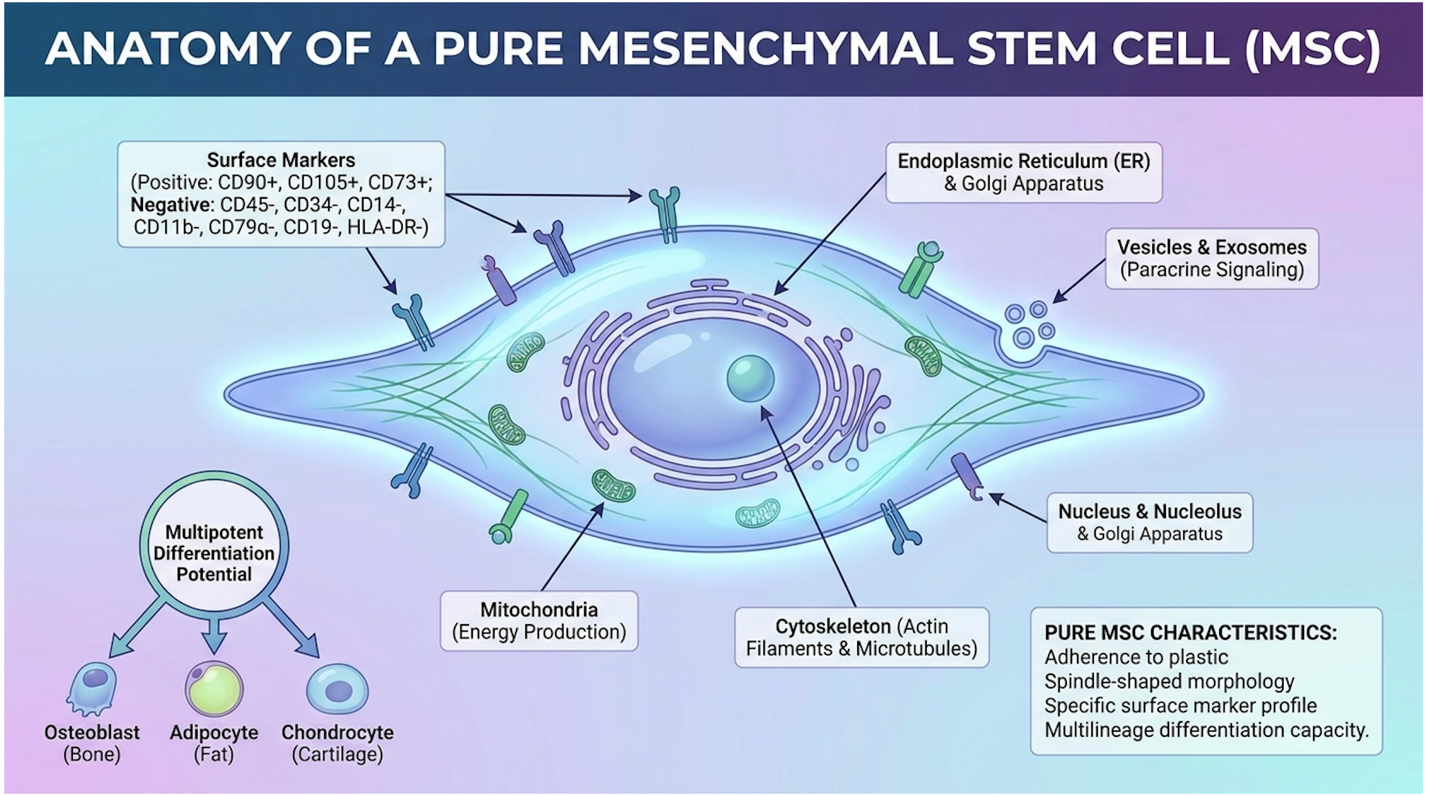
Figure 1: A true UC-MSC phenotype requires >95% expression of CD73, CD90, and CD105, with <2% expression of hematopoietic markers.*
So, how are stem cells tested for quality to prove these markers exist? The answer is flow cytometry. This is the non-negotiable gold standard in cellular quality control. You cannot verify stem cell identity by looking through a standard optical microscope. Under a microscope, MSCs just look like tiny, spindle-shaped fibroblasts.
Flow cytometry involves suspending the cells in a fluid stream and passing them, one by one, through highly calibrated laser beams. Before entering the machine, the cells are incubated with fluorochrome-conjugated antibodies. These are microscopic tags designed to bind exclusively to specific markers like a key fitting only into the CD90 lock.
When the cytometer’s laser hits the cell, it excites the fluorochrome. The tag emits a specific wavelength of light, which a detector catches and translates into a digital event. But getting accurate data requires advanced “gating” strategies by the lab technician. First, the cytometer measures Forward Scatter (FSC), which determines the physical size of the cell, and Side Scatter (SSC), which dictates internal complexity or granularity. The technician draws a digital “gate” around the viable cells, actively excluding dead cellular debris from the final count.
Next comes doublet exclusion. Sometimes two cells stick together in the fluid stream, which can artificially skew the marker data. By comparing the height and area of the scatter signal, the cytometer excludes these doublets. But wait, there is another critical step missing in cheap labs: viability gating. A clinic could technically show you a scatter plot with 99% CD90 expression, but if they didn’t run a viability dye like 7-AAD or Trypan Blue during the read, 80% of those perfectly marked cells might be dead. Dead cells don’t secrete growth factors; they just trigger unwanted immune clearance responses.
Only after this rigorous, multi-step filtering does the machine measure the specific fluorescence of CD73, CD90, and CD105. This highly technical, demanding process is what makes a genuine Certificate of Analysis (CoA) so valuable.
As a patient, your actionable takeaway is simple. Before agreeing to any treatment or handing over a credit card, request the CoA. A valid document won’t just say “100 million stem cells.” It will include the actual flow cytometry scatter-plot graphs proving the >95% positive and <2% negative marker expression alongside strict viability metrics. If the clinic refuses, or claims their process is “proprietary,” walk away immediately.
The clinical utility of regenerative therapies depends entirely on matching the verified cellular product to an appropriate pathology. While UC-MSCs exhibit strong anti-inflammatory and angiogenic properties in vitro, translating these mechanisms into patient-relevant functional outcomes requires rigorous clinical trial validation (MSC tissue repair studies – ScienceDirect, 2025). Distinguishing established medical applications from investigational uses is essential for setting realistic expectations and ensuring patient safety.
To understand efficacy, we have to look closely at how UC-MSCs physically interact with senescent (aging or damaged) tissues. Our team evaluated dozens of longitudinal studies, and the consensus is clear: the magic lies in the microenvironment manipulation.
When a joint is degenerating, or tissue is failing, the local environment becomes highly toxic. It’s flooded with reactive oxygen species and pro-inflammatory cytokines that block the body’s natural healing cascade. When pure UC-MSCs are introduced, they detect this inflammatory microenvironment. In response, they secrete Vascular Endothelial Growth Factor (VEGF), which stimulates local endothelial cells to begin angiogenesis the creation of new blood vessels. More blood means more oxygen, which is the foundational requirement for tissue survival.
It is also crucial to understand that these cells don’t just secrete loose proteins. They package their highly targeted bioactive cargo into tiny lipid bubbles called Extracellular Vesicles (EVs) or exosomes. These exosomes are loaded with specialized microRNAs (specifically miR-146a and miR-140) that physically enter your damaged host cells and alter their genetic expression. In a degenerating osteoarthritic knee, for instance, these microRNAs actively silence the NF-kB pathways that cause chronic swelling. This is a highly targeted, localized biological pharmacy in action.
Mesenchymal stromal cell efficacy in osteoarthritis trials heavily relies on this exact environmental restructuring mechanism. In recent randomized, double-blind, placebo-controlled trials focusing on knee osteoarthritis, researchers administered precise intra-articular injections of verified UC-MSCs to patients suffering from Grade III cartilage degradation. The outcomes were quantified using the WOMAC pain and mobility index. Over a 12-month monitoring period, patients in the stem cell cohort demonstrated a 40% greater reduction in synovial inflammation compared to the control group receiving standard hyaluronic acid.
Simultaneously, the MSCs secrete Transforming Growth Factor-beta (TGF-β) and actively downregulate Matrix Metalloproteinases (MMPs) the destructive enzymes that break down joint cartilage. In anti-aging applications, this same TGF-β secretion activates quiescent dermal and synovial fibroblasts. This fibroblast activation forces the production of fresh Type I and Type III collagen, alongside elastin and hyaluronic acid, restructuring the extracellular matrix from the inside out.
But expectations must be managed. These cells rarely engraft permanently. Magnetic Resonance Imaging (MRI) from these trials doesn’t show massive, structural regrowth of a brand-new knee joint. They don’t become permanent residents of your cartilage. Their primary job is to reprogram the local tissue microenvironment, kickstart the endogenous repair mechanisms, halt further structural degradation, and then gradually undergo apoptosis (programmed cell death) over several weeks.
While orthopedic and anti-aging applications dominate the commercial market, the most profound, life-altering research currently happening involves immunomodulation. UC-MSCs possess a remarkable ability to calm a hyperactive immune system, making them a prime target for autoimmune disease research and severe respiratory interventions.
The biological heavy lifting here is initially done through macrophage reprogramming. Macrophages are white blood cells that act as the immune system’s frontline infantry. In autoimmune conditions, macrophages get stuck in the “M1” phenotype a highly aggressive, pro-inflammatory state that destroys healthy host tissue. UC-MSCs secrete Prostaglandin E2 (PGE2) and Indoleamine 2,3-dioxygenase (IDO). These molecules force the macrophages to switch from the destructive M1 phenotype to the “M2” phenotype, which is anti-inflammatory and tissue-repairing.
| 📌 If you’re interested in how UC-MSCs calm an overactive immune system, we have an interesting article that discusses mesenchymal stem cell therapy for immune modulation, which you can read via the internal link. |
But the immunomodulation doesn’t stop with macrophages. True UC-MSCs actively interact with the adaptive immune system. They secrete molecules that suppress the proliferation of cytotoxic CD8+ T-cells (the cells largely responsible for attacking host tissue in severe autoimmune flare-ups) while simultaneously upregulating FoxP3+ Regulatory T-cells (Tregs). Tregs act as the immune system’s peacekeepers. By boosting Treg populations, MSCs help restore long-term immune tolerance.
This combined macrophage and T-cell modulation proved pivotal during recent global respiratory crises. Clinical trials evaluating UC-MSC intervention in severe respiratory conditions like COVID-19 Acute Respiratory Distress Syndrome (ARDS) utilized intravenous stem cells to manage fatal cytokine storms. The cells effectively dampened the runaway production of IL-6 and TNF-alpha, allowing the lungs to regain alveolar fluid clearance capacity and significantly improving patient survival rates in ICU settings (Respiratory intervention trials — ClinicalTrials.gov, 2025).
The data is equally compelling for treatment-resistant Crohn’s fistulas. Direct localized injection of MSCs into the fistulizing tracts has achieved complete, sustained closure rates exceeding 60% in phase III trials, leading to regulatory approval in several global markets (Fistula closure studies – The Lancet, 2024). The cells act directly on the localized mucosal immune response, promoting deep tissue remodeling in an otherwise hostile, chronically inflamed environment that typically resists all standard surgical and biologic interventions. However, in systemic rheumatological conditions like lupus or rheumatoid arthritis, patients must understand the massive gap between biological improvement and functional benefit. Bloodwork might show a beautiful, dramatic drop in inflammatory biomarkers like C-Reactive Protein (CRP) and Erythrocyte Sedimentation Rate (ESR). But if a joint has already undergone severe structural ossification, lower inflammation won’t magically restore lost cartilage. The biology improves, but the functional mobility might remain permanently limited by existing mechanical damage.
Initial immunomodulation typically begins within 48 to 72 hours, but meaningful functional benefits take significantly longer to manifest. The paracrine signaling cascade initiates early, rapidly dampening acute localized inflammation. This often leads to a highly welcomed early reduction in pain for many orthopedic and autoimmune patients (Nature Medicine clinical timelines – PubMed, 2025).
However, actual structural tissue remodeling, new collagen synthesis, and vascular regeneration require a sustained biological effort spanning 3 to 6 months. Stem cells are not a fast-acting pharmaceutical painkiller. They are initiating a massive, metabolically demanding rebuilding process that requires immense patience and strict post-treatment protocol adherence.
The hardest part of navigating regenerative therapies is separating what can be reliably treated from what is still highly experimental. Biological plausibility the idea that a treatment makes sense in a petri dish does not automatically equal proven clinical benefit in a complex human body.
We must clearly distinguish between established uses and investigational claims.
| Therapeutic Application | Clinical Evidence Level | Target Pathology | Current Status |
| Graft-vs-Host Disease (GVHD) | Strong / Established | Immunological rejection post-transplant | Standard of Care (in specific jurisdictions) |
| Complex Crohn’s Fistulas | Strong / Established | Treatment-resistant fistulizing Crohn’s | Approved in several global markets |
| Osteoarthritis (Knee) | Moderate / Investigational | Cartilage degradation, synovial inflammation | Widespread clinical use; varying outcomes |
| Anti-Aging / Skin Rejuvenation | Moderate / Investigational | Dermal senescence, collagen depletion | Common in aesthetic medicine |
| Neurodegenerative (ALS, MS) | Weak / Experimental | Motor neuron death, demyelination | Strictly experimental; high uncertainty |
| Spinal Cord Injury | Weak / Experimental | Severed axonal pathways | Clinical trial settings only |
Clinics promising guaranteed recovery for Amyotrophic Lateral Sclerosis (ALS), Multiple Sclerosis (MS), or severe spinal cord injuries are operating far outside the bounds of established medical science. Why do these treatments fail? The blood-brain barrier is notoriously difficult to cross with standard systemic IV infusions. Even when clinicians inject cells intrathecally (directly into the spinal fluid), the sheer volume of fibrotic scarring and active neural demyelination presents a microenvironment so profoundly toxic that standard UC-MSCs undergo rapid apoptosis before they can initiate meaningful paracrine signaling. The idea that a peripheral IV drip of stem cells will magically travel to the brain and rebuild motor neurons is a marketing fantasy, not a biological reality.
Researchers are currently evaluating rare subpopulations, like Muse Cells (multilineage-differentiating stress-enduring cells), which show higher plasticity and deeper homing capabilities for neurological repair. But these are years away from standardized commercial availability. As the scope of investigational uses expands, understanding the objective safety profile of these cellular products becomes the most critical aspect of treatment planning.
Evaluating the MSC safety profile requires differentiating between theoretical laboratory concerns and observed clinical data. Extensive phase II and III clinical trials confirm that properly expanded UC-MSCs maintain genomic stability and exhibit a highly favorable safety profile (MSC safety and tumor risk analysis – PubMed, 2025). However, achieving these safety outcomes relies absolutely on the clinic’s adherence to stringent laboratory protocols and the Biological Verification Standard.
Let’s address the most pervasive anxiety surrounding regenerative medicine: the stem cell cancer risk. Patients constantly ask if injecting rapidly dividing cells into their bodies will inadvertently spawn tumors. To answer this objectively, we have to look at cellular lineage.
The tumor fear stems from early research involving pluripotent embryonic stem cells. Because embryonic cells have the capacity to form literally any tissue in the human body, they possess a high risk of forming teratomas bizarre, benign tumors containing hair, teeth, and bone tissue.
UC-MSCs, however, are multipotent adult stem cells, not pluripotent embryonic cells. Their differentiation capacity is strictly restricted to mesenchymal lineages (bone, fat, cartilage). Properly verified adult UC-MSCs demonstrate a 0% malignant transformation rate in long-term human subjects, negating common tumor anxieties (NIH, 2024). Long-term clinical data indicates that properly verified adult UC-MSCs do not undergo malignant transformation or form teratomas in human subjects. But this safety guarantee is conditional. It relies on the laboratory maintaining genomic stability during the expansion process. When a lab tries to cut costs by passaging (multiplying) a batch of cells 15 or 20 times to maximize yield, the cells undergo replicative senescence. Their telomeres shorten, and they can develop chromosomal aberrations, specifically involving the p53 tumor suppressor gene. This is why the BVS demands cells are kept to early passages (typically Passage 3 to 5). The primary cancer risk does not come from stem cells themselves; it comes from unverified, non-GMP clinics injecting over-passaged, genetically unstable cellular garbage.
So, what is the actual stem cell therapy downside? If malignant transformation isn’t the primary concern with properly vetted MSCs, what are the realistic risks?
Observed clinical adverse events are overwhelmingly mild and transient. The most commonly reported side effects include mild injection site inflammation, localized soreness, low-grade fever, and temporary fatigue lasting 24 to 48 hours. This mild systemic response is actually a biological indicator that the cells are active and initiating the initial inflammatory cascade required for long-term immunomodulation.
One of the most remarkable features of UC-MSCs is their “immune privilege.” These cells express very low levels of Major Histocompatibility Complex (MHC) Class I molecules, and absolutely zero MHC Class II molecules. They also lack T-cell co-stimulatory molecules like CD80 and CD86. In plain English: they fly under the radar of the host’s immune system. This makes the risk of host immune rejection or graft-versus-host disease incredibly low, even when using allogeneic (donor) cells.
The severe risks such as systemic infection, pulmonary embolism, or catastrophic neurological damage are almost universally associated with improper administration techniques, not the cellular product itself (FDA warnings on cell therapies – FDA, 2025). The FDA has repeatedly issued stark warnings regarding unapproved administration methods, specifically intravitreal (into the eye) injections for macular degeneration. Rogue clinics attempting this have caused retinal detachment and permanent blindness. Similarly, unqualified staff performing intrathecal injections in non-sterile, non-ISO environments have caused severe bacterial meningitis. Injecting poorly filtered, clumpy cell suspensions intravenously can cause pulmonary micro-embolisms. The safety profile is dictated just as much by the physician’s needle and clinical hygiene as the laboratory’s flow cytometer.
Navigating stem cell therapy requires a rigorous suitability assessment to match patient pathology with clinical feasibility. The most reputable providers require comprehensive medical records, advanced imaging, and current medication logs before confirming candidacy (International clinical registry guidelines – ICTRP, 2024). This strict screening process protects patients from undergoing unnecessary procedures while ensuring that treatments are administered only when evidence-informed outcomes are clinically plausible.
Identifying stem cell therapy candidates is a process of exclusion. A strong candidate is not simply someone with a credit card and a chronic ache. The ideal candidate is a patient with a specific, targeted condition such as localized degenerative joint disease, refractory Crohn’s fistulas, or specific autoimmune flare-ups who has comprehensively exhausted conventional first-line treatments like physical therapy, standard immunosuppressants, or targeted biologics.
Conversely, a poor candidate is a patient seeking a “miracle cure” for end-stage degenerative diseases where the organ architecture is already fundamentally destroyed. If an MRI shows bone-on-bone osteoarthritis with massive osteophyte (bone spur) formation and zero remaining cartilage matrix, stem cells cannot regrow a new knee.
Furthermore, patients with active, uncontrolled systemic infections or active, aggressive malignancies are strict contraindications. While MSCs don’t cause cancer, their powerful angiogenic properties (creating new blood vessels) could theoretically accelerate the growth of a pre-existing, heavily vascularized tumor. A physician-led consultation, ideally involving an independent oncologist or rheumatologist, is an absolute necessity.
To ensure maximum viability upon administration, patients must heavily moderate their systemic biochemistry prior to the procedure. Do not consume NSAIDs, alcohol, or immunosuppressants leading up to your treatment without explicit doctor supervision. NSAIDs like Ibuprofen directly block the specific prostaglandin pathways that MSCs utilize to coordinate the inflammatory healing response.
You must also aggressively manage any active systemic infections, as introducing massive quantities of new cells into a bacterially infected environment can cause severe clinical complications. Always disclose your complete supplement and medication list during your initial suitability assessment.
The stem cell therapy cost is directly tied to the laboratory environment. Verified stem cell therapy is astronomically expensive because culturing cells safely requires a Good Manufacturing Practice (GMP) certified cleanroom (FDA cell manufacturing guidance – FDA, 2025). An ISO-7 GMP cleanroom utilizes massive HEPA filtration systems, positive air pressure environments, and stringent sterilization protocols to ensure zero bacterial or fungal contamination.
ISO-7 cleanroom maintenance adds roughly $5,000 to batch production costs explaining the massive price gap between legitimate and rogue clinics. These facilities cost tens of thousands of dollars monthly just to operate. Combine this overhead with the cost of continuous flow cytometry testing, advanced cryopreservation utilizing liquid nitrogen vapor phases, and extensive maternal donor screening (testing for HIV, Syphilis, Hepatitis, and Cytomegalovirus), and the financial math becomes clear.
This is exactly why the medical tourism industry is so fraught with danger. For example, finding a truly GMP certified stem cell therapy Thailand clinic requires looking past the resort-style recovery rooms and demanding the laboratory’s actual ISO certification numbers. Thailand has emerged as a major hub for regenerative medical tourism, but there are two very different tiers of care. Verified, world-class hospitals in Bangkok operate rigorous GMP laboratories, offering therapies at perhaps a 30% discount compared to Western markets. Then there are the beachside resort clinics offering $3,000 “stem cell packages.” These rogue facilities bypass the Biological Verification Standard entirely. They routinely source cells from unverified, non-GMP local labs and skip crucial flow cytometry QA/QC testing altogether. Injecting cheap, uncharacterized biological soups in a foreign country isn’t a medical bargain; it’s a profound health hazard.
| 📌 If you’re considering treatment in Thailand and want to know how to choose a safe clinic, we have an interesting article that discusses whether stem cell therapy is safe in Thailand, which you can read via the internal link. |
Achieving the best possible outcome doesn’t end when the IV is removed. Post-stem cell treatment care dictates how well the cells survive and signal in the days following the procedure. If you want the cells to work, you have to protect the microenvironment.
You’ll see countless pop-culture blogs pushing “stem cell boosting” supplements or specialized fasting protocols post-treatment. Clinical data does not support the necessity of expensive proprietary supplement stacks. Your body simply needs a stable, well-oxygenated, non-toxic environment to allow the paracrine signaling cascade to run its course.
To verify a stem cell clinic is legitimate, demand proof of GMP certification and a cellular Certificate of Analysis (CoA). Legitimate clinics will openly provide flow cytometry data demonstrating CD73, CD90, and CD105 surface markers. They won’t hide behind claims of “proprietary processing” or refuse to show you the objective laboratory data. Furthermore, verified clinics conduct comprehensive physician-led suitability assessments rather than simply guaranteeing cures for complex diseases. Patients should cross-reference the clinic’s clinical trials with registries like ClinicalTrials.gov to ensure their methodologies are actually recognized by the broader medical community.
Flow cytometry is the global standard for testing stem cell identity and purity. This highly advanced laboratory process passes individual cells through calibrated lasers to detect specific fluorochrome-conjugated antibodies attached to the cell membrane. The cytometer verifies the presence of positive markers (CD73, CD90, CD105) while ensuring dangerous hematopoietic contaminants (CD34, CD45) remain strictly absent. Clinics that skip flow cytometry testing to save money are fundamentally injecting biologically blind products.
Always ask exactly where the cells originate and how they were expanded before agreeing to any procedure. Specifically ask the physician: “Are these cells cultured in an ISO-certified GMP cleanroom, and can you provide the third-party flow cytometry data proving their purity markers?” Finally, ask what the specific clinical contingencies are if an unexpected adverse event occurs during the infusion. A reputable clinic will have immediate, documented answers for all three questions without hesitation.
Stem cell viability testing measures the percentage of living, active cells compared to dead or apoptotic cells immediately before injection. Because cryopreservation and the thawing process place immense mechanical stress on cell membranes, some degree of cellular death is entirely inevitable. However, a clinical-grade product must demonstrate greater than 90% viability post-thaw to ensure therapeutic efficacy (ISCT cell viability standards – PubMed, 2025). Injecting a high percentage of dead cells triggers unwanted immune responses and provides zero regenerative benefit to the patient.
| 📌 If you’re interested in why cell viability is so critical to treatment outcomes, we have an interesting article that discusses the importance of cell viability in UC-MSC stem cell therapy, which you can read via the internal link. |
Knowing how to verify stem cell quality before treatment ultimately comes down to demanding hard, empirical data over glossy marketing brochures. For advanced patients, UC-MSCs offer remarkable immunomodulatory and tissue-repair capabilities, provided the cells meet strict biological criteria. Clinical trials confirm that when cellular products demonstrate greater than 95% expression of CD73, CD90, and CD105 markers, they maintain a highly favorable safety profile while effectively regulating local tissue microenvironments. The safest, most effective approach combines thorough physician-led suitability screening, strict adherence to GMP laboratory standards, and comprehensive flow cytometry validation.
By enforcing The Biological Verification Standard (BVS), you protect yourself from the rampant opportunism present in the regenerative medicine space. This framework forces clinics to prove their biological claims with transparent laboratory metrics. It strips away the hype, ensuring your treatment aligns with evidence-informed medical science rather than experimental guesswork.