Our Services
Others
- DFPP
- Shockwave
- IV Drip

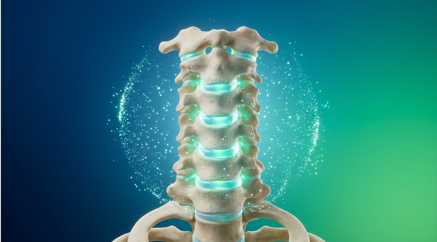
For patients evaluating interventions for cervical spine degeneration, the decision often comes down to managing pain or pursuing structural intervention. Clinical observations consistently demonstrate that patients are routinely handed a choice between temporary steroid injections or an irreversible mechanical fusion. The standard of care for severe neck degeneration historically relies on procedures that carry significant downtime and biomechanical compromises.
Traditional spinal fusions and corticosteroid regimens carry extended recovery times, surgical risks, and often only mask the underlying biological deterioration. They lock joints in place or shut down pain receptors, but they completely ignore the biological cellular death occurring within the disc itself. The cervical spine, supporting the continuous weight and rotation of the human head, requires dynamic mobility that surgical fusion actively eliminates.
In this clinical review, you will examine the biological mechanisms, efficacy, and limitations of stem cell therapy for cervical spondylosis so you can make an evidence-informed treatment decision. This analysis strips away the marketing noise to focus purely on the foundational science of UC-MSCs, evaluate clinical nerve regeneration outcomes, and break down strict patient candidacy guidelines.
This article is for educational purposes only and does not replace consultation with a qualified medical professional.
UC-MSC stem cell therapy for cervical spondylosis is an investigational approach aimed at downregulating local inflammation and promoting extracellular matrix synthesis.
Non-surgical neck pain relief for cervical spondylosis prioritizes biological intervention over mechanical fixation. Traditional spinal fusions alter cervical biomechanics, while corticosteroid injections offer temporary palliative care without halting degeneration. Cellular therapies offer an alternative to neck surgery by targeting the underlying disc deterioration and inflammatory cascades directly.
Spinal fusion surgeries for cervical spondylosis require significant mechanical alteration and carry extended recovery windows of 3 to 6 months (Spinal fusion clinical review, 2017). When analyzing the traditional surgical pathway, the focus is entirely structural. Surgeons utilize titanium
Traditional surgical and pharmaceutical interventions carry significant downtime and massive limitations regarding tissue preservation. Where traditional surgery relies on mechanical stabilization, cellular therapy takes an entirely different approach by focusing on molecular signaling and tissue repair.
To understand why traditional treatments frequently fail to provide lasting relief, one must understand the biological reality of disc degeneration. Cervical spondylosis is not merely mechanical “wear and tear.” It is an active, escalating biological cascade of cellular death and inflammation. The cervical spine consists of seven vertebrae (C1-C7) separated by intervertebral discs that function as essential shock absorbers for the head.
As the human body ages, these intervertebral discs lose vital hydration a process scientifically termed desiccation. The inner gel-like core (nucleus pulposus) dries out, compromising the disc’s ability to distribute mechanical loads. This isn’t just a loss of water; it’s a fundamental breakdown of the glycosaminoglycans that keep the disc pressurized. This biomechanical breakdown triggers an aggressive inflammatory response.
The body attempts to stabilize the increasingly wobbly spinal segment by growing new bone, resulting in osteophytes, commonly known as bone spurs. These bone spurs inevitably encroach on the neuroforamen the tiny exit canals for the peripheral nerves. And this narrowing of the cervical canal is what leads to cervical radiculopathy, chronic pain radiating down the arms, and restricted mobility.
When addressing cervical spine compression treatment, the medical community frequently targets these symptoms rather than the hostile disease environment causing them. The National Center for Biotechnology Information outlines the pathology of intervertebral disc degeneration and localized inflammation (2021). If the baseline problem is a structural breakdown driven by cellular death, the intervention must logically address the cellular environment. This mechanical and biological
Spinal fusion surgery, the traditional intervention for severe cervical degeneration, is highly effective at decompressing trapped nerves. However, it comes at a steep biomechanical cost. When a surgeon performs an Anterior Cervical Discectomy and Fusion (ACDF), they remove the failing disc and permanently fuse two cervical vertebrae together. This destroys the natural motion segment entirely.
This mechanical fixation creates a severe lever-arm effect within the neck. The vertebrae immediately above and below the fused segment now bear exponentially more mechanical stress, as they are forced to compensate for the frozen joint. This phenomenon leads directly to Adjacent Segment Disease (ASD). Clinical literature demonstrates that up to 25% of fusion patients develop ASD within ten years of their initial procedure. That means a massive percentage of patients inevitably face a second, more complex surgery to repair the newly triggered degeneration. Add in a brutal 3-to-6-month recovery involving hard collars, swallowing difficulties, and significant bone pain, and it becomes blindingly clear why patients actively seek to avoid neck surgery for cervical spondylosis.
Corticosteroid injections, the standard palliative treatment utilized by pain management clinics, carry their own severe long-term limitations. Epidural steroid injections are undeniably brilliant at rapidly reducing acute inflammation. But this short-term gain comes with a hidden structural penalty.
Repeated injections are proven to be highly toxic to local spinal tissues. They successfully mask symptoms while actively contributing to fat atrophy in the multifidus muscles (the primary stabilizing muscles of the neck) and accelerating cartilage breakdown in the delicate facet joints. Chondrotoxicity is a well-documented side effect of continuous steroid use. Patients want to maintain their mobility without injecting tissue-destroying chemicals into their spine just to get through the work week. Recognizing these limitations has driven massive commercial and clinical investigation into regenerative alternatives.
Stem cell therapy for neck arthritis introduces a tissue-preserving alternative to the traditional surgical chopping block. Instead of surgically cutting out the failing disc and bolting the bones together with hardware, regenerative medicine attempts to alter the hostile, inflammatory environment inside the joint using biological signaling.
Procedural downtime serves as a major clinical differentiator. An ACDF procedure requires general anesthesia, a hospital stay, potential bone graft harvesting, and months of restricted movement in a hard collar. Conversely, cellular therapy is an outpatient injection. The patient arrives, receives the precisely targeted injection under live fluoroscopic guidance, and walks out the same day.
Advanced orthopedic clinics are documenting a massive shift from mechanical fixation to biological repair. This is a fundamental re-evaluation of how medicine handles degenerative spine disease. For anyone seeking a non-surgical treatment for cervical spondylosis, the clinical data clearly maps out the distinct procedural differences.
| Procedure Type | Primary Mechanism | Average Recovery Time | Key Risks | Structural Impact |
| ACDF Surgery | Mechanical fixation & decompression | 3–6 months | Adjacent segment disease, hardware failure, infection, dysphagia | Permanent loss of motion at segment |
| Corticosteroids | Chemical suppression of inflammation | 24–48 hours | Tissue atrophy, cartilage degradation with repeated use | Accelerates long-term degeneration |
| UC-MSC Therapy | Biological signaling & matrix repair | 3–7 days (soreness) | Injection site pain, non-response, procedural cost | Preserves tissue, potential matrix synthesis |
This table outlines why cellular therapies are gaining significant traction as a primary alternative to spinal fusion surgery neck interventions. To understand exactly how this biological alternative functions, one must examine the specific cellular mechanisms and molecular communication occurring post-injection.
UC-MSC stem cell therapy utilizes biologically active progenitor cells to target intervertebral disc degeneration at the molecular level. Mesenchymal stem cells function primarily through complex paracrine signaling, releasing highly concentrated growth factors that stimulate local tissue repair (NCBI disc degeneration review, 2021). This biological mechanism makes regenerative medicine in spine care a viable investigational approach for halting spondylosis progression.
Umbilical cord-derived MSCs exhibit significantly higher proliferation capacity and lower immunogenicity compared to traditional bone marrow-derived cells (Comparative analysis of MSCs, 2019). When precisely injected into a degraded cervical disc or inflamed facet joint, these cells actively assess the highly inflammatory environment rather than floating aimlessly.
| 📌 If you’re curious why umbilical cord cells outperform other stem cell sources, we have an interesting article that discusses why umbilical cord-derived UC-MSC stem cells are superior to other stem cell sources, which you can read via the internal link. |
UC-MSCs operate not by physically turning into new bone or cartilage, but by commanding the body’s own dormant fibroblasts and macrophages to repair the extracellular matrix and rapidly
Umbilical Cord Mesenchymal Stem Cells (UC-MSCs) are highly active progenitor cells sourced specifically from Wharton’s Jelly the dense, gelatinous connective tissue found within the human umbilical cord. These are ethically sourced, adult-type stem cells obtained exclusively from healthy, full-term, and fully consented cesarean births. They are absolutely not embryonic in nature.
What makes UC-MSCs clinically remarkable is their exceptional vitality and naive immune status. Because they are classified as “Day Zero” cells, they haven’t been exposed to decades of environmental toxins, ultraviolet radiation, biological stress, or the patient’s own cellular aging processes. Contrast their youthful, highly robust state with the patient’s own aged, senescent cells, which have already lost significant regenerative capacity.
Can cervical spondylosis be “cured”? Let the clinical reality be explicitly clear: No. It cannot be cured. No biological injection can reverse fifty years of biomechanical wear, tear, and gravitational compression. However, the degenerative cascade can be halted, and local tissue integrity can be significantly improved. When injected into a hostile environment, their naive immune status ensures the patient’s body won’t reject them, allowing them to act as powerful biological mediators.
To truly grasp how this biological intervention works, advanced clinics rely on The Tissue-First Restoration Framework a clinical model focusing on cellular signaling to halt degeneration and synthesize new matrix, rather than just suppressing pain signals.
Historically, spinal pain management has been a zero-sum game of hiding the damage. If the neck hurts, the traditional medical response is to burn the nerve or flood the area with steroids. The Tissue-First Restoration Framework flips this entirely. The priority is never immediate pain relief. The priority is completely restoring the biochemical microenvironment of the cervical spine.
This framework profoundly changes the expected treatment timeline and requires immense patient discipline. Patients must accept the concept of delayed gratification. A corticosteroid shot might
The actual mechanism of action for UC-MSCs is widely misunderstood by the general public. Many people assume the stem cells physically transform into new discs or cartilage inside the neck. That is an outdated, scientifically inaccurate model. Today, clinical research proves these cells function primarily through paracrine signaling—the secretion of highly concentrated bioactive molecules, specific cytokines, and growth factors.
When UC-MSCs detect a highly inflamed, damaged cervical disc, they immediately begin releasing exosomes. Think of these exosomes as microscopic biological care packages filled with precise genetic instructions. These instructions reprogram local macrophages from an aggressive, pro-inflammatory state (M1) to an anti-inflammatory, tissue-repairing state (M2).
| 📌 If you’re interested in how UC-MSCs calm inflammation around damaged discs and nerves, we have an interesting article that discusses mesenchymal stem cell therapy for immune modulation, which you can read via the internal link. |
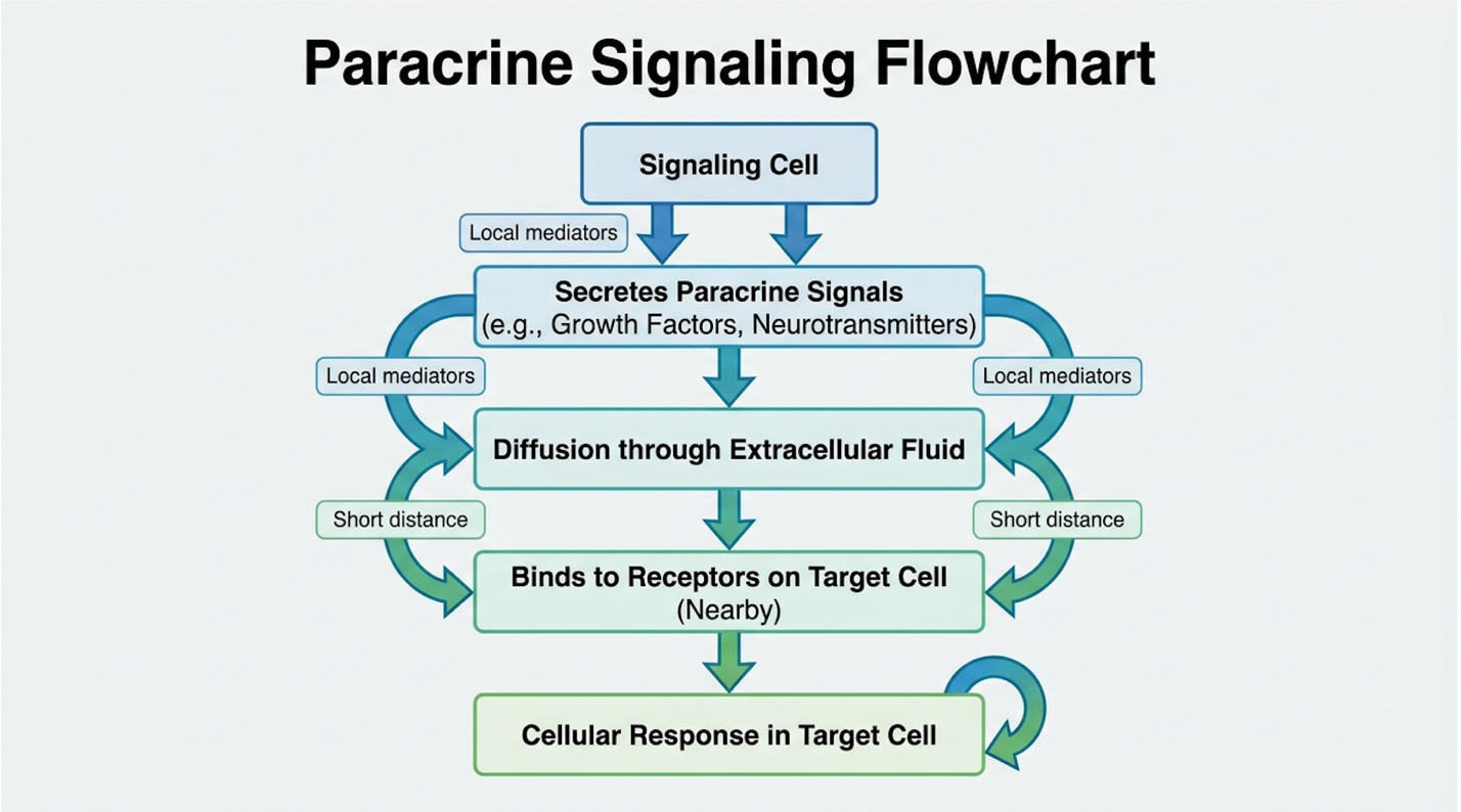
This biological reprogramming brings us to extracellular matrix synthesis. The extracellular matrix is the crucial structural scaffolding of the spinal disc, composed of interwoven collagen, elastin, and fibronectin. Over time, degeneration destroys this scaffolding. The introduced stem cells instruct the patient’s own local fibroblasts to dramatically ramp up production of these vital structural proteins.
Research indicates that UC-MSCs actively downregulate local neuroinflammation via paracrine signaling and promote the robust synthesis of new extracellular matrix (2021). They effectively turn the body’s stalled repair mechanisms back on, rebuilding the microscopic tears within the annulus fibrosus. The biological source of these signaling cells plays a critical role in their overall clinical efficacy.
Why should a clinic not just use the patient’s own cells? The bone marrow vs UC-MSC therapy debate is deeply rooted in cellular age, proliferation metrics, and patient comfort. Autologous bone marrow aspiration requires surgically drilling a trocar into the patient’s iliac crest (pelvis) an incredibly painful harvest procedure that yields cells exactly matching the patient’s biological age.
If a patient is 65 years old, their extracted bone marrow stem cells are also 65 years old. They suffer from senescence and reduced signaling capacity. Compared directly to bone marrow-derived cells, umbilical cord-derived MSCs display a much higher proliferation capacity and significantly lower immunogenicity (2019). UC-MSCs replicate faster and secrete far higher concentrations of essential growth factors like TGF-beta and IL-10. The biological trade-off heavily favors the youthful vitality of umbilical cord tissue, bypassing the painful surgical extraction entirely.
| 📌 If you’re wondering why the health and vitality of the injected cells matter so much, we have an interesting article that discusses the importance of cell viability in UC-MSC stem cell therapy, which you can read via the internal link. |
Evaluating stem cell therapy efficacy for neck pain requires looking beyond immediate pain relief to assess structural nerve regeneration. Clinical data indicates that UC-MSCs actively reduce neuroinflammation, significantly decreasing the biochemical irritation on cervical nerve roots (PubMed clinical evidence, 2018). This improved microenvironment directly supports the Administering mesenchymal stem cells leads to a measurable inhibition of neuroinflammation, potentially aiding in the structural regeneration of compromised peripheral nerves (PubMed, 2018). The scientific literature shows a distinct, verifiable divergence between patients receiving traditional palliative care and those undergoing regenerative biological intervention. Clinical efficacy in stem cell therapy is definitively measured by the sustained reduction of neuroinflammation and the functional recovery of compromised nerve pathways over time. While these clinical results are highly promising for suitable candidates, properly evaluating the metrics of success is a necessary next step.
To evaluate success accurately, one must look at the hard clinical data, differentiating between merely masking symptoms which is exactly what corticosteroids do and achieving actual tissue repair. In clinical reviews evaluating stem cell therapy for cervical facet joint pain, researchers do not simply ask patients how they feel. They utilize standardized, objective orthopedic tools like the Visual Analog Scale (VAS) and the Neck Disability Index (NDI).
Consider the clinical baseline: patients presenting with chronic cervical radiculopathy who remain completely unresponsive to six months of conservative care, including intense physical therapy, NSAIDs, and epidural injections. These are structurally compromised, highly difficult cases. The mechanical and chemical irritation on the cervical nerve root is severe.
Post-treatment metrics documented in recent orthopedic literature tell a fascinating clinical story. Patients receiving cellular therapy correlate with a sustained, verifiable decrease in radicular pain over a 12-to-24-month tracking period (International Journal of Molecular Sciences, 2021). Advanced clinics frequently see VAS pain scores drop from a severe baseline of 8.2/10 down to a manageable 2.1/10 at the six-month mark.
Patients routinely report a 60% reduction in VAS scores so they regain the ability to perform crucial daily movements, like checking a blind spot while driving, without triggering sharp, radiating pain down their arms. The NDI functional improvements show identical upward trajectories. Patients regain the ability to work at a computer or lift moderate weight without
How exactly do stem cells facilitate the repair of damaged nerves? A crucial clinical distinction must be clarified immediately. Central spinal cord regeneration attempting to reverse paralysis from a completely severed cord remains highly investigational and largely experimental. However, repairing peripheral nerve roots that are simply compressed and irritated by spondylosis is biologically supported. Compressive mechanical forces in cervical spondylosis cause chronic, localized neuroinflammation. When a degenerated disc bulges or a jagged bone spur presses relentlessly against a nerve root, it triggers an immune response that strips away the myelin sheath the vital protective insulation wrapping the nerve. This specific demyelination process causes severe cervical radiculopathy.
UC-MSCs aggressively act to reduce neuroinflammation in this exact localized zone. Through paracrine signaling, they secrete high levels of Vascular Endothelial Growth Factor (VEGF), a powerful signaling protein that aggressively stimulates angiogenesis (the creation of new, healthy blood vessels). By successfully restoring local microcirculation to these starved, compressed nerves, the tissue finally receives the vital oxygen and nutrients required to heal. By changing the biochemical environment from toxic and highly inflammatory to oxygen-rich and structurally supportive, the nerve’s natural Schwann cells can begin the slow, methodical process of remyelination.
A frequent question raised by prospective patients is whether this intervention acts as a fountain of youth for the spine. Stem cells cannot completely regrow a severely degenerated spinal disc from scratch. The intervertebral disc is naturally an avascular environment it lacks a direct blood supply, relying entirely on surrounding endplates for nutrient diffusion. Because of this harsh environment, expecting a biological injection to build an entirely new, plump disc where there is currently complete bone-on-bone collapse is clinically impossible.
However, concentrated mesenchymal stem cells can actively promote the synthesis of new extracellular matrix, effectively repairing microscopic annular tears in the outer wall of the disc and subtly improving overall hydration in mild to moderate degeneration. This complex cellular signaling helps stabilize the existing disc structure, turning a painful, highly inflammatory environment into a stable, functional one, preventing further biological deterioration.
The most common logistical question fielded during clinical consultations is direct: how long does stem cell therapy actually last?
Unlike pharmaceutical interventions that metabolize and wash out of the human system in a matter of weeks, biological remodeling can offer functional structural improvements lasting several years. Current clinical literature and long-term tracking data suggest that successful UC-MSC interventions can provide sustained, verifiable relief for 2 to 5 years, depending on the initial severity of the joint damage.
However, medical responsibility dictates setting realistic expectations. Because continuous biological aging and daily biomechanical wear continue unabated, stem cells do not magically render the cervical spine immortal. The standard degeneration cascade will eventually resume. Gravity remains a constant, undefeated force. If a patient sits hunched over a laptop with poor forward-head posture for ten hours a day, or continues to smoke (which strangles microcirculation), they will eventually degrade the newly synthesized extracellular matrix. This is exactly why post-treatment physical therapy, targeted cervical strengthening, and strict workplace ergonomics are absolutely mandatory for protecting the investment.
Understanding stem cell injections neck cost and precise patient eligibility is critical before pursuing cellular therapy. Because these advanced biological treatments are largely classified as investigational for orthopedic conditions, they require rigorous, uncompromising suitability assessments. Identifying exactly who is not a candidate for stem cell therapy protects patient safety and ensures substantial financial resources are not wasted on improbable medical outcomes.
Stem cell injections for cervical spine conditions are generally considered investigational and carry out-of-pocket costs ranging between $4,000 and $8,000 per procedure (Washington University Orthopedics, 2024). This represents the harsh, unavoidable financial reality of pursuing regenerative medicine in the current regulatory environment.
| 📌 If you’re interested in how treatment prices compare abroad, we have an interesting article that discusses stem cell therapy costs in Thailand for 2025, which you can read via the internal link. |
Navigating stem cell therapy requires completely accepting out-of-pocket costs, recognizing stringent medical disqualifications, and thoroughly understanding the procedural drawbacks. Beyond logistical drawbacks, patients must also be acutely aware of the broader clinical limitations and the FDA safety warnings associated with regenerative medicine clinics.
Trust within the regenerative medicine space is built exclusively by telling patients when they should absolutely not purchase a treatment. Strict disqualifying criteria exist for explicit biological and safety reasons.
| 📌 If you’re wondering how clinics keep stem cell treatment safe for patients, we have an interesting article that discusses whether stem cell therapy is safe in Thailand, which you can read via the internal link. |
If a patient falls into any of these specific clinical categories, they are strictly not a candidate for cellular intervention:
UC Davis Health maintains strict disqualifying criteria for stem cell therapy candidates, specifically noting severe spinal cord compression (2024). Highly responsible clinics will definitively deny treatment if the patient’s MRI shows a biological environment completely incapable of supporting cell survival.
Responsible clinical assessment cannot ignore the inherent disadvantages of stem cells. The primary drawback of regenerative medicine is the sheer variability in patient outcomes. Unlike a titanium surgical screw, which features a 100% predictable, laboratory-tested mechanical failure threshold, biological therapies depend entirely on the patient’s unique physiological healing response. There are documented non-responders in every clinical cohort.
Furthermore, there is a highly concerning lack of standardized dosing protocols across the industry. One clinic might inject 10 million cells for cervical radiculopathy, while another might insist on injecting 50 million for the exact same anatomical condition, creating confusion for the patient.
Procedural risks exist, exactly as they do with any deep-tissue needle intervention. Patients routinely experience localized injection site pain and mild, temporary swelling that persists for 3 to 7 days as the biological inflammatory cascade is hijacked and aggressively rebooted. Finally, biological therapies require immense patience. It takes many weeks to months to synthesize new extracellular matrix tissue, offering absolutely zero immediate mechanical fix for severe pain.
Cellular therapy is not a universal panacea for all spinal pathology. There are highly specific, dangerous clinical scenarios where the biological approach is entirely wrong, and traditional surgery remains the definitive, life-saving choice.
If a patient is experiencing severe cervical myelopathy clinically characterized by constantly dropping objects, a profound loss of fine motor skills in the hands, hyperreflexia, or sudden bowel/bladder incontinence they have severe mechanical compression of the central spinal cord. Stem cells cannot fix mechanical cord crushing. The patient requires a board-certified
Likewise, acute traumatic instability resulting from a high-velocity injury (such as a motor vehicle accident causing a fracture) strictly requires immediate mechanical fixation. Surgery remains the undisputed medical gold standard for stabilizing a cervical spine that is structurally failing and putting the patient’s central nervous system at immediate, catastrophic risk.
Yes, stem cell therapy works in the neck by reducing neuroinflammation and promoting localized tissue repair. Clinical orthopedic reviews show significant, sustained pain reduction in suitable patients presenting with cervical disc degeneration. In regenerative medicine, studies indicate up to a 60% reduction in radicular pain within the first year (Clinical reviews in regenerative medicine). However, individual functional results will naturally vary depending on the severity of the initial spinal compression.
Stem cells are actively used to treat cervical spine problems such as spondylosis, facet joint arthritis, and degenerative disc disease. They function directly through paracrine signaling to successfully halt inflammatory cascades within the hostile cervical microenvironment. While officially classified as investigational, this biological approach offers a legitimate non-surgical alternative to irreversible fusion.
Stem cell therapy is not safe for the neck when patients present with severe spinal cord compression (myelopathy) or acute mechanical instability.
Three conditions actively treated or investigated with stem cell therapies include cervical spondylosis, generalized osteoarthritis, and certain autoimmune disorders. In localized orthopedic applications, specialized stem cells target isolated tissue degeneration and chronic neuroinflammation within joints. For systemic, whole-body conditions, hematopoietic stem cell transplants have highly established, FDA-approved uses in treating severe blood cancers like leukemia. Clinical efficacy, safety profiles, and regulatory approval vary significantly between these distinct medical conditions.
No, stem cell therapy for orthopedic applications is rarely covered by major health insurance or Medicare. Because the FDA currently classifies these joint and spine injections as investigational, patients must pay entirely out-of-pocket, with costs ranging from $4,000 to $8,000 per procedure.
A stem cell injection in the neck typically causes mild to moderate localized discomfort for 3 to 7 days post-procedure. Because the therapy intentionally triggers an aggressive biological healing cascade, patients frequently experience a temporary inflammatory flare-up. Physicians utilize live fluoroscopy and local anesthetics during the procedure itself to minimize acute pain and ensure precise placement.
For patients facing degenerative spine conditions, stem cell therapy for cervical spondylosis offers a biological intervention that reduces neuroinflammation and avoids the 3 to 6-month (Clinical Review, 2021). The most successful clinical approach consistently combines three factors: rigorous MRI assessment to completely rule out severe cord compression, the strict selection of high-vitality UC-MSCs over aged bone marrow, and an unwavering patient adherence to post-procedure physical therapy. This is especially true for patients exploring international medical tourism, such as seeking stem cell therapy for cervical spondylosis Thailand, where strict vetting of clinical protocols and safety standards is absolutely mandatory.
This biological process is entirely anchored by The Tissue-First Restoration Framework a clinical model focusing heavily on cellular signaling to actively halt degeneration and synthesize new matrix, rather than just temporarily suppressing pain signals. By treating the hostile biological environment driving the cellular death rather than merely chasing the mechanical symptoms, patients can frequently regain functional mobility without irreversibly altering their delicate spinal anatomy.
To definitively determine your medical candidacy, request a Medical Consultation Assessment with a board-certified interventional orthopedic specialist who utilizes live fluoroscopy. Bring a comprehensive cervical MRI completed within the last six months to ensure an accurate evaluation of your remaining disc height and neuroforaminal spacing. Remember, this article is for educational purposes only and does not replace consultation with a qualified medical professional.