Our Services
Others
- DFPP
- Shockwave
- IV Drip

For oncology and rheumatology patients evaluating advanced therapeutic options, the combination of Natural Killer (NK) cells and Umbilical Cord Mesenchymal Stromal Cells (UC-MSCs) represents a highly specific investigational frontier. And let’s be blunt navigating the medical tourism market for these therapies is exhausting. When patients look into NK cell therapy for cancer Thailand, they are routinely bombarded with commercial hype that completely obscures the strict biological realities of HLA matching, immune rejection, and active clinical trial statuses.
That hype ends here. Clinical reviews routinely demonstrate how misunderstanding the foundational science costs patients time, money, and potentially their safety.
This evidence-informed guide evaluates the exact biological mechanisms, current clinical evidence, and safety protocols surrounding NK cell and UC-MSC synergy. We examine allogeneic sourcing, the biochemical interactions of immune modulation, current applications in oncology, and the necessary clinical monitoring for patient safety. We won’t sugarcoat the limitations, nor
This article is for educational purposes only and does not replace consultation with a qualified medical professional. Always seek localized, evidence-informed suitability assessments before pursuing any cellular therapy.
Investigating NK cell therapy for cancer Thailand requires understanding how sequential cellular administration impacts immune modulation versus targeted cytotoxicity.
Umbilical cord tissue banking provides the foundation for off-the-shelf NK cell therapy by supplying highly proliferative, non-HLA restricted progenitor cells. Unlike autologous extraction, this allogeneic approach allows advanced clinics in Thailand to maintain standardized cellular inventories. This drastically reduces the logistical delays inherent in patient-specific cell processing while mitigating severe immune rejection risks (ResearchGate analysis, 2026). Frankly, relying on sluggish autologous bone marrow in urgent oncology scenarios is becoming an outdated logistical bottleneck.
Wharton’s Jelly MSCs demonstrate a 5-fold higher proliferation capacity than bone marrow providing an abundant off-the-shelf progenitor source that completely bypasses traditional HLA-matching limitations.
| 📌 If you’re curious why umbilical cord cells outperform bone marrow and other sources, we have an interesting article that discusses why umbilical cord-derived UC-MSC stem cells are superior to other stem cell sources, which you can read via the internal link. |
Sourcing viable stem cells is usually the first massive bottleneck in cellular therapy. When we talk about umbilical cord tissue banking, we are specifically focused on Wharton’s Jelly. This gelatinous mucopolysaccharide substance inside the umbilical cord is a biological goldmine for MSCs. Why? Because these are “Day Zero” cells. They haven’t been exposed to decades of environmental toxins, UV radiation, or the basic biological wear-and-tear that degrades adult bone marrow. Their telomere length he protective caps on the ends of their DNA is remarkably long, which translates directly to their replication lifespan in the laboratory.
| 📌 If you’re interested in why keeping cells alive and healthy is so critical to treatment success, we have an interesting article that discusses the importance of cell viability in UC-MSC stem cell therapy, which you can read via the internal link. |
In verified clinical protocols, the difference in proliferation kinetics is staggering. Traditional autologous bone marrow aspirations are not only notoriously painful for the patient—often requiring localized drilling into the iliac crest but they also yield a lower starting concentration of MSCs. Those adult cells hit senescence (cellular aging) much faster in culture. Wharton’s Jelly MSCs, conversely, are harvested non-invasively from discarded tissue post-birth and divide rapidly in the lab.
Wharton’s Jelly MSCs maintain a rapid population doubling time of 24 to 30 hours, compared to 72 hours for bone marrow so clinics can generate clinical-grade therapeutic doses exponentially faster for oncology patients. They pump out a vastly superior array of growth factors, particularly Vascular Endothelial Growth Factor (VEGF) and basic fibroblast growth factors, which are critical for tissue repair.
This logistical advantage changes everything for clinical application. If an oncology patient needs immediate intervention, waiting three to four weeks to culture their own bone marrow cells simply isn’t viable. Banked UC-MSCs offer an off-the-shelf solution. The cells are already extracted, expanded, cryopreserved in liquid nitrogen at highly controlled negative temperatures (-196°C), and rigorously tested for endotoxins and infectious diseases. The clinical team just has to thaw and administer them at the point of care. But the cold-chain banking infrastructure only works if the patient’s body actually accepts the cells.
Here’s the thing about your immune system: it is ruthlessly efficient at recognizing and destroying anything it doesn’t own. This recognition relies heavily on Human Leukocyte Antigen (HLA) matching. Your cells display highly specific HLA-A, HLA-B, and HLA-DR proteins that act as biological barcodes.
Standard autologous therapies avoid this rejection by using your own cells, but that severely limits the patient pool and treatment speed. Allogeneic cell advantages—using donor cells—historically crashed into the wall of immune rejection. If the donor’s HLA didn’t perfectly match the recipient, you’d trigger a massive immune response. Historically, mismatched allogeneic bone marrow transplants carry a graft-versus-host disease (GVHD) risk exceeding 30% to 40%—so researchers have pivoted entirely toward immune-evasive cellular sources for systemic therapies. GVHD isn’t just a minor side effect; it’s a life-threatening condition where the donated immune cells literally attack the recipient’s organs, including the liver, skin, and gastrointestinal tract.
Non-HLA restricted MSCs bypass this entirely. UC-MSCs are incredibly unique because they exhibit what immunologists call “immune privilege.” They completely lack Major Histocompatibility Complex (MHC) Class II expression on their cell surface. Even more crucially, they do not express the co-stimulatory molecules CD80 and CD86.
Think of MHC II and CD80/86 as the secondary ID badges the immune system demands to see before launching an attack. Because UC-MSCs don’t carry these specific badges, the host’s T-cells essentially ignore them. They act as stealth cells, quietly navigating the bloodstream without sounding the alarm. This biological reality completely rewrites the logistics of cellular therapy. It allows clinics to administer high-dose, systemically infused UC-MSCs across mismatched populations without triggering deadly GVHD flares.
While MSCs handle the immune modulation, treating aggressive diseases requires aggressive effector cells. This brings us to the immense challenge of primary CAR-T cell sources renewable supply.
Standard CAR-T therapy relies on extracting a patient’s own T-cells, genetically engineering them in a bioreactor, and putting them back. But in patients who have undergone heavy chemotherapy, those extracted T-cells are often exhausted. They simply lack the vitality, telomere length, and metabolic capacity to persist and fight in vivo. Allogeneic Natural Killer cells solve this critical manufacturing bottleneck. Derived primarily from umbilical cord blood (UCB) or induced pluripotent stem cells (iPSCs), NK cells don’t require the strict HLA matching that T-cells do because they lack a highly specific T-cell receptor (TCR).
This means a single healthy donor cord can theoretically produce thousands of standardized, highly potent NK cell doses. These cells undergo rigorous laboratory standardization to ensure optimal expression of activating receptors like NKG2D and CD16, guaranteeing uniform therapeutic vitality before they ever reach a patient. Maintaining this exact receptor density is what separates verified clinical-grade biologics from unregulated medical tourism offerings.
| 📌 If you’re curious how laboratories use surface markers to verify cell quality, we have an interesting article that discusses the CD73 marker and how important it is for stem cell therapy, which you can read via the internal link. |
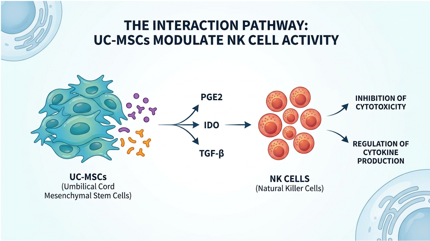
Figure 1: UC-MSCs alter the microenvironment through specific cytokine signaling pathways without disabling NK effector functions.
The Synergistic Benefits of Combining NK Cells with UC-MSCs lie in a paradox of immune regulation: suppressing systemic inflammation while maintaining targeted tumor destruction. Through precise MSC-NK interaction mechanisms, stromal cells reprogram the local microenvironment, promoting tissue repair without neutralizing the aggressive cytolytic functions required for oncology applications (NIH PMC Research, 2026). It is biologically fascinating that stromal cells can force an entire tissue bed into a resting state while simultaneously leaving targeted assassins like NK cells completely unrestricted.
In-vitro models show MSCs reduce pro-inflammatory TNF-alpha by 60% so aggressive targeted cytotoxicity remains preserved exclusively for malignant cells without broad collateral damage.
When you infuse UC-MSCs into a highly inflamed patient, the cells act like biological thermostats. They sense the inflammatory cytokine load in the tissue and respond by releasing massive amounts of specific anti-inflammatory proteins, primarily Indoleamine 2,3-dioxygenase (IDO) and Prostaglandin E2 (PGE2).
One of the most profound mechanisms evaluated in modern immunology is phenotypic macrophage reprogramming. Macrophages are white blood cells that generally exist in two contrasting states: M1 (pro-inflammatory, tissue-damaging, alarm-sounders) and M2 (anti-inflammatory, tissue-repairing, clean-up crew). UC-MSCs force a hard switch in the local environment, driving localized M1 macrophages to adopt the M2 phenotype. They simultaneously inhibit Toll-like receptor signaling, shutting down the biochemical alarms that cause chronic inflammation.
| 📌 If you’re interested in how UC-MSCs rebalance the immune system to calm chronic inflammation, we have an interesting article that discusses mesenchymal stem cell therapy for immune modulation, which you can read via the internal link. |
Furthermore, the release of IDO physically depletes tryptophan in the local microenvironment, converting it into kynurenine. This creates a metabolic dead-zone. MSCs can suppress pro-inflammatory TNF-alpha secretion by roughly 60% within 48 hours so the tissue bed is stabilized rapidly without permanently deactivating the aggressive cytolytic functions of co-administered NK cells. This dead-zone effectively starves hyperactive, inflammatory T-cells, arresting them in the G0/G1 phase of the cell cycle and forcing the surrounding tissue into a resting state.
This brings us to The MSC-NK Synergy Matrix. This is the specific biochemical framework where MSCs downregulate pro-inflammatory tissue destruction while actively preserving the targeted cytolytic functions of co-administered NK cells. Most clinicians initially assume broad immunosuppression would disable NK cells. If you quiet the immune system to heal tissue, shouldn’t that also turn off the tumor-killing immune cells? The Matrix shows us exactly why that assumption is biologically flawed.
The clinical concern is entirely valid: do MSCs suppress NK cells? If they do, combining them for cancer treatment would be catastrophic.
Biology, however, provides a brilliant workaround via the IL-12 and IL-18 stimulation response. When NK cells are resting, MSCs can and do suppress their proliferation. But in a clinical setting, NK cells aren’t resting. They are either pre-activated in the lab or co-stimulated in the body by specific interleukins primarily IL-12, IL-15, and IL-18.
When NK cells receive this specific interleukin signaling, their STAT4 and ERK/MAPK signaling pathways lock open. They become highly resistant to the suppressive signals of MSCs like PGE2. Remember that tryptophan-depleted dead-zone created by IDO? While T-cells starve in that environment, NK cells primed with IL-15 bypass this metabolic starvation entirely. They maintain their mechanistic target of rapamycin (mTOR) activity, keeping their glycolysis and oxidative phosphorylation rates exceptionally high despite the local suppressive factors. The NK cell effectively puts on blinders to the MSC’s “calm down” signals.
This means targeted cytotoxicity is preserved exclusively against malignant or virally infected cells. The NK cells maintain their ability to dump perforin and granzymes into tumors, ripping the cancer cells apart, even while the MSCs are right next door forcing the rest of the tissue bed into a quiet, anti-inflammatory state. This dual-action microenvironment is what makes the combination so aggressively researched.
To truly grasp how these cells operate simultaneously without cancelling each other out, we need to look at their specific cellular outputs. The table below outlines the functional bifurcation that allows this therapy to work seamlessly.
| Cellular Component | Primary Mechanism of Action | Target Cytokines/Markers | Clinical Effect in Synergy |
| UC-MSCs | Immune modulation, macrophage reprogramming, tissue scaffolding | Secretion of PGE2, IDO, TGF-β; Downregulation of TNF-α | Reduces systemic inflammation and fibrotic tissue damage; prepares a receptive tissue bed. |
| Natural Killer (NK) Cells | Direct cytolysis of stressed/malignant cells without prior sensitization | Upregulation of Perforin, Granzyme B, IFN-γ | Identifies and destroys senescent or tumor cells using activating receptors (NKG2D). |
| The Interaction (Synergy) | Compartmentalized immune response via IL-12/15/18 pathways | CD56+ CD16+ NK phenotypes maintained | Clears pathological cells (NK) while simultaneously repairing structural tissue damage (MSC). |
What you see here is a distinct biological division of labor. The MSCs act as the medics, stabilizing the environment by secreting Transforming Growth Factor-beta (TGF-β) and reducing fibrotic scarring. The NK cells act as the targeted assassins, secreting Interferon-gamma (IFN-γ) right at the tumor site. Neither interferes with the other’s core directive, provided the administration timing and cellular activation states are perfectly calibrated in the laboratory prior to infusion.
The application of NK cells in immunotherapy represents a critical advancement for oncology, particularly when enhanced through CAR-NK cell synergy. By engineering these effector cells to recognize specific tumor antigens, clinicians can overcome the immune-evasive tactics of malignant cells, offering a potent alternative to traditional blood cancer immunotherapy treatments
Radiotherapy upregulates tumor stress ligands by up to 40% stripping away malignant camouflage so infused NK cells can aggressively clear previously treatment-resistant solid masses.
Traditional CAR-T cell therapy is an absolute powerhouse for treating certain blood cancers like B-cell acute lymphoblastic leukemia. But it comes with terrifying baggage that patients must carefully weigh.
When evaluating NK cell therapy vs chemotherapy, patients must understand that while chemotherapy broadly attacks all rapidly dividing cells (causing systemic toxicity, alopecia, and severe neutropenia), engineered cellular therapies selectively target antigen-expressing tumors. However, standard CAR-T therapy carries massive risks of Cytokine Release Syndrome (CRS) and severe neurotoxicity, clinically known as ICANS (Immune Effector Cell-Associated Neurotoxicity Syndrome). Standard CAR-T therapy carries a massive risk of severe neurotoxicity occurring in nearly 30% to 40% of cases so researchers are aggressively pivoting toward CAR-NK cells which practically eliminate this life-threatening cytokine release syndrome. The massive Interleukin-6 (IL-6) release from CAR-T can destroy the blood-brain barrier’s integrity, leading to life-threatening brain swelling.
Furthermore, manufacturing autologous CAR-T takes weeks. For a patient with a rapidly progressing malignancy, waiting a month for cell expansion is agonizing, and sometimes fatal. This is exactly why CAR-NK cell synergy is dominating modern immunological research. By taking healthy, allogeneic NK cells often from cord blood and engineering them with Chimeric Antigen Receptors (CARs), you get the targeting precision of CAR-T without the lethal side effects.
Why? Because NK cells produce an entirely different profile of cytokines when they activate. They primarily secrete IFN-γ and GM-CSF, rather than the massive bursts of IL-6 that trigger life-threatening CRS and endothelial activation in T-cell therapies. Shorter hospital stays, lower ICU
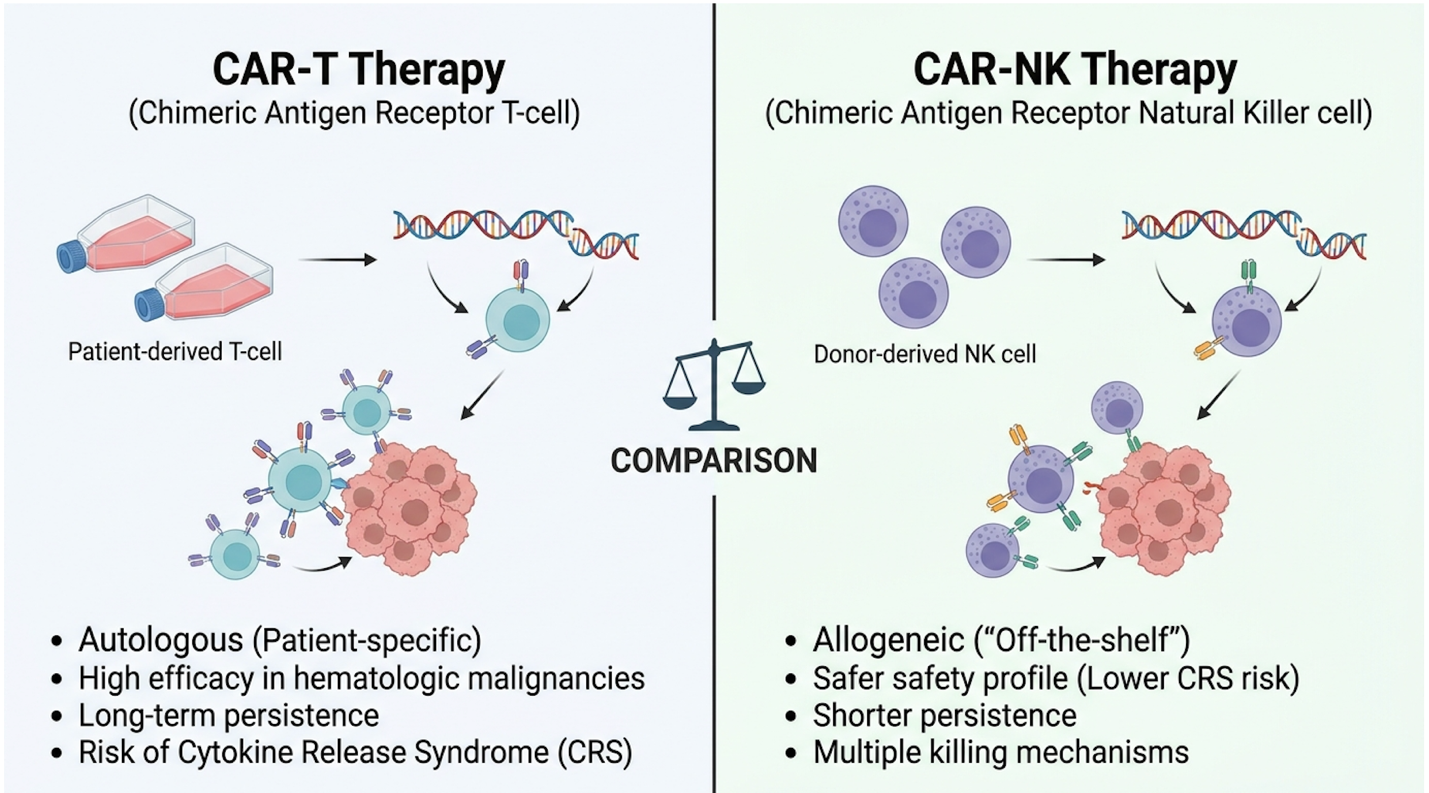
Figure 2: CAR-NK therapies offer a distinct safety profile advantage regarding severe cytokine release syndrome (CRS).
We rarely see cellular therapies operate in a vacuum. In modern oncology, the magic happens in adjunctive applications. The NK cell and radiotherapy combination is a perfect example of turning a localized treatment into a systemic immune trigger.
Combining NK cell therapy with traditional radiotherapy has demonstrated synergistic potential by increasing tumor cell susceptibility to immune-mediated clearance (NIH PubMed Central, 2026).
Standard localized radiotherapy severely damages tumor DNA, but it also heavily alters the Tumor Microenvironment (TME). When tumor cells are irradiated, the massive DNA damage triggers the cell’s internal DNA damage response (DDR) pathway. Under this extreme biological stress, the tumor cells drastically upregulate specific stress ligands on their surface specifically MICA and MICB proteins.
To an NK cell, MICA and MICB are massive neon signs screaming “kill me.” The activating receptor on the NK cell, NKG2D, binds directly to these stress ligands. Liquid cancers, like leukemia, circulate in the blood, making them highly accessible. Solid tumors, however, build fortress-like fibrotic microenvironments. Using radiotherapy just days prior to an NK cell infusion effectively primes the solid tumor, breaking up its fibrotic stroma and stripping away its camouflage, making it highly visible to the immune system.
The synergy gets even deeper when we introduce modern pharmacology. If you want to understand why experts get excited about targeted oncology, look at Antibody-Dependent Cellular Cytotoxicity (ADCC).
ADCC is a process where monoclonal antibodies with NK cells work sequentially to execute tumor cells. It works like this: a patient is given a monoclonal antibody drug (like Rituximab for lymphomas marking the CD20 antigen, or Trastuzumab for HER2+ breast cancer). These synthetic antibodies circulate in the blood and bind directly to specific antigens on the surface of the tumor. They flag the tumor. But the antibody itself doesn’t always kill the cancer cell—it mostly just marks it for destruction.
Enter the NK cell. NK cells naturally express a highly specialized receptor called CD16 (specifically CD16a or FcγRIIIa). This receptor is perfectly designed to grab the tail end of those monoclonal antibodies. Once the NK cell’s CD16 receptor locks onto the antibody attached to the tumor, it triggers an immediate, lethal release of cytotoxic granules right into the cancer cell membrane.
This specific stem cell and NK cell combination therapy creates a dual-targeting framework. You are essentially providing the targeting system (the antibody) and the heavily armed payload (the NK cell) at the exact same time, overcoming potential limitations found in patients with naturally occurring CD16a polymorphisms. These multi-layered protocols require strict oncological oversight, highly specialized drug timing, and intense post-infusion monitoring.
Mesenchymal stromal cells drive regenerative medicine away from localized structural repair toward serving as a systemic-level intervention in aging. By regulating the local immune microenvironment and clearing reactive oxygen species, these advanced therapies aim to mitigate the biological deterioration that drives age-related chronic disease (Cytoniche technical review, 2026). True anti-aging isn’t found in superficial cosmetic fillers; it demands the systemic clearance of pathological senescence at the deepest biological level.
Senescent cells can comprise up to 15% of aged tissue so deploying sequential MSC-NK protocols holistically eliminates these toxic drivers of chronic joint degradation.
When clinics move away from oncology and into systemic regeneration, the administration protocol changes entirely. You don’t just mix everything in an IV bag and hope for the best. The sequential strategy of mesenchymal stromal cells and NK cells is a masterclass in biological timing.
Why does timing matter? Because if you infuse highly active NK cells directly into a highly inflamed, fibrotic tissue bed (like an aging lung or a severely degraded arthritic joint), the hostile microenvironment can actually suppress the NK cells before they do their job.
The protocol dictates that UC-MSCs are administered first, usually on Day One. This initial step is entirely about damage control. The MSCs home in on inflammatory signals, downregulate systemic inflammation, restore mitochondrial membrane integrity, and “quiet” the fibrotic microenvironment. They begin laying down a fresh extracellular matrix (ECM).
Then, roughly three to five days later, the NK cells are infused.
Because the MSCs have already neutralized the hostile, suppressive environment and begun remodeling the ECM, the NK cells arrive in optimal conditions. Their job in regenerative medicine isn’t to kill tumors it’s to identify and lyse senescent cells.
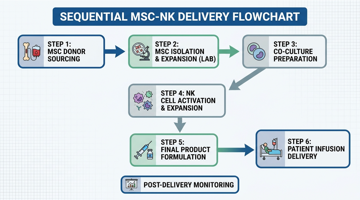
Figure 3: Proper clinical protocol dictates administering MSCs to quiet inflammation before deploying NK cells to clear senescent tissue.
Traditional cosmetic anti-aging is largely superficial. You freeze a muscle to stop a wrinkle, or you inject hyaluronic acid fillers. None of that addresses why the tissue is failing biologically. A true systemic-level intervention in aging targets the biological drivers of decline, specifically cellular senescence.
When human cells reach their Hayflick limitvthe maximum number of times they can divide they are supposed to undergo apoptosis (programmed cell death). Senescent cells, however, are often referred to as “zombie” cells. They stop dividing, but they absolutely refuse to die. Worse, they secrete a highly toxic cocktail of inflammatory cytokines, proteases, and chemokines known as the Senescence-Associated Secretory Phenotype (SASP).
Aged tissue environments can accumulate senescent ‘zombie’ cells at rates exceeding 10% to 15% of total cellular volume so targeted NK cell infusion is required to selectively lyse these toxic cells before structural rebuilding can begin. SASP is highly destructive because it includes heavy outputs of Interleukin-6 (IL-6), Interleukin-8 (IL-8), and Matrix Metalloproteinases (MMPs),
By infusing highly active, young, allogeneic NK cells, clinics aim to jumpstart this clearance process. The NK cells track down the beta-galactosidase positive senescent cells and lyse them. Once the zombie cells are cleared, delaying age-related decline becomes vastly more achievable because the tissue is no longer actively poisoning itself.
Beyond just clearing the garbage, the stroma must rebuild the infrastructure. This is where UC-MSCs provide aggressive, sustained tissue protection.
MSCs are essentially biological factories. Through paracrine signaling, they secrete massive amounts of bioactive molecules, including VEGF and Hepatocyte Growth Factor (HGF). These specific factors actively stimulate angiogenesis (the creation of new, healthy blood vessels), ensuring the cleared tissue gets the oxygen and nutrient perfusion it needs to survive and thrive.
Furthermore, MSCs directly stimulate quiescent dermal fibroblasts and osteoblasts. They force the local tissue to ramp up the synthesis of collagen type I, fibronectin, and elastin, rebuilding the architectural scaffolding of the joint or organ. To combat any remaining tissue degradation, MSCs actively secrete Tissue Inhibitors of Metalloproteinases (TIMPs). These TIMPs directly bind to and neutralize the destructive MMP enzymes left behind by senescent cells, actively stopping tissue breakdown so new structural matrices can form.
But I need to ground this in reality. While these tissue protection mechanisms are heavily documented in laboratory settings and early human trials, biological plausibility does not instantly equal a guaranteed clinical outcome. Patients must seek evidence-informed suitability assessments to determine if their specific state of tissue degradation is actually viable for this level of intervention.
CAR-T cell therapy utilizes genetically modified T-cells from the patient to target specific cancers, whereas NK cell therapy utilizes Natural Killer cells that can be sourced from healthy donors without causing graft-versus-host disease. This allogeneic advantage makes NK therapies faster to produce and deploy as off-the-shelf treatments. For patients facing rapidly progressing malignancies, this time-to-treatment advantage is often a deciding clinical factor. However, both remain highly specialized interventions requiring rigorous specialist evaluation and clinical monitoring to ensure patient safety and suitability.
Umbilical cord tissue bypasses immune rejection because its Mesenchymal Stromal Cells (UC-MSCs) lack major histocompatibility complex (MHC) class II expression and crucial co-stimulatory molecules. This unique lack of biological identification prevents the host’s immune system from mounting an attack. Furthermore, the absence of CD80 and CD86 means no secondary activation signals are sent to host T-cells. This biological profile makes them inherently immune-evasive, acting as a “stealth” cell in the body. Consequently, these cells can be administered to patients without the strict HLA-matching requirements necessary for traditional bone marrow transplants, though clinical monitoring is always mandated.
Yes, combining NK cell therapy with localized radiotherapy demonstrates significant clinical synergy in emerging oncology research. Radiotherapy alters the tumor microenvironment and upregulates stress ligands on the surface of malignant cells. This biological stress response makes the tumor cells highly visible and susceptible to targeted destruction by administered NK cells. This powerful combination potentially improves the clearance of radiation-resistant solid tumors under strict oncological supervision.
The sequential strategy in regenerative medicine involves administering UC-MSCs first, followed by NK cells, to optimize tissue repair. The MSCs act initially to reduce systemic inflammation and remodel the fibrotic microenvironment into a healing state. Without this initial stromal intervention, the highly inflammatory environment could prematurely exhaust the infused NK cells
No, stem cell and cellular combination therapies do not guarantee disease reversal or symptom resolution. While research shows strong biological plausibility for immune modulation and tissue repair, outcomes are highly dependent on the patient’s specific diagnosis, condition severity, and cellular response. Biology is inherently unpredictable, and systemic conditions often require multi-modal treatment plans. Patients must demand transparent efficacy data specific to their exact pathology rather than relying on generalized marketing claims. Patients must undergo rigorous, evidence-informed suitability assessments with a qualified clinician to set realistic expectations for functional outcomes.
Off-the-shelf cell therapies derived from properly banked umbilical cord tissue have demonstrated favorable safety profiles regarding immune rejection and graft-versus-host disease. However, “safe” does not mean risk-free. Cold-chain logistics, proper cryopreservation, and immediate pre-infusion viability testing are critical components of a safe administration protocol. The safety of any cellular intervention relies entirely on the clinic’s laboratory standards, proper cell culturing techniques, and stringent patient screening. Investigational treatments require ongoing clinical oversight to monitor for any adverse immune reactions or localized complications.
| 📌 If you’re wondering how to evaluate a clinic’s safety standards before treatment, we have an interesting article that discusses whether stem cell therapy is safe in Thailand, which you can read via the internal link. |
For patients investigating advanced oncological and systemic interventions, NK cell therapy for cancer Thailand, when synergized with UC-MSCs, represents a paradigm of targeted immune modulation. Clinical evidence indicates that allogeneic sourcing bypasses HLA restriction, allowing these cells to alter the tumor microenvironment and support adjunctive treatments like radiotherapy. The most effective approach relies on strict, evidence-informed laboratory protocols and condition-specific application.
The efficacy of this combination relies entirely on The MSC-NK Synergy Matrix the precise biochemical balance where stromal cells reduce destructive inflammation without deactivating the tumor-killing capacity of NK cells. Understanding this mechanism empowers patients to look past commercial hype and focus on verifiable biological rationales.
Because cellular medicine outcomes vary drastically by diagnosis, patients should compile their medical records, imaging, and laboratory findings to seek a formal suitability assessment. Discuss your treatment history with a qualified regenerative medicine clinician to establish realistic functional goals and ensure rigorous clinical monitoring.