Our Services
Others
- DFPP
- Shockwave
- IV Drip

https://vegastemcell.com/articles/the-importance-of-cell-viability-in-uc-msc-stem-cell-therapy/
A diagnosis of Autism Spectrum Disorder (ASD) marks the beginning of a demanding, lifelong journey for a family Applied Behavior Analysis, occupational training, speech counseling, specialized educational frameworks, and a daily balancing act between celebrating breakthroughs and managing sensory overload, communication barriers, and chronic digestive issues. For decades, the standard clinical approach has treated autism largely as a fixed neurological blueprint, managed through behavioral adaptation and, when needed, medication for anxiety, hyperactivity, or sleep disturbance. Those supportive therapies matter but they work on the surface. They don’t directly address the brain inflammation, immune dysregulation, and reduced blood flow increasingly documented underneath the behaviors. This is where research into stem cell therapy in Thailand, using Umbilical Cord Mesenchymal Stem Cells (UC-MSCs), has entered the conversation.
The brain has its own specialized immune cells called microglia. In a healthy state, they stay in a quiet, ramified shape acting as housekeepers clearing cellular waste and supporting healthy neural connections.
A body of neurological research has documented that autism involves chronic, low-grade neuroinflammation and immune dysregulation more than a simple structural wiring difference. In some individuals with ASD, microglia appear to get stuck in a hyperactivated state, releasing pro-inflammatory cytokines like Interleukin-6 (IL-6) and Tumor Necrosis Factor-alpha (TNF-α). This chronic signaling is believed to disrupt normal electrical communication between brain regions contributing, researchers theorize, to sensory sensitivities, speech delays, and emotional processing difficulties.
A meaningful share of individuals on the autism spectrum experience chronic intestinal hyperpermeability commonly called leaky gut where the tight junctions of the intestinal lining degrade and the gut microbiome shifts out of balance.
This breakdown is theorized to allow bacterial endotoxins called lipopolysaccharides (LPS) to enter systemic circulation, eventually reaching the Blood-Brain Barrier (BBB). Endotoxins are believed to loosen tight-junction proteins including Claudin-5 that normally hold brain blood vessels together, increasing vascular permeability and allowing inflammatory signals into the central nervous system, where they may keep microglia locked in a reactive state. This gut-immune-brain pathway is an active, genuinely interesting area of research, though it’s worth noting much of the mechanistic detail comes from broader neuroimmunology research rather than autism-specific human trials.
UC-MSCs are ethically harvested from umbilical cord tissue specifically Wharton’s Jelly following healthy, full-term births and thorough donor screening. Because they’re day-zero cells, they display strong proliferative capacity and release a higher volume of anti-inflammatory proteins and growth factors than a patient’s own adult-derived stem cells. They’re also immunoprivileged, lacking the HLA Class II surface markers that would trigger immune rejection, meaning they can be administered without donor matching or anti-rejection medication.
UC-MSCs don’t work by manually rebuilding nerve networks cell-for-cell. They work through paracrine signaling releasing exosomes carrying molecular messengers that influence surrounding cells.
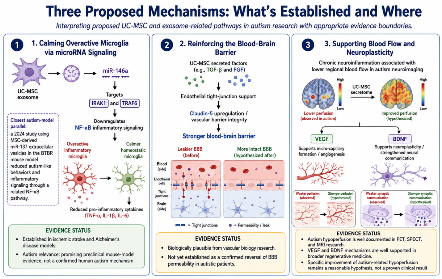
1. Calming Overactive Microglia via microRNA Signaling
One of the more scientifically interesting UC-MSC exosome components is microRNA-146a (miR-146a), a well-documented anti-inflammatory microRNA shown to suppress the NF-κB inflammatory cascade by targeting IRAK1 and TRAF6 the signaling machinery that drives microglia to keep producing inflammatory cytokines. It’s important to be precise about where this evidence actually comes from: miR-146a’s microglia-calming effect via this pathway has been demonstrated in ischemic stroke and Alzheimer’s disease models, not autism specifically. The closest autism-specific parallel comes from a 2024 study using a different MSC-derived microRNA, miR-137, delivered via extracellular vesicles in the BTBR mouse model of autism which found that MSC-miR137-EVs eased autism-like behaviors and reduced inflammatory signaling through a related NF-κB pathway. This is a genuinely promising line of research, but it remains preclinical, mouse-model evidence rather than confirmed human mechanism.
2. Reinforcing the Blood-Brain Barrier
UC-MSCs secrete vascular and tissue growth factors, including Transforming Growth Factor-beta (TGF-β) and Fibroblast Growth Factor (FGF), which are studied for their role in supporting endothelial tight junctions including Claudin-5 and vascular barrier integrity generally. Direct evidence of this specific effect reversing BBB permeability in autistic patients is not yet established; this remains a biologically plausible extension of vascular biology research rather than an autism-specific confirmed outcome.
3. Supporting Blood Flow and Neuroplasticity
Chronic neuroinflammation is associated with reduced blood flow in specific brain regions a pattern well-documented in autism neuroimaging research using PET, SPECT, and MRI. UC-MSCs produce Vascular Endothelial Growth Factor (VEGF), studied for supporting new micro-capillary formation, alongside Brain-Derived Neurotrophic Factor (BDNF), which supports neuroplasticity the brain’s capacity to form and strengthen communication pathways. Both mechanisms are well-supported in broader regenerative medicine research; their specific effect on documented autism-related hypoperfusion is a reasonable, evidence-grounded hypothesis rather than a proven clinical result.
| Biological Target | Cellular Pattern in ASD | UC-MSC Mechanism | Evidence Status |
| Activated Microglia | Chronic neuroinflammation, elevated IL-6 | Exosomal microRNA signaling calms NF-κB activation | Established in stroke/Alzheimer’s models; autism evidence is preclinical (mouse) |
| Endothelial Linings | Increased BBB permeability | TGF-β/FGF support tight-junction integrity | Established in general vascular biology; autism-specific data limited |
| Intestinal Mucosa | Hyperpermeability, LPS leakage | Supports gut barrier repair | Theoretical/emerging; active research area |
| Neural Synapses | Reduced connectivity, hypoperfusion | VEGF and BDNF support blood flow and plasticity | Well-supported generally; autism-specific trials limited |
Real-World Expectations: Tracking Functional Progress
Stem cell therapy is not an overnight cure, and it doesn’t instantly change a person’s personality or erase years of developmental difference in a single treatment. The realistic goal is creating a calmer internal biological environment that allows behavioral and speech therapy to work more effectively alongside it.
Families who respond to UC-MSC protocols often describe gradual changes unfolding over two to six months, including better tolerance of sensory input, expanding vocabulary and communication attempts, longer attention spans with fewer repetitive stimming behaviors, and more stable digestive function. These are consistent, worth-taking-seriously patterns in parent-reported experience not a guaranteed outcome, and not yet backed by the same volume of controlled human trial data as the underlying biological mechanisms described above.
Stem cell therapy in Thailand has grown around infrastructure suited to delivering UC-MSC protocols responsibly certified laboratories, physicians trained in both neurology and regenerative medicine, and clinics that integrate biological therapy with behavioral and developmental support rather than presenting it as a standalone fix.
This mechanism-focused overview connects to other areas worth exploring: UC-MSC therapy and gut microbiome biomarkers for autism, the neuroplasticity critical period and treatment timing for autism, cerebral blood flow research in autism spectrum disorder, and co-occurring GI and sleep symptoms in autism each covering a related piece of this broader biological picture in more depth.
Is the miR-146a mechanism proven to work this way in autism specifically?
Not yet in humans. miR-146a’s role in calming microglial inflammation via NF-κB suppression is well documented in stroke and Alzheimer’s research. The closest autism-specific evidence uses a different microRNA (miR-137) in a mouse model, showing a related but distinct mechanism. This remains promising preclinical research, not confirmed human evidence.
Does UC-MSC therapy repair the blood-brain barrier in autism?
The underlying growth factors (TGF-β, FGF) are studied for supporting vascular barrier integrity generally. Direct evidence of this reversing blood-brain barrier permeability specifically in autistic patients isn’t yet established, it’s a biologically reasonable hypothesis under active investigation.
How long before families notice changes after treatment?
Reported changes typically unfold gradually over two to six months, reflecting how biological repair and remodeling processes actually work rather than an immediate effect.
Does stem cell therapy replace behavioral therapy for autism?
No. The goal is a calmer internal biological environment that makes existing behavioral and speech therapy more effective not a replacement for that ongoing work.
Why do families consider stem cell therapy in Thailand for autism?
Stem cell therapy in Thailand combines physicians trained in neurology and regenerative medicine, certified laboratory standards, and integrated care models connecting UC-MSC therapy with established developmental support.
Moving From Passivity to Informed, Proactive Support
Understanding both what’s scientifically established and what remains preclinical or theoretical helps families evaluate UC-MSC stem cell therapy with genuinely informed expectations rather than either dismissing or overselling the science. If you’re trying to understand how this research applies to your child’s specific situation, book a consultation with Vegastemcell for an assessment grounded in accurate, current evidence.
For more infomation you can reach me directly on WhatsApp or Email. Here’s our official link:
WhatsApp: +66982509599
Email: vegastemcell@gmail.com
Zhang, Z., Zou, X., Zhang, R., et al. (2021). Human umbilical cord mesenchymal stem cell-derived exosomal miR-146a-5p reduces microglial-mediated neuroinflammation via suppression of the IRAK1/TRAF6 signaling pathway after ischemic stroke. Aging (Albany NY). https://www.ncbi.nlm.nih.gov/pmc/articles/PMC7880318/ (Mouse ischemic stroke model — not autism.)
Nakano, M., Kubota, K., Kobayashi, E., et al. (2020). Bone marrow-derived mesenchymal stem cells improve cognitive impairment in an Alzheimer’s disease model by increasing the expression of microRNA-146a in hippocampus. Scientific Reports. https://www.ncbi.nlm.nih.gov/pmc/articles/PMC7330036/ (Mouse Alzheimer’s model — not autism. Source of the general miR-146a/IRAK1/TRAF6/NF-κB mechanism description.)
Synergistic effect of mesenchymal stem cell-derived extracellular vesicle and miR-137 alleviates autism-like behaviors by modulating the NF-κB pathway. (2024). Journal of Translational Medicine. https://www.ncbi.nlm.nih.gov/pmc/articles/PMC11089771/ (BTBR mouse autism model; uses miR-137, not miR-146a. This is the closest available evidence to the article’s core claim, and it does not match the original source document’s specific mechanism.)
Perets, N., Oron, O., Herman, S., Elliott, E., & Offen, D. (2020). Exosomes derived from mesenchymal stem cells improved core symptoms of genetically modified mouse model of autism Shank3B. Molecular Autism. https://molecularautism.biomedcentral.com/articles/10.1186/s13229-020-00366-x (Shank3B mouse autism model — another genuine autism-specific preclinical data point.)
Neuroimaging studies of cerebral perfusion in autism spectrum disorder using SPECT, PET, and MRI-based methods — consistent with findings referenced in the companion article on cerebral blood flow in autism, documenting reduced perfusion in frontal, temporal, and limbic regions correlated with symptom severity.
Immunomodulatory Mechanisms and Therapeutic Potential of Mesenchymal Stem Cells. Stem Cell Reviews and Reports. https://link.springer.com/article/10.1007/s12015-023-10539-9