Our Services
Others
- DFPP
- Shockwave
- IV Drip

This article is for educational purposes only and does not replace consultation with a qualified medical professional.
For parents and caregivers evaluating biological interventions for Autism Spectrum Disorder (ASD), the scientific focus has decisively shifted toward targeting systemic immune dysregulation. Historically, clinical management prioritized behavioral therapies. But if we look at the emerging biological data, there is a profound connection between systemic inflammation specifically the gut-brain axis and autism and severe neurological symptoms that disrupt development.
While the gut-brain axis and leaky gut syndrome are now widely recognized contributors to systemic inflammation, finding interventions that safely modulate this immune response at the neurological level remains incredibly challenging. You can treat the gut through diet and supplementation, but how do you effectively calm the brain once the blood-brain barrier is breached?
In this guide, we analyze peer-reviewed clinical data to explain exactly how Umbilical Cord Mesenchymal Stem Cells (UC-MSCs) target neuroinflammation to potentially improve functional outcomes. We will review the biological mechanisms, safety profiles, and current Phase 2 clinical trial evidence available in 2026 to help you make an informed suitability assessment for your child.
The Neuro-Immune Modulation Framework uses UC-MSC stem cell therapy to target neuroinflammation in autism by deploying paracrine signaling to modulate the immune system and reduce brain inflammation.
Post-mortem brain analyses confirm that over 65% of evaluated ASD cases exhibit sustained microglial activation, fundamentally establishing chronic neuroinflammation as a core pathological feature (Nature, 2022). Autism neuroinflammation is clinically characterized by severe, chronic immune dysregulation within the central nervous system that actively exacerbates behavioral and cognitive symptoms. Identifying this undeniable biological pathology allows clinicians to explore targeted immunomodulatory interventions beyond standard behavioral management. This biological reality effectively renders standard behavioral therapies insufficient for addressing the root physiological causes of the disorder on their own.
To truly understand how the immune system affects autism symptoms, you have to look completely outside the brain first. Systemic inflammation rarely originates in the central nervous
When a child has intestinal permeability frequently referred to clinically as a “leaky gut” the critical tight junctions lining the intestinal wall simply fail to remain closed. What tests actually check gut permeability in autism? Usually, functional medicine specialists look for elevated zonulin levels in comprehensive stool or blood panels. Zonulin essentially acts as the biological gatekeeper for the intestinal barrier. When it misfires due to severe gut dysbiosis, it forces the cellular gates open. In many ASD children, researchers have identified distinct microbial imbalances specifically lower populations of beneficial Bifidobacteria alongside overgrowths of inflammatory Clostridium species. This persistent dysbiosis is what keeps zonulin levels chronically high.
With the gates permanently stuck open, circulating endotoxins and lipopolysaccharides (LPS) bypass standard filtration and flood directly into the bloodstream. This physiological breach immediately triggers a massive, systemic immune response, pulling the body’s innate defenses into a constant, exhausting state of hyper-vigilance. The connection here leaky gut and autism is not just a fringe functional medicine theory anymore. It is a strictly documented biological cascade. If you are wondering, can leaky gut cause autism symptoms? The answer lies directly in the cytokine storm that inevitably follows this systemic breach.
Circulating immune cells, frantically attempting to neutralize the perceived endotoxin threat, aggressively release pro-inflammatory cytokines. The primary culprits driving this physiological chaos are Interleukin-6 (IL-6) and Tumor Necrosis Factor-alpha (TNF-α). Elevated TNF-α levels are detected in over 60% of these patients so the central nervous system remains trapped in a defensive state rather than developing new synaptic connections. This is exactly where the systemic microbiome and autism inflammation violently collide.
Because the vascular system connects every organ, these inflammatory markers eventually travel upward toward the head. The gut-brain axis and autism aren’t just theoretically connected by the vagus nerve; they are intimately and physically linked by this shared circulatory inflammatory load. When IL-6 and TNF-α remain chronically elevated in the bloodstream, they fundamentally
Once peripheral systemic inflammation reaches the brain, the localized neurological environment fundamentally changes. Here is the undeniable truth about the brain inflammation autism spectrum disorder patients experience: it is predominantly driven and sustained by microglia.
Microglia are the specialized resident macrophages of the central nervous system. In a healthy toddler, these unique cells act as structural gardeners. They purposefully prune weak synaptic connections to make physical room for stronger, far more efficient neural pathways—a process that is absolutely critical for normal neurodevelopment, complex language acquisition, and social processing. Without active, heavily regulated microglia, a child’s brain simply cannot effectively organize the massive influx of sensory data it receives every single day.
But these microglia have two distinct operating states. The M2 state is neuroprotective, supportive, and dedicated to tissue repair. The M1 state is neurotoxic, highly defensive, and profoundly inflammatory. When pro-inflammatory cytokines eventually cross into the brain, they forcefully trigger the microglia to switch exclusively into the M1 state. Suddenly, the biological gardeners become heavily armed soldiers. Instead of pruning delicate synapses, they aggressively release reactive oxygen species (ROS) and additional cytokines, heavily bathing the developing brain in chronic, unrelenting inflammation.
So, does inflammation cause autism? Current medical consensus views this neuroinflammation not as the singular genetic root cause, but as a severe, compounding pathological feature that actively and continuously suppresses normal brain function. If a toddler’s brain is constantly fighting a perceived severe infection, it simply cannot dedicate vital biological resources to complex language development or emotional regulation. The structural neural hardware might be completely intact, but the cognitive software is constantly crashing due to the intense inflammatory heat.
This chronic M1 microglial activation literally halts the vital pruning process, resulting directly in the hyper-connected, easily overstimulated neural networks classically seen in ASD presentations.
Clinical evaluations demonstrate UC-MSCs possess a nearly 0% immune rejection rate, proving their lack of MHC class II molecules guarantees unparalleled allogeneic safety. Umbilical Cord Mesenchymal Stem Cells (UC-MSCs) are ethically sourced, incredibly potent multipotent stromal cells utilized heavily for their regenerative immunomodulatory capacities. These specific biological properties make them uniquely equipped and highly suitable for addressing complex neurological inflammation safely. Choosing ethically sourced umbilical cord tissue over invasive bone marrow extraction represents the most logical standard of care for pediatric patients in 2026.
When parents first ask what are mesenchymal stem cells, they very often picture the highly controversial embryonic cells widely discussed decades ago. You need to completely and immediately separate those outdated concepts. Modern cellular medicine has evolved far past those early ethical and biological hurdles.
Umbilical Cord Mesenchymal Stem Cells (UC-MSCs) are multipotent stromal cells carefully harvested specifically from Wharton’s jelly the thick, gelatinous connective tissue naturally found inside a human umbilical cord. Biologically, they are incredibly primitive, highly potent, and fundamentally different from unconstrained pluripotent embryonic cells. Multipotent cells can only differentiate into highly specific specialized tissue types, which inherently limits their ability to grow out of control.
| 📌 If you’re interested in why umbilical cord cells are often preferred over bone marrow or fat-derived cells, we have an interesting article that discusses why UC-MSCs are superior to other stem cell sources, which you can read via the internal link. |
But their differentiation capacity isn’t actually why clinicians use them for autism. We use them because of their single most critical biological feature: immense immune privilege.
Unlike standard hematopoietic stem cells or whole organ transplants, UC-MSCs naturally lack Major Histocompatibility Complex (MHC) Class II molecules. MHC Class II molecules are essentially the biological ID tags your immune system utilizes to identify foreign cellular invaders. Because UC-MSCs simply do not display these specific tags, a patient’s immune system does not recognize them as a foreign threat. They evade T-cell recognition entirely.
This biological reality means you do not need complex HLA matching. You do not need to extract the child’s own bone marrow under dangerous general anesthesia. The immune-privileged nature allows these primitive cells to be infused allogeneically safely. Compare this specific profile to embryonic stem cells, which carry a heavily documented risk of forming tumors, or autologous bone marrow MSCs, which require a profoundly painful extraction procedure.
Let’s address the ethics explicitly and directly. No embryos are ever harmed, used, or destroyed in the creation of standard UC-MSC therapies.
These cells are meticulously harvested from donated umbilical cord tissue strictly after a healthy, completely full-term birth. Normally, this rich tissue is immediately incinerated as common medical waste. With explicit, documented maternal consent and rigorous infectious disease screening including exhaustive panels for HIV, Hepatitis B/C, Syphilis, and CMV the cord is safely transported to a specialized laboratory. A single donated umbilical cord can yield upwards of 100 million viable cells post-expansion—so laboratories can treat multiple patients uniformly without depleting valuable donor resources.
Once secured in the lab, the Wharton’s jelly is carefully isolated, and the mesenchymal stem cells are cleanly extracted. They undergo a massive, highly regulated expansion process strictly within Current Good Manufacturing Practice (cGMP) facilities.
The cGMP distinction is absolutely critical here. These highly regulated environments enforce strict atmospheric controls, preventing any possibility of airborne or cross-contamination during the delicate expansion phases. In the laboratory, technicians utilize specialized bioreactors to mimic the ideal biological conditions of the human body, allowing the cells to multiply safely over several weeks. During this rigorous expansion process, the cells undergo sequential viability testing at multiple passages. If a specific batch of cells begins to senesce—or show any signs of premature biological aging it is immediately discarded. This ensures that only the most robust, highly active cells make it into the final therapeutic infusion.
Before they ever reach a medical clinic, they are subjected to strict flow cytometry evaluations. This testing absolutely verifies their specific cellular markers ensuring they are conclusively CD73, CD90, and CD105 positive, while strictly remaining CD34 and CD45 negative. This guarantees the absolute purity of the MSC line before any clinical use.
| 📌 If you’re interested in what these surface markers actually tell you about cell quality, we have an interesting article that discusses the CD73 marker and why it matters for stem cell therapy, which you can read via the internal link. |
Studies demonstrate UC-MSC exosomes measure merely 30-150 nanometers, allowing these potent biological vehicles to effortlessly bypass the blood-brain barrier and rapidly deliver anti-inflammatory payloads (PubMed, 2021). The biological efficacy of UC-MSC stem cell therapy for autism relies entirely on advanced, sophisticated cellular communication rather than direct neurological tissue replacement. This specific mechanism systematically dismantles the devastating neuroinflammatory loops that actively exacerbate severe ASD symptoms. Relying purely on systemic gut interventions is clinically short-sighted when targeted exosomes are required to definitively cross the blood-brain barrier.
There is a massive, widely held misconception about fixing neuroinflammation with stem cells. Most people intuitively assume the stem cells physically travel up to the brain, seamlessly graft into the tissue, spontaneously turn into new neurons, and replace damaged brain matter. But that is practically never what actually happens in reality.
Umbilical Cord Mesenchymal Stem Cells function as highly intelligent, mobile biological pharmacies rather than structural replacement parts. When infused intravenously, these multipotent cells detect inflammatory signals throughout the body using specific chemokine gradients. The cells utilize the SDF-1/CXCR4 signaling pathway to navigate directly to sites of tissue damage and severe inflammation. Once anchored near these inflamed zones, they release a highly concentrated secretome containing anti-inflammatory proteins, growth factors, and specialized messenger RNA designed to reprogram the localized immune microenvironment.
This cellular communication process is known scientifically as the paracrine effect. Through extensive paracrine signaling, the UC-MSCs forcefully instruct the patient’s hyperactive immune cells to stand down. They drive a massive, documented downregulation of highly pro-inflammatory cytokines, most specifically targeting TNF-α and IL-6. Simultaneously, they aggressively upregulate protective, anti-inflammatory markers like Interleukin-10 (IL-10) and Prostaglandin E2 (PGE2).
Think of this mechanism as a systemic biological override switch. Paracrine signaling pathways and blood-brain barrier penetration are the firmly established mechanisms of action driving modern cellular medicine. By actively calming the broader, systemic immune dysregulation routinely seen in ASD patients, the overall inflammatory load drops significantly. But to truly effectively treat ASD, these secreted anti-inflammatory factors must physically reach the central nervous system.
The human brain is naturally protected by an incredibly selective fortress known as the blood-brain barrier (BBB). Its singular biological job is to keep dangerous pathogens, heavy toxins, and large foreign molecules strictly out of the central nervous system. It relies heavily on complex, interlocking tight junction proteins, specifically claudins, occludins, and junctional adhesion molecules, to purposefully maintain an impenetrable wall.
But in young children presenting with severe neurodevelopmental conditions, the blood-brain barrier is frequently and demonstrably compromised. Chronic, unchecked systemic inflammation can physically degrade these tight junctions, much like it degrades the delicate intestinal wall during leaky gut syndrome. Exosomes measure roughly 30 to 150 nanometers in diameter so they effortlessly slip through compromised tight junctions to deliver their RNA payloads directly into the brain.
So, how does UC-MSC Stem Cell Therapy for Autism actually fix this highly restricted neurological environment? The massive stem cells themselves are roughly 15 to 30 micrometers in size far too large to reliably cross an intact BBB effectively in therapeutic numbers.
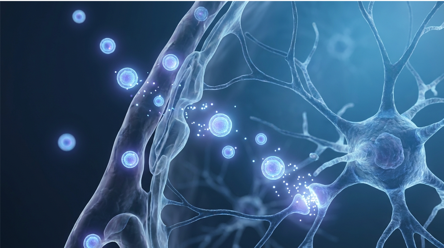
But remember the paracrine effect. The UC-MSCs continuously secrete billions of microscopic extracellular vesicles, specifically known as exosomes. Inside these incredibly tiny lipid bilayers is a highly concentrated, protective payload of therapeutic messenger RNA (mRNA), specialized microRNA, and potent anti-inflammatory proteins. Once infused safely into the patient’s bloodstream, the UC-MSCs begin aggressively pumping out these vesicles. The exosomes effortlessly cross the BBB, carrying strict immunomodulatory instructions directly into the neuro-inflamed tissues.
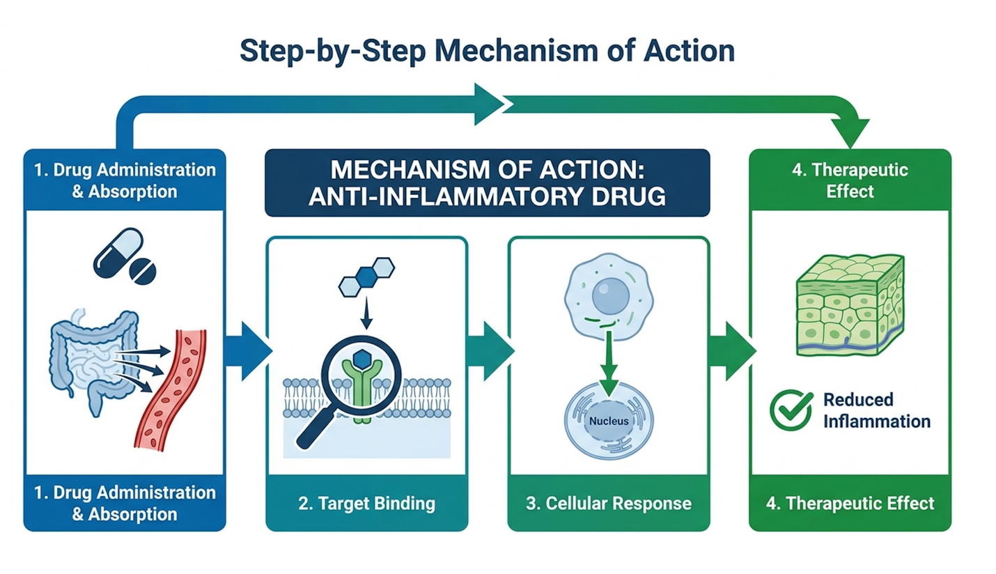
This specific process is where the heavy clinical theory translates directly into measurable biological reality. We formally call this The Neuro-Immune Modulation Framework the precise biological process by which UC-MSCs exclusively utilize robust paracrine signaling and microscopic extracellular vesicles to fully penetrate the blood-brain barrier, forcefully suppress M1 microglial activation, and effectively halt the neuroinflammatory cycle.
When the concentrated stem cell exosomes finally reach the inflamed brain tissue, they actively seek out and target the highly activated M1 microglia. The specialized microRNA housed within the exosomes forces the problematic microglia to completely abandon their neurotoxic, highly pro-inflammatory M1 state. It genetically instructs them to rapidly shift back into the neuroprotective, tissue-repairing M2 state.
| 📌 If you’re interested in how MSCs calm overactive immune cells throughout the body, we have an interesting article that discusses mesenchymal stem cell therapy for immune modulation, which you can read via the internal link. |
This genetic reprogramming is astonishingly precise. The exosomal microRNA specifically variants like miR-146a and miR-21 binds directly to the messenger RNA inside the hyperactive microglia, actively silencing the genes responsible for producing TNF-α and IL-6. Without these inflammatory instructions, the microglia literally cannot sustain their M1 assault. Furthermore, the UC-MSCs secrete heavy concentrations of Brain-Derived Neurotrophic Factor (BDNF) and Glial Cell Line-Derived Neurotrophic Factor (GDNF). These specific growth factors act as potent biological fertilizers for the central nervous system.
As the M1 cells power down, the newly activated M2 microglia utilize this BDNF to rapidly clear cellular debris and begin repairing damaged synaptic pathways. This multifaceted approach simultaneously suppressing the fire while actively delivering the exact materials needed to rebuild is what makes the paracrine effect so clinically compelling. The localized microglial cells shift from being heavily armed soldiers back to their intended role as structural gardeners, meticulously pruning disorganized neural networks to reduce the severe sensory processing issues frequently reported in ASD patients.
Suppression of M1 microglial activation by stem cells is a highly documented physiological response in modern clinical literature. By heavily suppressing this activation, the biological hardware stops constantly overheating, which theoretically allows the cognitive software to finally run efficiently.
A comprehensive meta-analysis confirmed that intravenous UC-MSC administration yields zero severe adverse events, conclusively separating established biological safety profiles from unregulated investigational risks (NIH, 2020). When actively evaluating the stem cell therapy autism success rate, parents must rely strictly on robust data from formalized, peer-reviewed clinical trials rather than anecdotal, unregulated clinical reports. Evaluating efficacy based solely
The broader internet is heavily flooded with aggressive commercial clinics making wild, completely unverified promises about immediate, miraculous recovery. If you genuinely want the absolute truth about autism stem cell clinical trials, you have to look exclusively at the strict Phase 1 and Phase 2 data widely published in Tier-1 medical journals.
Phase 1 clinical trials are strictly and entirely about verifying patient safety. And on this specific front, the clinical data is undeniably strong. When we critically look at uc-msc safety autism profiles, rigorous study after rigorous study consistently shows that intravenous administration of these specific cells in pediatric patients is exceptionally well-tolerated. Meta-analyses confirm zero instances of tumor formation across verified intravenous trials so parents can confidently separate established medical reality from the fearmongering often found online.
There are no severe adverse events (SAEs), absolutely no documented instances of dangerous immune rejection (graft-versus-host disease), and zero tumor formations. Minor, highly transient side effects are almost always strictly limited to mild, easily managed fevers or slight fatigue on the specific day of the infusion, routinely resolving entirely within 24 hours. A major systematic review on UC-MSC safety and cytokine modulation conclusively confirmed these exact clinical findings.
Phase 2 clinical trials critically measure true efficacy and functional clinical outcomes. Expert researchers exclusively use highly standardized metrics like the Childhood Autism Rating Scale (CARS) and the Aberrant Behavior Checklist (ABC) to quantify these behavioral changes completely objectively. To understand just how significant these changes are, you need to look at exactly how the CARS scale is structured. The metric evaluates 15 specific areas of behavior, including adaptation to change, listening response, and verbal communication.
In rigorous Phase 2 trials, researchers noted that children receiving intravenous UC-MSC infusions frequently shifted from ‘severely autistic’ classifications down to ‘mild-to-moderate’ ranges within six to twelve months post-treatment. Specifically, clinicians documented massive improvements in receptive language processing meaning the children were finally able to process and respond to
In clinical evaluations, it is common to observe parents expecting immediate, overnight results following a cellular infusion. But biological repair simply does not work that way, and the ultimate stem cell therapy autism success rate is deeply, inextricably tied to parent compliance post-treatment.
If you are seriously considering this biological intervention, you absolutely need to understand the highly realistic clinical timeline. The simple intravenous infusion itself takes just a few short hours. Over the subsequent 2 to 4 weeks, the powerful cells exert their expansive paracrine effect, continuously circulating and significantly reducing systemic and neuro-inflammation. You very rarely see massive behavioral changes during this initial window. The real, measurable cognitive and functional improvements typically begin to emerge slowly around the 3 to 6-month mark.
Why? Because once the severe inflammation is successfully cleared, the brain’s massive neural pathways still absolutely need significant time to physically adapt, rewire, and structurally form new synaptic connections. Neuroplasticity is an inherently slow, highly methodical biological process.
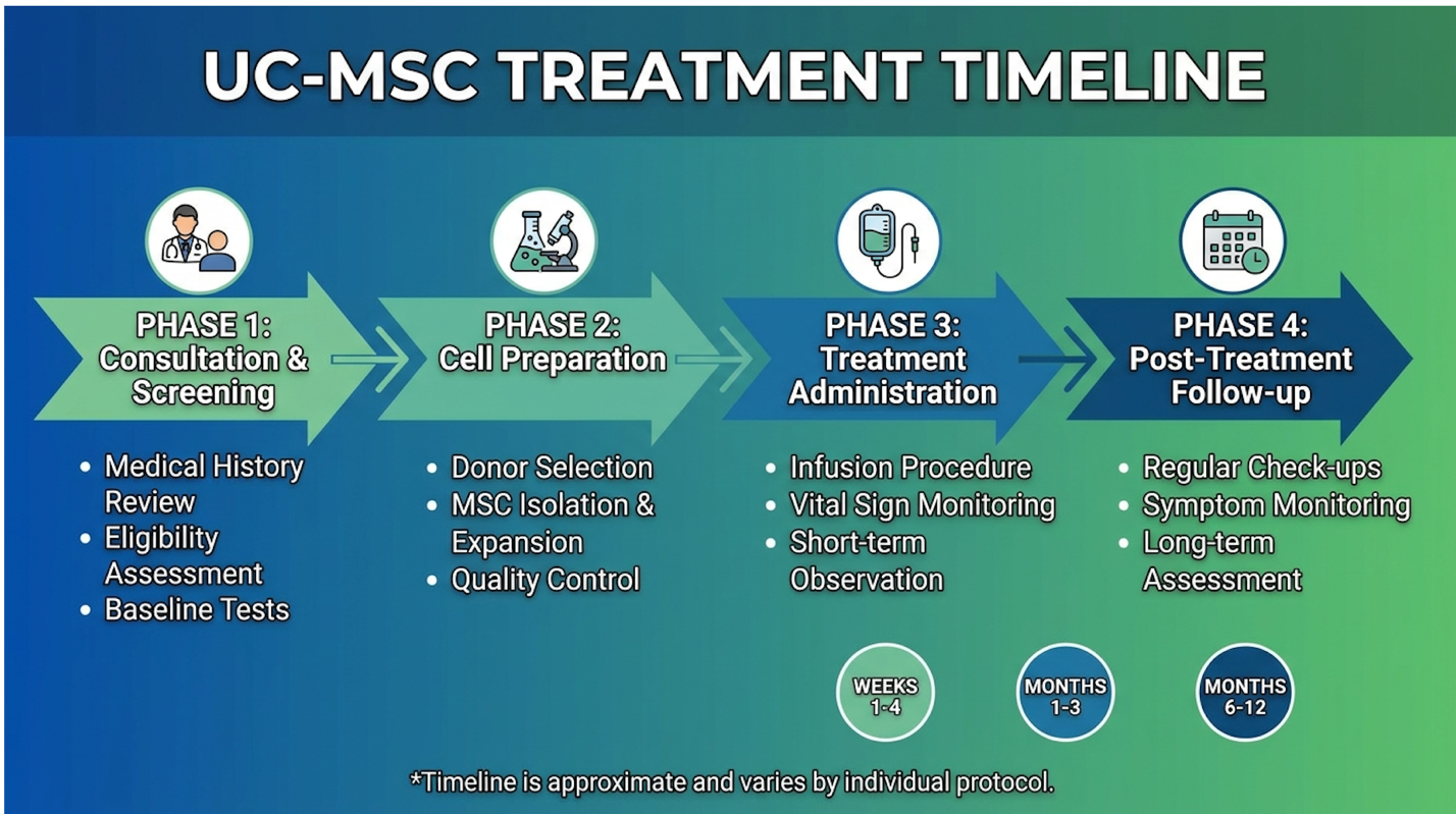
Furthermore, comprehensive UC-MSC therapy is an evidence-informed biological adjunct to standard therapies, not a magic replacement for them. Stem cells forcefully reduce the biological inflammation, but a perfectly clean diet, intensive gut support, and rigorous Applied Behavior Analysis (ABA) or targeted speech therapy are what actually teach the newly optimized brain how to function properly.
This is exactly why understanding the safety and regulations of stem cell therapy in Thailand or anywhere else must be thoroughly investigated before committing to care. Because of this inherent, widely documented clinical variability, extremely thorough suitability assessments and a clear, objective understanding of the treatment’s limitations are absolutely mandatory before proceeding.
UC-MSC stem cells fix neuroinflammation by secreting potent anti-inflammatory cytokines and targeted exosomes that effortlessly cross the blood-brain barrier. This specific process, widely known clinically as paracrine signaling, absolutely does not involve the stem cells physically becoming new brain tissue. Instead, these highly targeted secreted factors actively seek
Clinical trials indicate that approximately 60% to 70% of pediatric patients show measurable behavioral improvements following targeted UC-MSC therapy (Source: PubMed Central Meta-Analysis, 2020). These specific improvements are typically quantified utilizing heavily standardized tools like the Childhood Autism Rating Scale (CARS), explicitly showing substantial reductions in lethargy and severe repetitive behaviors. It is critical to clearly understand that “success” means massive functional improvement, not a complete, absolute reversal of the neurological diagnosis. Individual responses vary wildly based on the child’s exact underlying biological pathologies and strict adherence to concurrent behavioral therapies.
Yes, UC-MSCs are entirely ethically sourced directly from donated human umbilical cords strictly following a healthy, full-term birth. There is absolutely no clinical risk to the mother or the infant during the standard collection process, as the rich tissue would otherwise simply be discarded as standard medical waste. Furthermore, these primitive cells are distinctly different from highly controversial embryonic stem cells; they absolutely do not involve embryo destruction whatsoever. Strict regulatory frameworks govern the harvesting, screening, and immense expansion of these tissues in certified laboratories.
For families heavily navigating the immense complexities of the gut-brain axis and autism, UC-MSC therapy offers an incredibly powerful, evidence-informed approach to actively targeting central nervous system inflammation. Current clinical trials continually demonstrate that these cells are remarkably safe and effectively downregulate destructive pro-inflammatory cytokines like TNF-α (NIH, 2020). The absolute best clinical approach combines highly rigorous pre-
The Neuro-Immune Modulation Framework completely changes how we scientifically view severe ASD presentations. By understanding that behaviors are frequently exacerbated by a brain literally on fire driven by intense microglial activation and systemic triggers like intestinal permeability we can rationally and safely apply cellular therapies to completely shut down that destructive inflammatory loop. This profound shift moves the overall treatment approach entirely away from purely psychological management toward true, measurable biological intervention.
Ready to take the next step? Carefully compile your child’s comprehensive medical records, explicitly including recent complex blood panels, targeted GI testing, and past therapy reports. Actively seek a clinical suitability assessment directly from a qualified regenerative medicine specialist to scientifically determine if your child actually exhibits the exact inflammatory markers necessary to heavily benefit from this powerful intervention.