Our Services
Others
- DFPP
- Shockwave
- IV Drip


“This comprehensive literature review explores the anatomical architecture of the synovio-entheseal complex and its pathophysiological mechanisms as the primary epicenter of inflammation and bone remodeling in psoriatic arthritis”
Psoriatic arthritis is a chronic heterogeneous disease of the musculoskeletal system, with manifestations in multiple clinical domains, including synovitis of peripheral joints, axial spondyloarthropathy, dactylitis, and enthesitis. Although traditional diagnostic paradigms have emphasized intra articular synovial membrane inflammation as the major driver of joint disease, growing anatomical and imaging evidence points to the synovio entheseal complex as the primary target and initiating epicenter of musculoskeletal pathology in psoriatic arthritis. It is an anatomical unit distributing biomechanical loading through the fibrocartilaginous entheses, periosteal linings, adjacent bursae, fat pads, and adjacent synovium. This complex preserves joint homeostasis. However, repetitive mechanical loading leads to microdamage of avascular enthesal fibrocartilage in genetically predisposed individuals, activating stromal cells and resident fibroblasts. Mechanical strain results in activation of resident dendritic cells, macrophages, and innate immune populations to secrete interleukin 23. Local interleukin 23 signaling to resident unconventional lymphocytes (γδ T cells and type 3 innate lymphoid cells) initiates an interleukin 23 interleukin 17 cascade. Pro-inflammatory mediators originating from the entheses spill over into a hyper-vascularized synovial sublining provoking endothelial activation and neoangiogenesis. Primary inflammation at the enthesis thus leads to secondary synovitis, tenosynovitis, and joint swelling. Additionally, enthesal bone changes, with altered communication between osteoblasts and osteoclasts at the enthesal bone, lead to erosions of cortical bone and subsequent abnormal osteoproliferative bone repair (enthesophytes/syndesmophytes). This review discusses the current evidence for the synovio entheseal complex in the pathogenesis of psoriatic arthritis.
30% of psoriasis patients exhibit psoriatic arthritis-an inflammatory musculoskeletal disease(D’Ignazio et al., 2026). Phenotypes of psoriatic arthritis include peripheral polyarthritis, peripheral oligoarthritis, distal interphalangeal joint disease, axial spondyloarthritis, dactylitis, and enthesitis(Belasco & Wei, 2019; Sundanum, Orr, & Veale, 2023). Psoriatic arthritis has traditionally been classified as a group of primary synovial disease with rheumatoid arthritis. However, histopathology and imaging differentiate the two. In rheumatoid arthritis, it’s a hyperplasia of synovial membranes induced by autoantibodies, whereas in psoriatic arthritis, it’s an inflammation of entheses; neoangiogenesis in the sublining; and erosions with new bone formation(Tenazinha, Barros, Fonseca, & Vieira-Sousa, 2022).
As discussed in the previous review, genetic predisposition is critical, and HLA-C06:02 and HLA-B27 define clinical phenotypes and tissue tropisms. In skin psoriasis the epidermis in hyperplastic due to HLA-C*06:02, whereas mechanical stress points associate with musculoskeletal disease. Mechanical stress at sites of attachment primes tissue for interleukin 23 production and activates cells, as discussed in the preceding review. Imaging studies have further built on this to demonstrate that the musculoskeletal expression of psoriatic arthritis is not a synovial-only event. Instead, pathogenesis is related to underlying immune and stromal events at the synovio enthesial complex at sites of high mechanical stress(Dennis McGonagle, Abacar, & Kirkham, 2024; Yıldız & Aydıngöz, 2024).
The synovio entheseal complex model unifies entheses with contiguous synovial tissues and accounts for the presentation of enthesitis as joint swelling and dactylitis(Smerilli, Di Matteo, Cipolletta, Grassi, & Filippucci, 2021). Understanding the microanatomy, strain responses, cellular networks, and molecular signaling of the complex is important in disease initiation and the development of interceptive therapies(Kavanaugh, Helliwell, & Ritchlin, 2016). Here we review the synovio entheseal complex as the key target in psoriatic arthritis with reshaping in anatomy and biomechanics, stromal priming, immune networks, vascularization, and osteoimmunological consequences.
An enthesis is the site of attachment of tendons, ligaments, joint capsules, or fascia to bone. Histologically, entheses are divided into fibrocartilaginous and fibrous entheses. Fibrous entheses connect muscle tendons to metaphyseal surfaces of bone with a thick cortex. Fibrocartilaginous enthesis, however, attaches tendons and ligaments to sites of epiphyseal and apophyseal bone where the cortex is thinner. Psoriatic arthritis primarily involves inflammation at fibrocartilaginous entheses(Smerilli et al., 2021; Yıldız & Aydıngöz, 2024).
The classical views were replaced by the enthesis organ concept. The enthesis organ is made up of tissues that dissipate shear forces at the soft tissue-bone interface. The subunits include the insertion tendon, entheseal fibrocartilage, sesamoid fibrocartilage, periosteal fibrocartilage, fat pads, retroenthesis bursae, subchondral bone marrow, and adjacent synovial linings. The synovio entheseal complex is the coupling of avascular fibrocartilage with adjacent vascularized synovial tissue(D. McGonagle et al., 1998; Watad, Cuthbert, Amital, & McGonagle, 2018).
Articular and functional entheses are central components of the synovio entheseal complex. Functional entheses are located at friction surfaces (e.g., pulleys where tendons turn) and not on bone. Examples: flexor digitorum tendons at annular pulleys in fingers; extensor central slips at proximal interphalangeal joints; peroneus longus tendon at the cuboid trochlea. Fibrocartilaginous entheses also occur at synovial joints lined by fibrocartilage and hyaline articular cartilage (e.g., sacroiliac and symphysis pubis joints)(Benjamin & McGonagle, 2001).
Figure 1 illustrates the classic architecture of a fibrocartilaginous insertional synovio entheseal complex as a continuous biomechanical unit consisting of periosteal fibrocartilage, retrocalcaneal bursa, and adjacent synovial lining as a buffer of tensile and compressive forces. Typically, the underlying synovial subunit provides metabolic and lubricant substrates and applies immune surveillance of the fibrocartilage. But such proximity provides a route for diffusion of mechanical microdamage and local cytokine signals at the enthesis into the neighboring synovial cavity.
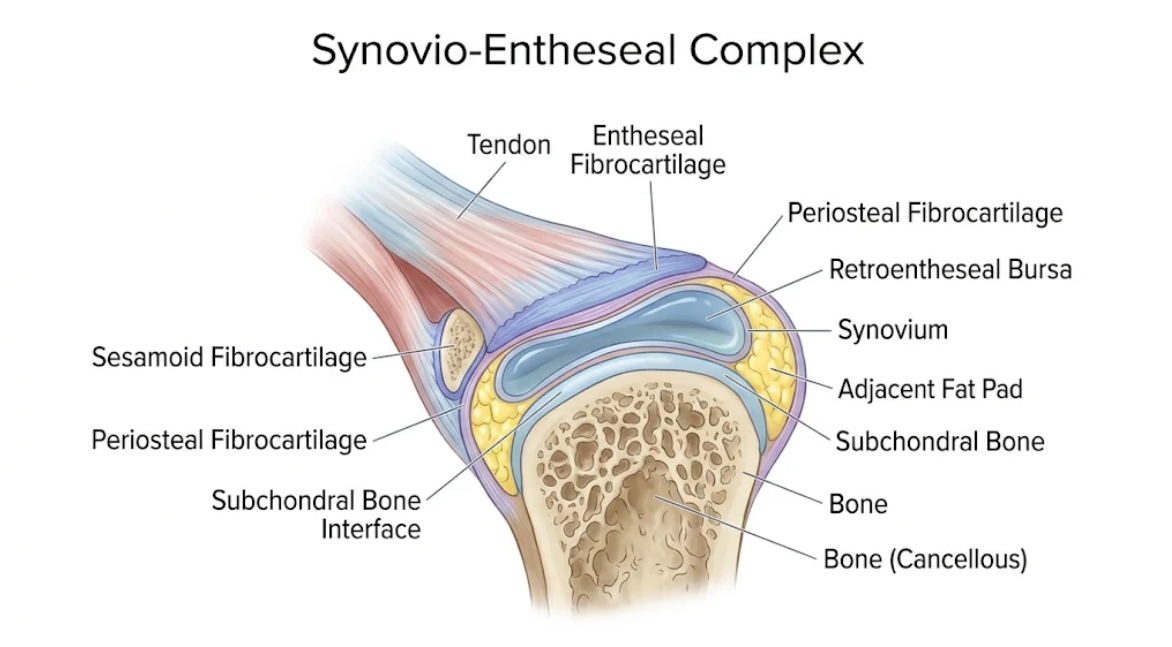
Figure 1. The Synovio-Entheseal Complex at the Tendon-Bone Interface. Anatomical diagram illustrating the integration of the tendon, associated fibrocartilages, synovium-lined bursa, and subchondral bone.
The enthesis organ as a stress transducer of muscle power on the skeleton. Fibrocartilaginous entheses are subjected to tensile pull, compression and shear friction on joint movement(Sundanum et al., 2023). Entheseal fibrocartilage is composed of a type II collagen- and aggrecan-rich matrix, which is resistant to deformation. However, repetitive mechanical loading and biomechanical stress induce microdamage at the insertion site of soft tissue to bone(van Kuijk & Tak, 2011).
Entheseal fibrocartilage is avascular and not as reparative as vascular tissues. Microdisruption of the extracellular matrix exposes matrix components and produces damage associated molecular patterns(Sun, Fu, Yan, & Dang, 2026). Enthesitis requires mechanical strain, as this has been experimentally demonstrated in animal models. In mouse models, mechanical unloading by tail suspension abrogates enthesitis and synovitis(Sobolewski, Walecka, & Dopytalska, 2017).
This mechanobiological susceptibility may explain the pattern of enthesitis in psoriatic arthritis. Hence the entheses of the lower extremities, which in particular are exposed to weight bearing impact (plantar fascia, Achilles tendon, patellar and quadriceps insertions), are a frequent site of enthesitis. Furthermore, dactylitis seems to be related to microdamage associated with biomechanical stress at some functional entheses, such as the pulleys of the flexor digitorium tendons in attempts to grip an object repetitively. As such, mechanical stress acts as a non-immune first hit, at which immune-mediated inflammation is facilitated at the synovio-entheseal complex(Abacar, Zabotti, Watad, & McGonagle, 2026; Corte et al., 2024).
Injury to the synovio entheseal complex causes reprogramming of stromal resident cells. Early joint pathology involves resident fibroblasts. Fibroblasts upregulate tissue remodeling markers such as fibroblast activation protein in response to strain and damage(Corte et al., 2026).
Early arthritis psoriatic arthralgia: pilot study on the uptake of fibroblast activation protein inhibitors in entheseal lesions by gallium-68 positron emission tomography. Fibroblast activation inhibitor uptake at stressed sites in a prospective cohort predicts increased risk of subsequent psoriatic arthritis. These imaging observations position mesenchymal activation upstream of leukocyte recruitment and sonographic destruction, raising the possibility that stromal cell priming is an early event in disease initiation(Corte et al., 2026; Corte et al., 2024).
Activated, primed fibroblasts secrete an altered secretome, including proinflammatory cytokines (e.g., interleukin 6), matrix metalloproteinases (e.g., MMP3, MMP13), and chemokines, which recruit circulating immune cells. Entheseal stromal fibroblasts also communicate with resident immune cells and endothelial cells. Early inflammation of the enthesis. Mechanical strain induces microdamage in fibrocartilage enthesis that leads to cascading early enthesis pathology, modulating local cytokine secretion by resident cells of synovium, and recruiting innate immune effector cells (Figure 2). Activated fibroblasts also regulate periosteal extracellular matrix remodeling(Croft et al., 2019).
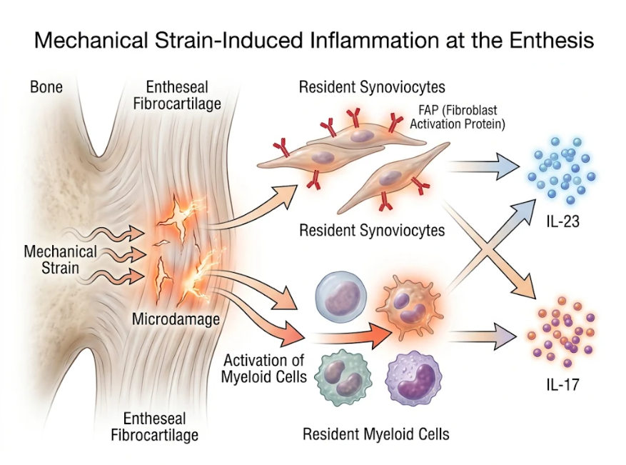
Figure 2. Mechanical Strain-Induced Inflammation at the Enthesis. Schematic illustrating how mechanical strain causes microdamage at the entheseal fibrocartilage. This process activates resident myeloid cells and FAP-expressing synoviocytes, culminating in the release of cytokines IL-23 and IL-17.
Importantly, the microenvironment of the human enthesis contains resident immune cells that can respond to stress and disruption. These include resident myeloid (dendritic) cells and macrophages, neutrophils, and classical CD4 and CD8 T cells and unconventional lymphocyte populations. Unconventional T cells (γδ T cells, mucosal associated invariant T cells, type 3 innate lymphoid cells) expressing interleukin 23 receptor are found at the enthesis(Reinhardt et al., 2016; Stavre et al., 2022).
Mechanical stress and stromal priming stimulate excess interleukin 23 production by resident macrophages and myeloid cells. Interleukin 23 associates with its receptors on resident entheseal lymphocytes and activates downstream signaling. This results in production of major effector cytokines (interleukin 17A, interleukin 17F, interleukin 22, tumor necrosis factor α). Importantly, enthesal γδ T cells secrete interleukin 17A in the absence of interleukin 23 receptor transcription and are therefore always primed for immune activation(Jacques et al., 2014; Reinhardt et al., 2016).
As described above, interleukin 17A and tumor necrosis factor alpha have synergistic effects in the recruitment of neutrophils and induction of pro inflammatory gene expression in synoviocytes. Increased interleukin 17A further promotes CD8+ T cell recruitment to the inflamed site. Several studies have demonstrated increased interleukin 17 producing CD8+ T cells in psoriatic joint tissue that correlate with disease activity score and acute phase reactants and, in some reports, with measures of erosive disease in imaging as confirmed by histological studies(Dennis McGonagle et al., 2024; Sun et al., 2026; Tenazinha et al., 2022).
The entheses fibrocartilage is avascular, but parts of the synovio entheseal complex (notably the adjacent synovial sublining and bursal membranes) are richly vascular. Activated entheseal stroma can release local cytokines and strongly upregulate pro angiogenic factors, such as vascular endothelial growth factor, basic fibroblast growth factor, and angiopoietin 2(van Kuijk & Tak, 2011).
These microvascular changes in the synovial sublining are more marked in psoriatic arthritis than in other inflammatory joint diseases. Under the microscope, the capillaries are dilated, with vascular contacts and the endothelium, protruding and in increased numbers. The endothelial cells are hypertrophied, vessels dilated, rough endoplasmic reticulum dilated, and numerous capillaries are seen. Endothelial cells overexpress the angiogenic downregulator micro-RNA 125a, which disrupts glycolysis and vessel formation(van Kuijk & Tak, 2011; Wade et al., 2019). Simultaneously, endothelial activation up-regulates expression of adhesion molecules at cellular membranes (i.e. intercellular adhesion molecule 1, vascular cell adhesion molecule 1, and E selectin) capable of tethering and extravasating circulating leukocytes in a transendothelial route(van Kuijk & Tak, 2011).
The enthesis is in continuity with the adjacent synovial cavity, facilitating rapid propagation of cytokine signals and activated cells from the enthesis to the vascularized synovial sublining of the synovio entheseal complex. Map of inflammation in the synovio entheseal complex Major cytokines diffuse, and microdamage at the fibrocartilaginous enthesis impinges on the synovium (Figure 3) resulting in secondary synovitis in the adjacent hypervascular synovial sublining. This propagation of signal results in secondary synovial lining hyperplasia, sublining leukocytic infiltration, joint effusion and peripheral synovitis. This explains the bizarre clinical picture of an articularly symptomatic and swollen joint disease that begins at focal sites of enthesitis(Abacar et al., 2026; Dennis McGonagle et al., 2024; Tenazinha et al., 2022).
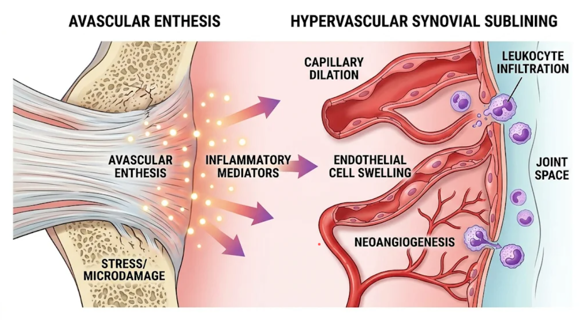
Figure 3. Propagation of Entheseal Inflammation to the Adjacent Synovium. Cross-sectional diagram illustrating how biomechanical stress at the avascular enthesis triggers the release of inflammatory mediators. These mediators induce vascular changes (capillary dilation, neoangiogenesis) and leukocyte infiltration within the hypervascular synovial sublining.
The dual osteoimmunological nature of psoriatic arthritis distinguishing it from rheumatoid arthritis and osteoarthritis is the hallmark of the disease in terms of entheseal cortical bone resorption and osteoproliferative new bone formation. That is the enthesis attached to bone and site of disrupted homeostasis of bone remodeling(Sundanum et al., 2023).
At the interface of inflamed synovium and bone marrow, osteoclasts are activated, leading to bone erosion at the synovio entheseal complex. Interleukin 17A synergizes with tumor necrosis factor to activate synoviocytes and stromal cells to upregulate immunoreactivity for receptor activator of nuclear factor kappa B ligand (RANKL) and downregulation of osteoprotegerin(Fitzgerald & Winchester, 2009). RANK-expressing osteoclast precursors in the circulation home to subchondral bone margins where they are induced by RANKL binding to differentiate into osteoclasts and cause focal bone resorption. In addition, dendritic cell specific transmembrane protein (DC-STAMP, which is upregulated in psoriatic bone marrow) enhances osteoclast fusion and erosive destruction(Belasco & Wei, 2019).
Conversely, the aberrant Wnt signaling pathway in osteoblasts and local interleukin 22 signaling favors new bone formation. Interleukin 22 induces osteoblast differentiation and mineralization of the periosteum to form enthesophytes at sites of insertion and syndesmophytes at axial ligaments. In addition, a transcriptome analysis of psoriatic synovium revealed local over-expression of WNT3A, bone morphogenetic protein receptor type II (BMPR2), and transforming growth factor beta receptor 1 (TGFβR1) that mediate endochondral ossification and matrix formation. BMPs (e.g., BMP4) drive periosteal appositional bone formation and account for radiographic fluffy periostitis and mouse ears deformities. It is the double bone remodeling in the osteoimmunological environment of the synovioentheseal complex(Belasco & Wei, 2019; Sundanum et al., 2023; Yıldız & Aydıngöz, 2024).
Anatomical, imaging, cellular, and molecular data point to the synovio entheseal complex as the site and the main target of disease in psoriatic arthritis. A view of psoriatic arthritis focused exclusively on hyperplasia of the intra articular synovial lining does not account for the clinical, sonographic, and osteoimmunological features of the disease. The enthesitis model proposes the enthesis organ and its synovial membrane as a single pathogenetic unit accounting for peripheral synovitis, dactylitis, axial spondyloarthritis, nail dystrophy, and bone remodeling.
Magnetic resonance imaging, at high resolution, and ultrasonography reveal subclinical entheseal inflammation in patients with psoriasis before overt clinical arthritis(Dennis McGonagle et al., 2024). Ultrasound demonstration of enthesitis with thickening, hypoechogenicity, power Doppler hypervascularity, and cortical bone erosions constitutes objective evidence of early disease involvement beyond the sensitivity of physical examination(Smerilli et al., 2021). Moreover, molecular imaging of 68Ga-labeled fibroblast activation protein inhibitor uptake identifies tissue resident mesenchymal cell activation prior to leukocyte influx and structural damage, providing a window for early disease interception(Corte et al., 2026).
The synovio entheseal complex paradigm has important clinical diagnostic and therapeutic implications. A delayed diagnosis (> 6 months) correlates with erosive joint disease, loss of function, and poor quality of life(Sundanum et al., 2023). Risk assessment at an early stage can be done in psoriatics with nonspecific joint pain by imaging of the key digital and lower extremity entheseal sites in the synovio entheseal complex. Therapeutically targeting biologic agents directed against either IL-23 or IL-17A or TNF-α may act on mediators of the cell and osteoimmunologic pathways of the synovio entheseal complex(Belasco & Wei, 2019; Sun et al., 2026). Although tumor necrosis factor inhibitors reduce synovial inflammation and osteoclasts, direct modulation of the interleukin 23 interleukin 17 axis via targeting entheseal resident lymphocyte activation resolves enthesitis, dactylitis, and cutaneous psoriasis(Sundanum et al., 2023). Ongoing studies of stromal immune interactions and tissue resident memory lymphocyte circuits in the entheseal microenvironment will refine interceptive therapies to stop the progression from psoriasis to psoriatic arthritis.
The synovio entheseal complex is the main anatomical unit and the major target of the pathogenesis of psoriatic arthritis. Pathological events occur at avascular fibrocartilaginous entheses, where mechanical shear stress leads to microdamage of the extracellular matrix. This mechanical disruption primes fibroblasts, induces fibroblast activation protein expression, and generates damage associated molecular patterns. Activated resident myeloid cells secrete interleukin 23 that acts as an immune amplifier by inducing the production of interleukin 17A, -22, and tumor necrosis factor alpha by the innate lymphoid cells (γ δ T cells and type 3 innate lymphoid cells) that reside at the enthesis. Pro-inflammatory mediators rapidly diffuse from the enthesis into the adjacent vascularized synovial sublining leading to endothelial swelling, neoangiogenesis, and subsequent clinical synovitis. At the same time, osteoimmunological crosstalk at the enthesis bone anchorage fuels parallel RANKL mediated bone erosion and interleukin 22 and Wnt mediated osteoproliferative new bone formation. The anatomical and site- and molecular bile of the synovio entheseal complex redefine psoriatic arthritis and rationalize the strategy of image diagnosis and therapeutic means at this stage to avoid irreversible structural joint destruction.
Abacar, K., Zabotti, A., Watad, A., & McGonagle, D. (2026). Does effective therapy of psoriasis prevent the development of psoriatic arthritis? EULAR Rheumatol Open, 2(2), 100121. doi:10.1016/j.ero.2026.01.005
Belasco, J., & Wei, N. (2019). Psoriatic Arthritis: What is Happening at the Joint? Rheumatology and Therapy, 6(3), 305-315. doi:10.1007/s40744-019-0159-1
Benjamin, M., & McGonagle, D. (2001). The anatomical basis for disease localisation in seronegative spondyloarthropathy at entheses and related sites. J Anat, 199(Pt 5), 503-526. doi:10.1046/j.1469-7580.2001.19950503.x
Corte, G., Atzinger, A., Noversa de Sousa, R., Mutlu, M. Y., Temiz, A., Bayat, S., . . . Fagni, F. (2026). Increased synovial and entheseal fibroblast activation detected by (68)Ga-FAPI-PET/CT is associated with the development of psoriatic arthritis in psoriasis patients with arthralgia. RMD Open, 12(1). doi:10.1136/rmdopen-2025-006567
Corte, G., Atzinger, A., Temiz, S. A., Noversa de Sousa, R., Mutlu, M. Y., Schoenau, V., . . . Fagni, F. (2024). Anatomical pattern of entheseal and synovial fibroblast activation in patients with psoriasis and its risk of developing psoriatic arthritis. RMD Open, 10(2). doi:10.1136/rmdopen-2024-004294
Croft, A. P., Campos, J., Jansen, K., Turner, J. D., Marshall, J., Attar, M., . . . Buckley, C. D. (2019). Distinct fibroblast subsets drive inflammation and damage in arthritis. Nature, 570(7760), 246-251. doi:10.1038/s41586-019-1263-7
D’Ignazio, E., Sahin, D., Baldi, C., Emery, P., McGonagle, D., Wakefield, R., . . . Di Matteo, A. (2026). The Role of Musculoskeletal Ultrasound in Psoriatic Arthritis: From Preclinical Detection to Treatment Monitoring. Curr Rheumatol Rep, 28(1). doi:10.1007/s11926-026-01225-z
Fitzgerald, O., & Winchester, R. (2009). Psoriatic arthritis: from pathogenesis to therapy. Arthritis Res Ther, 11(1), 214. doi:10.1186/ar2580
Jacques, P., Lambrecht, S., Verheugen, E., Pauwels, E., Kollias, G., Armaka, M., . . . Elewaut, D. (2014). Proof of concept: enthesitis and new bone formation in spondyloarthritis are driven by mechanical strain and stromal cells. Ann Rheum Dis, 73(2), 437-445. doi:10.1136/annrheumdis-2013-203643
Kavanaugh, A., Helliwell, P., & Ritchlin, C. T. (2016). Psoriatic Arthritis and Burden of Disease: Patient Perspectives from the Population-Based Multinational Assessment of Psoriasis and Psoriatic Arthritis (MAPP) Survey. Rheumatol Ther, 3(1), 91-102. doi:10.1007/s40744-016-0029-z
McGonagle, D., Abacar, K., & Kirkham, B. (2024). GRAPPA 2023 Debate: Is Psoriatic Disease Really a Primary Enthesitis That Drives Joint Synovitis? The Enthesitis Hypothesis 25 Years On. The Journal of Rheumatology, 51(Suppl 2), 101-105. doi:10.3899/jrheum.2024-0593
McGonagle, D., Gibbon, W., O’Connor, P., Green, M., Pease, C., & Emery, P. (1998). Characteristic magnetic resonance imaging entheseal changes of knee synovitis in spondylarthropathy. Arthritis Rheum, 41(4), 694-700. doi:10.1002/1529-0131(199804)41:4<694::Aid-art17>3.0.Co;2-#
Reinhardt, A., Yevsa, T., Worbs, T., Lienenklaus, S., Sandrock, I., Oberdörfer, L., . . . Prinz, I. (2016). Interleukin-23-Dependent γ/δ T Cells Produce Interleukin-17 and Accumulate in the Enthesis, Aortic Valve, and Ciliary Body in Mice. Arthritis Rheumatol, 68(10), 2476-2486. doi:10.1002/art.39732
Smerilli, G., Di Matteo, A., Cipolletta, E., Grassi, W., & Filippucci, E. (2021). Enthesitis in Psoriatic Arthritis, the Sonographic Perspective. Curr Rheumatol Rep, 23(9), 75. doi:10.1007/s11926-021-01039-1
Sobolewski, P., Walecka, I., & Dopytalska, K. (2017). Nail involvement in psoriatic arthritis. Reumatologia, 55(3), 131-135. doi:10.5114/reum.2017.68912
Stavre, Z., Bridgewood, C., Zhou, Q., Maeda, Y., Huang, T. T., Karman, J., . . . Gravallese, E. M. (2022). A role for neutrophils in early enthesitis in spondyloarthritis. Arthritis Res Ther, 24(1), 24. doi:10.1186/s13075-021-02693-7
Sun, Y., Fu, S., Yan, J., & Dang, N. (2026). Clinical, hematologic, and inflammatory factors associated with psoriatic arthritis in a Chinese hospital-based study. Front Immunol, 17, 1928389. doi:10.3389/fimmu.2026.1928389
Sundanum, S., Orr, C., & Veale, D. (2023). Targeted Therapies in Psoriatic Arthritis-An Update. Int J Mol Sci, 24(7). doi:10.3390/ijms24076384
Tenazinha, C., Barros, R., Fonseca, J. E., & Vieira-Sousa, E. (2022). Histopathology of Psoriatic Arthritis Synovium-A Narrative Review. Front Med (Lausanne), 9, 860813. doi:10.3389/fmed.2022.860813
van Kuijk, A. W., & Tak, P. P. (2011). Synovitis in psoriatic arthritis: immunohistochemistry, comparisons with rheumatoid arthritis, and effects of therapy. Curr Rheumatol Rep, 13(4), 353-359. doi:10.1007/s11926-011-0181-y
Wade, S. M., Ohnesorge, N., McLoughlin, H., Biniecka, M., Carter, S. P., Trenkman, M., . . . Fearon, U. (2019). Dysregulated miR-125a promotes angiogenesis through enhanced glycolysis. EBioMedicine, 47, 402-413. doi:10.1016/j.ebiom.2019.08.043
Watad, A., Cuthbert, R. J., Amital, H., & McGonagle, D. (2018). Enthesitis: Much More Than Focal Insertion Point Inflammation. Curr Rheumatol Rep, 20(7), 41. doi:10.1007/s11926-018-0751-3
Yıldız, A. E., & Aydıngöz, Ü. (2024). Pivotal role of the synovioentheseal complex in the imaging of arthritis and rheumatic diseases. Diagn Interv Radiol, 30(6), 409-418. doi:10.4274/dir.2024.242740