Our Services
Others
- DFPP
- Shockwave
- IV Drip


“This literature review summarizes recent postmortem, genetic, and neurophysiological findings to provide a multi-scale mechanistic exploration of how the imbalance between excitatory glutamatergic drive and inhibitory GABAergic control shapes the atypical neural circuits and diverse clinical symptoms of autism spectrum disorder.”
This literature review summarizes current scientific evidence for the neurobiological mechanisms of the Excitatory Inhibitory imbalance hypothesis in autism spectrum disorder. While structural templates are established in the prenatal environment, clinical symptoms are increasingly recognized to be driven by functional disruption at the synaptic level. The review recapitulates the principal findings regarding the two opposing forces: glutamate driven hyper excitation and gamma aminobutyric acid GABA mediated inhibitory deficits which upset the neurochemical balance. Specifically, it highlights documented structural alterations in prefrontal cortical layers including hyper dense excitatory synapses and a depletion of GABAergic interneurons. Moreover, the review describes the developmental delay of chloride transporter expression NKCC1 and KCC2 that impede the switch of GABA from excitatory to inhibitory neurotransmitter. It discusses how these microscopic synaptic failures propagate through neural circuits explaining how failing lateral inhibition and circuit desynchronization translate into sensory hypersensitivity social communication challenges and restricted repetitive behaviors. Finally, this review discusses how an understanding of these molecular pathways supports the move toward precision medicine and targeted treatments.
The human brain has been likened to a giant orchestra, with billions of neurons serving as individual instruments playing together in harmony. Two forces, excitation and inhibition, are finely balanced to tune the music of this neural symphony. Excitatory signals are the gas pedal, encouraging neurons to fire, while inhibitory signals are the brakes, tuning activity so that it doesn’t spiral into overwhelming chaos.(Wang et al., 2017) When this relationship is finely tuned, the brain easily processes sensory inputs, encodes memories and orchestrates social communication. But when it’s out of sync, the harmony is lost and the brain can’t make sense of the world.
In recent decades, researchers have turned to this equilibrium to gain insights into the biology of autism spectrum disorder (ASD). This question led to the Excitatory-Inhibitory (E/I) imbalance hypothesis, which proposes that the cardinal behavioral features of autism are caused by a change in this neurochemical ratio.(Rubenstein & Merzenich, 2003) More specifically, the hypothesis suggests that a relative increase in excitation or a decrease in inhibition or both results in hyperexcitable neural circuits and failure to suppress competing neural noise.(Casanova, Buxhoeveden, Switala, & Roy, 2002; Favorov & Kelly, 1994) Such instability propagates from microscopic synapses to macroscopic brain networks, influencing the perception and interaction of an individual with the environment.(Colomar et al., 2023; Madia, Sheikh, Pethe, Telange, & Agrawal, 2025)
To understand how this balance is perturbed in the postnatal brain, we must understand the developmental trajectory that precedes it. In our previous work we have described that early stages of embryonic development in autism spectrum disorder are characterized by atypical neural progenitor over-proliferation and altered neural migration, laying down a disorganized structural blueprint.(Beopoulos, Géa, Fasano, & Iris, 2022) However, the clinical symptoms are not merely the static consequence of this early template. These structural anomalies become apparent as active functional disturbances at the level of synaptic transmission during critical developmental windows as the brain matures. Here we continue our earlier reviews on the prenatal construction of the brain to the dynamic, postnatal neurochemical processes that regulate the day-to-day operation of the brain. In this literature review, we will review the synaptic mechanisms of glutamate-induced hyperexcitation and GABAergic inhibitory deficits to explore how molecular and cellular alterations disrupt the E/I scale. we explain how these microscopic synaptic failures cascade through neural circuits to create the sensory sensitivities, social communication deficits, and repetitive behaviors that characterize the clinical spectrum of autism.(Colomar et al., 2023) As illustrated in Figure 1, autism is characterized by a shift of the brain E/I balance toward hyper-excitability, as a consequence of the combined effect of excessive glutamatergic drive and reduced GABAergic control that is retained in normal development.
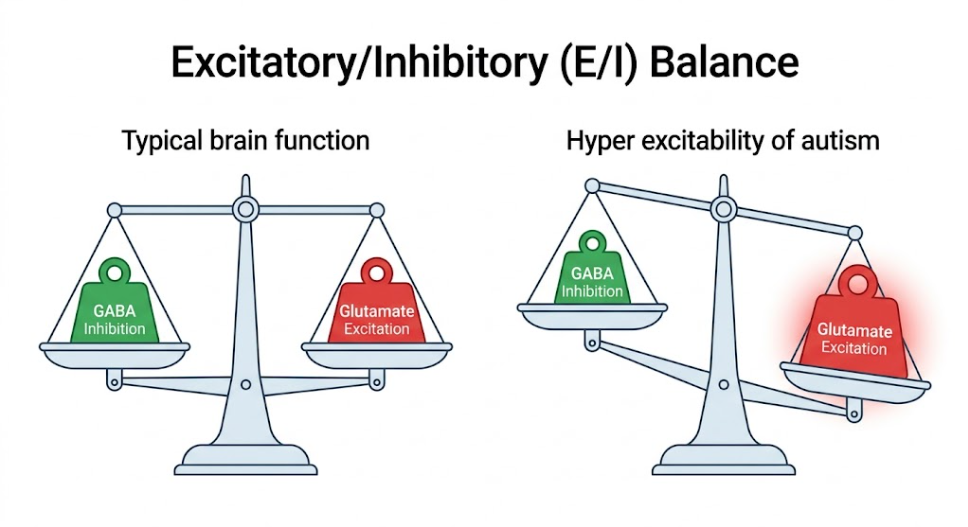
Figure 1. Excitatory/Inhibitory Imbalance. Demonstrating the shift toward hyperexcitability in autism compared to typical brain function.
To understand why the E/I ratio is important, we need to reviews the cellular physiology of the cerebral cortex. The neocortex is arranged in columns of neurons that are functionally similar. In such microcircuits, information transmission is based on two major classes of neurons: glutamatergic pyramidal neurons, which constitute about 80% of the cortical neurons and send long-distance excitatory signals, and GABAergic interneurons, which constitute the remaining 20% and provide local inhibitory control.(Wilson, Thompson, Rowse, & Freeth, 2023)
Excitatory and inhibitory synapses differ in structure and location. Glutamatergic synapses are asymmetric, and are located almost exclusively on dendritic spines, small protrusions along the dendrites of pyramidal cells.(Gao & Penzes, 2015; Vakilzadeh, Maseko, Bartely, McLennan, & Martínez-Cerdeño, 2024) Glutamate is released from the presynaptic terminals and binds to receptors embedded in the protein-rich, specialized postsynaptic density (PSD). The PSD is a large molecular scaffold that organizes neurotransmitter receptors, cell adhesion molecules and intracellular signaling enzymes for transduction efficiency.(Gao & Penzes, 2015; Vakilzadeh et al., 2024)
Conversely, inhibitory synapses are symmetric in structure, use gamma-aminobutyric acid (GABA) and are strategically located along the dendritic shafts, somata and axon initial segments of pyramidal neurons.(Hutsler & Zhang, 2010) Inhibitory interneurons are effective “gatekeepers” since they place inhibitory synapses in closer proximity to the cell body and the site of action potential generation.(Gao & Penzes, 2015) A neuron can receive thousands of excitatory signals via its far-flung dendrites, but just one well-timed inhibitory signal near the cell body can override them all and stop the neuron from firing. At the cellular level, E/I balance is homeostatic, with persistent excitation triggering mechanisms that downscale excitation or upscale inhibition in an effort to prevent neural activity from diverging from an optimal dynamic range.(Voineagu et al., 2011) Glutamate Hyper Excitation in Autism: Accelerator Glutamate is the main neurotransmitter for fast excitatory signaling. It is synthesized in the presynaptic terminals and loaded in vesicles by specialized vesicular glutamate transporters (VGlut1 and VGlut2).(Risher et al., 2014) When released, glutamate binds to ionotropic receptors (NMDA, AMPA and kainate) and to metabotropic receptors (mGluRs). AMPA receptors mediate fast currents, calcium permeable NMDA receptors are the substrate of synaptic plasticity, and metabotropic receptors modulate transmission through slower intracellular pathways.(Uzunova, Pallanti, & Hollander, 2016)
Excitotoxicity, which is toxic calcium overload and cell death caused by too much glutamate. Hence, glutamate has to be cleared fast from the synaptic cleft. This is mediated by the astrocytic high affinity transporters.(Alabdali et al., 2025) Then, the astrocytes scoop up the glutamate and transform it into a benign form, glutamine, via an enzyme called glutamine synthetase. Glutamine is then taken back up into the presynaptic neuron, where it is converted back to glutamate again and the glutamate-glutamine cycle is complete.(Nardi et al., 2023)
There is a lot of evidence that in autism this glutamatergic machinery is dysregulated creating a chronic hyper-excitation. Magnetic resonance spectroscopy (MRS) studies have indicated that there is an increase in the levels of glutamate and glutamine (Glx) in large cortical regions including the prefrontal and anterior cingulate cortexes.(R. G. Port, L. M. Oberman, & T. P. Roberts, 2019) This is supported by genetic studies which have identified mutations in the genes encoding the NMDA receptor subunits (e.g., GRIN2A and GRIN2B) that may result in abnormally long channel open times or impaired glutamate clearance.(Jung, Kim, Ko, & Um, 2023)
Structural evidence is important and comes from postmortem brain tissue analysis. Vakilzadeh et al. (2024) in a neuroanatomical study counted the number of excitatory synapses in the prefrontal cortex of autistic and control brains. presynaptic terminals immunolabelled for VGlut1 and postsynaptic sites immunolabelled for PSD95 (postsynaptic density protein 95).(Vakilzadeh et al., 2024) And out of the overlapping puncta came a striking discovery: a huge, multi-fold increase in the density of excitatory synapses in Layer 2 of the dorsolateral prefrontal cortex, specifically within Brodmann Area 9 (BA9) and Brodmann Area 47 (BA47), in autistic individuals. This local over-connectivity of structure forms the physical basis of hyper-excitation, structurally predisposing the prefrontal networks to over-respond to incoming inputs.(Vakilzadeh et al., 2024) The molecular architecture of these hyper-active excitatory synapses is shown in Figure 2. This illustrates the importance of VGlut1 in vesicle packaging, PSD95 scaffolding, and the excess receptor density driving the calcium mediated hyperexcitability.
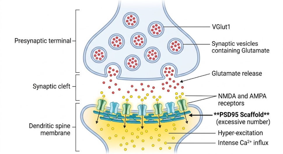
Figure 2. Synaptic Hyper-excitation Mechanism. Excess PSD95 scaffold proteins lead to an excess of NMDA and AMPA receptors. This structural defect causes a massive influx of Ca2+ ions and hyper-excitation following glutamate release.
Glutamate is the gas, gamma-aminobutyric acid (GABA) the main brake. GABA is synthesized from glutamate by the enzyme glutamic acid decarboxylase (isozymes GAD65 and GAD67) and stored in vesicles by the vesicular GABA transporter (VGAT). GABA modulates GABA release through ionotropic GABA-A (ligand-gated chloride channels) and metabotropic GABA-B receptors. GABA-A binding causes chloride to flow into the neuron, hyperpolarizing the membrane, and providing rapid inhibitory control.(Amina et al., 2021)
Precision of spatial and temporal GABAergic inhibition from local interneurons, particularly parvalbumin-positive (PV+) interneurons, is critical for accurate function. Basket cells synapsing on somata and chandelier cells synapsing on axon initial segments are PV+ interneurons.(Williams & Boksa, 2010) This strategic location confers powerful feedback inhibition that synchronizes firing to generate gamma-band oscillations (30-100 Hz) critical for cognitive flexibility. (Williams & Boksa, 2010)
In autism, this braking system is impaired at many levels. In postmortem studies, significant reductions in GAD65 and GAD67 were observed in the parietal cortex, cerebellum, and prefrontal regions. Moreover, there is a general downregulation of GABA-A receptor subunits (e.g. GABRB3, GABRA5) in vulnerable chromosomal regions.(Madia et al., 2025; Ren et al., 2015)
Vakilzadeh et al. (2024) labeled functional inhibitory synapses in postmortem tissue using VGAT and gephyrin (the scaffolding protein for GABA-A receptors at the postsynaptic membrane), and found a dramatic threefold reduction in the number of inhibitory synapses in both Layer 2 and Layer 5 in the prefrontal regions BA9, BA46 and BA47. This total collapse of the inhibitory infrastructure demonstrates that there is no physical wiring in the prefrontal cortex for proper braking.(Vakilzadeh et al., 2024)
Structurally the defect is visible in the living individual physiologically. Transcranial magnetic stimulation (TMS) studies of individuals with autism have assessed short-interval intracortical inhibition (SICI), a direct measure of GABA-A receptor function, and reported significant reductions in SICI in this group.(Masuda et al., 2019) EEG and MEG studies also consistently show reduced gamma-band oscillations to sensory stimuli, providing system-level evidence for a failing GABAergic brake.(R. G. Port, L. M. Oberman, & T. P. L. Roberts, 2019) The structural anatomy of a failing inhibitory synapse can be visualized, as illustrated in the Figure 3, showing dramatic reductions in VGAT-labeled presynaptic vesicles and gephyrin postsynaptic scaffolding leading to a failure of chloride-mediated membrane hyperpolarization.
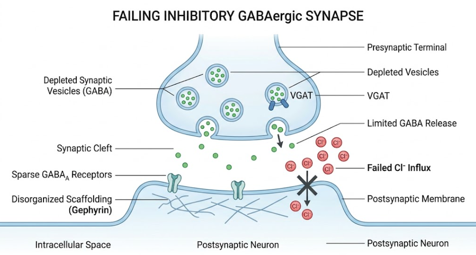
Figure 3. Impaired GABAergic Transmission. The presynaptic terminal exhibits severe depletion of GABA vesicles (VGAT). In Postsynaptic, disorganized Gephyrin scaffolding reduces GABA-A receptor density, resulting in a failed influx of negative chloride ions (Cl⁻).
To understand the occurrence of GABAergic dysfunction, one has to consider a very sensitive developmental mechanism: the developmental switch of GABA polarity. GABA functions as an excitatory neurotransmitter in the embryonic and early postnatal brain, stimulating neural stem cell proliferation, migration, and initial synapse formation.(Masuda et al., 2019) GABA can be either excitatory or inhibitory depending on the intracellular chloride concentration, which is regulated by two membrane cotransporters: Na-K-Cl cotransporter 1 (NKCC1, chloride importer) and K-Cl cotransporter 2 (KCC2, chloride exporter). In early development, immature neurons express high levels of NKCC1 and virtually no KCC2.(Ben-Ari, 2014; Fukuda et al., 1998) Intracellular chloride is high. GABA-A receptors cause chloride to move out of the cell, depolarizing the membrane and making it more likely that the neuron will fire. As the brain matures, NKCC1 is turned down and KCC2 is turned up, decreasing intracellular chloride. Hence GABA binding causes an influx of chloride into the cell, hyperpolarizing the membrane and resulting in an inhibitory effect.(Masuda et al., 2019)
In autism this critical developmental switch is delayed or impaired. In animal models, NKCC1 is not consistently downregulated and KCC2 is not upregulated properly, resulting in abnormal intracellular chloride concentration in neurons. This delay in chloride shift renders GABA weakly inhibitory or paradoxically excitatory during critical early windows.(Masuda et al., 2019) The mechanism provides an elegant explanation for why some children with autism sometimes react paradoxically to benzodiazepines (which usually enhance GABAergic inhibition) but can also cause severe agitation and hyperarousal in some autistic children. If the underlying chloride cotransporters are dysregulated and intracellular chloride is still high, then increasing the opening of GABA-A receptors will increase the outward depolarizing chloride current, hyperactivating neural circuits instead of calming them down.(Fukuda et al., 1998) The understanding of this chloride homeodynamics theory has opened up exciting therapeutic avenues. Bumetanide is a loop diuretic and a selective NKCC1 antagonist. Bumetanide blocks NKCC1, thus reducing intracellular chloride, and allowing GABA to exert its normal inhibitory effect. In clinical trials, Bumetanide has shown promise in children with autism with promising improvements in social communication and sensory processing, highlighting the important clinical relevance of investigating the fine biophysical processes regulating E/I balance.(Schulte, Wierenga, & Bruining, 2018)
How can such a microscopic imbalance of glutamate and GABA explain the rich and complex clinical phenotype of autism? To answer this question, we need to understand how E/I dysregulation changes the way information is processed in different brain regions and cognitive networks.(Hollestein et al., 2023)
Autism is a neurodevelopmental disorder that features impairments in social communication and interaction and restricted repetitive behaviors and interests.(Canitano & Palumbi, 2021) Atypical sensory processing is also a very common feature with more than 90% of autistic individuals affected. These symptoms can be interpreted as the compensatory or dysfunctional responses of neural networks with an impaired E/I ratio.(Johnson, Jones, & Gliga, 2015) Imagine a sensory hyper-sensitivity. In a healthy sensory cortex, local GABAergic interneurons exert strong lateral inhibition, sharpening the brain’s response to a particular sensory input and dampening irrelevant inputs in the surrounding area.(Casanova et al., 2002) Lateral inhibition fails when cortical inhibition is compromised. The sensory cortex is hyper-responsive and the brain is flooded with unrefined, un-gated sensory inputs. An ordinary environmental sound becomes a thunderous roar, a fluorescent light becomes a strobing beacon, and the soft texture of clothing becomes sandpaper.
This constant sensory overload creates a cascading effect on social communication. Social interactions are highly complex, rapidly changing and unpredictable, so the brain must process a huge stream of visual, auditory, and emotional cues in real-time.(Rubenstein & Merzenich, 2003) When sensory gating goes awry, the brain is already functioning at its limit of processing capacity and the very dynamic social world becomes overwhelming and exhausting. From an evolutionary perspective, the social withdrawal and avoidance characteristic of autistic people can be an active, protective strategy to escape highly complex and unpredictable environments and find refuge in quiet, predictable spaces.(Colomar et al., 2023) Restricted and repetitive behaviors can also be seen as adaptive mechanisms to cope with an unpredictable environment. The insistence on sameness and repetitive movements can provide very predictable structured sensory input. Alternatively, a brain with hyperexcitable neural circuits and high background noise might employ self-stimulatory behaviors as an active strategy to make order out of sensory chaos, dampening the distress of an unpredictable world.(Tyzio et al., 2014)
The findings summarized in this review suggest a major conceptual shift in our understanding of the pathophysiology of autism spectrum disorder. The E/I imbalance model of autism views autism as a dynamic, functional state of neural circuitry rather than as merely static structural anomalies that arise during early gestation.(Madia et al., 2025)
First, the spatial specificity of the synaptic changes has to be carefully taken into account. Post-mortem studies have shown layer-dependent changes in the prefrontal cortex. Instead we see hyper-dense excitatory connections in Layer 2 clustered with a widespread collapse of inhibitory synapses in Layers 2 and 5.(Vakilzadeh et al., 2024) This layer-specific pathology indicates a preferential disruption of cortical feed-forward processing and corticocortical communication that critically relies on these superficial and deep layers. The prefrontal cortex is engaged in executive function, social cognition, and working memory, all of which depend on highly coordinated, timed interactions between cortical layers. This selective loss of inhibitory control in these layers correlates directly with the executive and social difficulties observed clinically.(Vakilzadeh et al., 2024)
Second, reported genetic correlations in Hollestein et al. (2023) are helpful to disambiguate sources of specific symptom clusters. Our results suggest that ASD is not a single unique disorder, but that common differences in genes for glutamate are associated with particular social communication impairments, and GABA genes with sensory processing abnormalities. Or it might be a mixture of different and overlapping neurobiological dimensions.(Hollestein et al., 2023) This dimensional view is consistent with the clinical reality of the autism spectrum, where individuals have highly variable combinations of social, sensory, and behavioral phenotypes.(Colomar et al., 2023) Thus, therapeutic approaches targeting either glutamatergic or GABAergic system may not be equally effective in the treatment of all symptoms. Or they may respond to a glutamate modulator for social problems or a GABAergic agent for sensory overload.(Hollestein et al., 2023)
Third, we have to address the time dynamics of the E/I imbalance. The E/I ratio, far from being a fixed quantity, is a very fluid parameter that varies across the lifespan. The developmental switch in GABA polarity demonstrates how early-life disturbances in chloride transporters can trigger a cascade of developmental detours.(Masuda et al., 2019) If GABA stays excitatory during infancy for too long, this could potentially interfere with the critical period of synaptic pruning, resulting in the structural overconnectivity observed in adults.(Canitano & Palumbi, 2021) This sensitivity implies that the timing of therapeutic intervention is critically important. Treating a young infant or child with bumetanide to achieve correct chloride homeodynamics may reset brain development onto a normal course permanently. Treating an adult with the same drug may only provide temporary symptomatic relief.(R. G. Port et al., 2019)
However, the E/I hypothesis has its own limitations and controversies. One of the main criticisms is whether the E/I imbalance is the primary cause of autism or a secondary, compensatory response of a brain trying to compensate for other genetic or environmental insults. One of the greatest challenges in neurodevelopmental research today is to distinguish primary pathological changes from secondary adaptive responses.(Colomar et al., 2023; R. G. Port et al., 2019; Vakilzadeh et al., 2024)
To summarize, the Excitatory-Inhibitory imbalance hypothesis offers a consistent molecular-to-behavioral framework for the understanding of autism spectrum disorder. The persistent hyper-excitation caused by increased glutamatergic signaling, together with the structural and functional impairments of GABAergic braking, interfere with the brain’s capacity to filter and organize information. This failure of sensory gating and circuit synchronization reverberates outwards leading to sensory hypersensitivity, social-communication difficulties and repetitive behaviors. Moreover, the delayed developmental switch of GABA from excitatory to inhibitory, driven by dysregulated chloride cotransporters, suggests a critical developmental vulnerability that shapes postnatal connectivity. To go beyond behavior therapies and toward precision medicine, the detailed synaptic and molecular mechanisms must be understood. By identifying the unique biological subtypes of the autism spectrum, clinicians can tailor interventions—from glutamate antagonists and selective GABA modulators to targeted brain stimulation—to restore the delicate synaptic symphony, ultimately reducing distress and helping individuals with autism to navigate their world with greater ease and clarity.
Alabdali, A., Ben Bacha, A., Alonazi, M., Al-Ayadhi, L. Y., Alanazi, A. S. J., & El-Ansary, A. (2025). Comparative evaluation of certain biomarkers emphasizing abnormal GABA inhibitory effect and glutamate excitotoxicity in autism spectrum disorders. Front Psychiatry, 16, 1562631. doi:10.3389/fpsyt.2025.1562631
Amina, S., Falcone, C., Hong, T., Wolf-Ochoa, M. W., Vakilzadeh, G., Allen, E., . . . Martínez-Cerdeño, V. (2021). Chandelier Cartridge Density Is Reduced in the Prefrontal Cortex in Autism. Cereb Cortex, 31(6), 2944-2951. doi:10.1093/cercor/bhaa402
Ben-Ari, Y. (2014). The GABA excitatory/inhibitory developmental sequence: a personal journey. Neuroscience, 279, 187-219. doi:10.1016/j.neuroscience.2014.08.001
Beopoulos, A., Géa, M., Fasano, A., & Iris, F. (2022). Autism spectrum disorders pathogenesis: Toward a comprehensive model based on neuroanatomic and neurodevelopment considerations. Frontiers in Neuroscience, Volume 16 – 2022. doi:10.3389/fnins.2022.988735
Canitano, R., & Palumbi, R. (2021). Excitation/Inhibition Modulators in Autism Spectrum Disorder: Current Clinical Research. Front Neurosci, 15, 753274. doi:10.3389/fnins.2021.753274
Casanova, M. F., Buxhoeveden, D. P., Switala, A. E., & Roy, E. (2002). Minicolumnar pathology in autism. Neurology, 58(3), 428-432. doi:10.1212/wnl.58.3.428
Colomar, L., San José Cáceres, A., Álvarez-Linera, J., González-Peñas, J., Huertas Patón, A., Martín de Blas, D., . . . Parellada, M. (2023). Role of cortical excitatory/inhibitory imbalance in autism spectrum disorders from a symptom severity trajectories framework: a study protocol. BMC Psychiatry, 23(1), 213. doi:10.1186/s12888-023-04695-y
Favorov, O. V., & Kelly, D. G. (1994). Minicolumnar organization within somatosensory cortical segregates: I. Development of afferent connections. Cereb Cortex, 4(4), 408-427. doi:10.1093/cercor/4.4.408
Fukuda, A., Muramatsu, K., Okabe, A., Shimano, Y., Hida, H., Fujimoto, I., & Nishino, H. (1998). Changes in intracellular Ca2+ induced by GABAA receptor activation and reduction in Cl- gradient in neonatal rat neocortex. J Neurophysiol, 79(1), 439-446. doi:10.1152/jn.1998.79.1.439
Gao, R., & Penzes, P. (2015). Common mechanisms of excitatory and inhibitory imbalance in schizophrenia and autism spectrum disorders. Curr Mol Med, 15(2), 146-167. doi:10.2174/1566524015666150303003028
Hollestein, V., Poelmans, G., Forde, N. J., Beckmann, C. F., Ecker, C., Mann, C., . . . Naaijen, J. (2023). Excitatory/inhibitory imbalance in autism: the role of glutamate and GABA gene-sets in symptoms and cortical brain structure. Transl Psychiatry, 13(1), 18. doi:10.1038/s41398-023-02317-5
Hutsler, J. J., & Zhang, H. (2010). Increased dendritic spine densities on cortical projection neurons in autism spectrum disorders. Brain Res, 1309, 83-94. doi:10.1016/j.brainres.2009.09.120
Johnson, M. H., Jones, E. J., & Gliga, T. (2015). Brain adaptation and alternative developmental trajectories. Dev Psychopathol, 27(2), 425-442. doi:10.1017/s0954579415000073
Jung, H., Kim, S., Ko, J., & Um, J. W. (2023). Intracellular signaling mechanisms that shape postsynaptic GABAergic synapses. Curr Opin Neurobiol, 81, 102728. doi:10.1016/j.conb.2023.102728
Madia, D., Sheikh, M., Pethe, A., Telange, D., & Agrawal, S. (2025). Excitatory/Inhibitory balance in autism spectrum disorders: Integrating genetic, neurotransmitter and computational perspectives. AIMS Neurosci, 12(4), 635-675. doi:10.3934/Neuroscience.2025031
Masuda, F., Nakajima, S., Miyazaki, T., Yoshida, K., Tsugawa, S., Wada, M., . . . Noda, Y. (2019). Motor cortex excitability and inhibitory imbalance in autism spectrum disorder assessed with transcranial magnetic stimulation: a systematic review. Transl Psychiatry, 9(1), 110. doi:10.1038/s41398-019-0444-3
Nardi, L., Chhabra, S., Leukel, P., Krueger-Burg, D., Sommer, C. J., & Schmeisser, M. J. (2023). Neuroanatomical changes of ionotropic glutamatergic and GABAergic receptor densities in male mice modeling idiopathic and syndromic autism spectrum disorder. Front Psychiatry, 14, 1199097. doi:10.3389/fpsyt.2023.1199097
Port, R. G., Oberman, L. M., & Roberts, T. P. (2019). Revisiting the excitation/inhibition imbalance hypothesis of ASD through a clinical lens. Br J Radiol, 92(1101), 20180944. doi:10.1259/bjr.20180944
Port, R. G., Oberman, L. M., & Roberts, T. P. L. (2019). Revisiting the excitation/inhibition imbalance hypothesis of ASD through a clinical lens. British Journal of Radiology, 92(1101), 20180944. doi:10.1259/bjr.20180944
Ren, Z., Sahir, N., Murakami, S., Luellen, B. A., Earnheart, J. C., Lal, R., . . . Luscher, B. (2015). Defects in dendrite and spine maturation and synaptogenesis associated with an anxious-depressive-like phenotype of GABAA receptor-deficient mice. Neuropharmacology, 88, 171-179. doi:10.1016/j.neuropharm.2014.07.019
Risher, W. C., Patel, S., Kim, I. H., Uezu, A., Bhagat, S., Wilton, D. K., . . . Eroglu, C. (2014). Astrocytes refine cortical connectivity at dendritic spines. Elife, 3. doi:10.7554/eLife.04047
Rubenstein, J. L., & Merzenich, M. M. (2003). Model of autism: increased ratio of excitation/inhibition in key neural systems. Genes Brain Behav, 2(5), 255-267. doi:10.1034/j.1601-183x.2003.00037.x
Schulte, J. T., Wierenga, C. J., & Bruining, H. (2018). Chloride transporters and GABA polarity in developmental, neurological and psychiatric conditions. Neurosci Biobehav Rev, 90, 260-271. doi:10.1016/j.neubiorev.2018.05.001
Tyzio, R., Nardou, R., Ferrari, D. C., Tsintsadze, T., Shahrokhi, A., Eftekhari, S., . . . Ben-Ari, Y. (2014). Oxytocin-Mediated GABA Inhibition During Delivery Attenuates Autism Pathogenesis in Rodent Offspring. Science, 343(6171), 675-679. doi:doi:10.1126/science.1247190
Uzunova, G., Pallanti, S., & Hollander, E. (2016). Excitatory/inhibitory imbalance in autism spectrum disorders: Implications for interventions and therapeutics. World J Biol Psychiatry, 17(3), 174-186. doi:10.3109/15622975.2015.1085597
Vakilzadeh, G., Maseko, B. C., Bartely, T. D., McLennan, Y. A., & Martínez-Cerdeño, V. (2024). Increased number of excitatory synapsis and decreased number of inhibitory synapsis in the prefrontal cortex in autism. Cereb Cortex, 34(13), 121-128. doi:10.1093/cercor/bhad268
Voineagu, I., Wang, X., Johnston, P., Lowe, J. K., Tian, Y., Horvath, S., . . . Geschwind, D. H. (2011). Transcriptomic analysis of autistic brain reveals convergent molecular pathology. Nature, 474(7351), 380-384. doi:10.1038/nature10110
Wang, J., Fu, K., Chen, L., Duan, X., Guo, X., Chen, H., . . . Chen, H. (2017). Increased Gray Matter Volume and Resting-State Functional Connectivity in Somatosensory Cortex and their Relationship with Autistic Symptoms in Young Boys with Autism Spectrum Disorder. Front Physiol, 8, 588. doi:10.3389/fphys.2017.00588
Williams, S., & Boksa, P. (2010). Gamma oscillations and schizophrenia. J Psychiatry Neurosci, 35(2), 75-77. doi:10.1503/jpn.100021
Wilson, R. B., Thompson, A. R., Rowse, G., & Freeth, M. (2023). The experience of seeking, receiving, and reflecting upon a diagnosis of autism in the UK: A meta-synthesis of qualitative studies conducted with autistic individuals. Research in Autism Spectrum Disorders, 103, 102135. doi:https://doi.org/10.1016/j.rasd.2023.102135