Our Services
Others
- DFPP
- Shockwave
- IV Drip


“This literature review focuses on the deep cellular and molecular mechanisms that lead to the alteration in the cavernosal smooth muscle-to-collagen ratio and its contribution to penile structural failure.”
The predominant organic etiology of erectile dysfunction (ED) is corporal veno-occlusive dysfunction (CVOD), which is failure of the penile hemodynamic storage mechanism to trap outflowing blood. The clinical paradigm traditionally considers CVOD as a macroscopic venous leak, but recent studies show that it is mainly due to microscopic cellular and molecular lesions in the corpus cavernosum. In particular, the changing ratio of cavernosal smooth muscle to collagen provides the defining histopathological basis for veno-occlusive failure. Under physiological conditions, healthy contractile cavernosal smooth muscle cells (CSMCs) provide the compliance and volumetric expansion required for passive compression of the subtunical venules against the rigid tunica albuginea. Pathological processes including aging, diabetes, chronic ischemia and cavernosal nerve injury set in motion a cellular cascade involving loss of smooth muscle cells, programmed cell death (apoptosis) and phenotypic switching. Contractile CSMCs transdifferentiate to a synthetic, proliferative phenotype, downregulating markers of contractility (e.g., alpha-smooth muscle actin and calponin-1) and upregulating synthetic markers (e.g., osteopontin and vimentin). This synthetic switch leads to aberrant remodeling of the extracellular matrix, with excessive deposition of rigid Type I collagen at the expense of compliant Type III collagen. This literature review provides an in-depth analysis of the cellular transition, how it disrupts tissue compliance, increases cavernosal stiffness, and leads to irreversible structural failure of the penis. The review also highlights new therapeutic pathways to restore the smooth muscle-to-collagen equilibrium.
Erectile dysfunction (ED) is a major health care problem worldwide. It is estimated that the prevalence of ED is 16% in Western countries and 27% to 68% in Asian countries. The number of men affected with ED worldwide is expected to increase to over 322 million by 2025.(Hu et al., 2019) Penile erection is a neurovasculogenic mechanical event requiring a precise balance between blood inflow and limited outflow of blood. The physiological process of penile erection begins with the sexual stimuli that activate the parasympathetic nervous system. This activation causes the release of nitric oxide (NO) from the nonadrenergic, noncholinergic (NANC) cavernous nerve terminals and endothelial cells by the neuronal and endothelial nitric oxide synthases (nNOS and eNOS). Nitric oxide diffuses into the adjacent cavernosal smooth muscle cells (CSMC) to stimulate soluble guanylyl cyclase, increasing cyclic guanosine monophosphate (cGMP) levels.(Wang et al., 2025) This causes a decrease in intracellular calcium and smooth muscle relaxation. The rapid influx of arterial blood which results dilates the sinusoidal lacunar spaces, engorging the corpus cavernosum. This expansion passively wedges the outer trabeculae against the rigid tunica albuginea, compressing the subtunical venular plexus and reducing venous outflow to nearly zero. This mechanical trapping of the blood is the penile veno-occlusive mechanism.(Wang et al., 2025)
When this mechanism is damaged, the penis cannot maintain a sufficient rigidity for intercourse, a condition known as Corporal Veno-Occlusive Dysfunction (CVOD), or venogenic “venous leakage”. In our prior work on biomechanical failures of the penile hemodynamic storage system, we showed that veno-occlusion failure at a macroscopic level is not a fundamental anatomic abnormality of the exit veins per se, but rather a consequence of structural breakdown within the cavernosal spongy tissue and its surrounding fibrous sheath. In our previous paper we described the macro-physics of venous leakage, including longitudinal vessel stretching, orthogonal luminal narrowing, and sympathetic hyper-activity, but it also suggested significant cellular and molecular damage within the corpus cavernosum.(Ferrini, Gonzalez-Cadavid, & Rajfer, 2017) The present review extends the hemodynamic principles presented in our previous paper to a detailed analysis of the cellular and molecular dynamics of the change in the cavernosal smooth muscle to collagen ratio. In this paper, we specifically outline the mechanisms of cavernosal smooth muscle depletion, the apoptotic signaling cascades, the cellular phenomenon of contractile-to-synthetic phenotypic switching, and the subsequent pathological remodeling of the extracellular matrix (ECM). By investigating these microscopic transitions, we elucidate how sub-microscopic biochemical and phenotypic alterations give rise to tissue stiffness, non-compliance and ultimately to the failure of the penile veno-occlusive mechanism.(Qian et al., 2020)
The erectile tissue of the corpora cavernosa is composed of a specialized vascular network of intercommunicating sinusoidal spaces lined by endothelial cells and supported by a trabecular network of smooth muscle and connective tissue.(El-Sakka & Yassin, 2010) Under physiological, healthy conditions, the cellular and structural composition of this trabecular meshwork is tightly regulated to optimize compliance and tensile strength. Quantitative histomorphometric analysis indicates that the corpus cavernosum in normal young conditions is composed of about 40% to 52% cavernosal smooth muscle cells (CSMCs) and the remaining part is extracellular matrix, with a balanced network of Type I, Type III, and Type IV collagen fibers interspersed with elastic fibers.(Wespes et al., 1997) Cavernosal compliance, the ability of the spongy tissue to stretch and accommodate blood at low pressures during the early phases of tumescence, is mainly determined by the relative abundance of CSMCs. Active relaxation of this rich smooth muscle mass is required to favor expansion of the sinusoidal spaces. As these spaces fill in and expand, they produce the outward radial pressure needed to squeeze the thin-walled subtunical venules against the rigid, bi-layered collagenous textile of the tunica albuginea.(Ferrini et al., 2017)
Histopathological investigation with Masson’s trichrome is a classical method to identify and quantify these structural elements. Contractile smooth muscle fibers are purple or red and extracellular collagen fibers are blue. The staining revealed dense and very well-organized arrangement of healthy smooth muscle bundles in young and potent individuals, with thin and undulating blue collagen fibers providing structural support without restricting elasticity.(Qian et al., 2020) However, this microarchitectural equilibrium is severely disrupted in patients with CVOD. The proportion of smooth muscle decreases sharply: from 19% up to 36% in elderly patients with CVOD, and to 10% to 25% in patients with severe impotence.(El-Sakka & Yassin, 2010) This cell loss is accompanied by a reciprocal massive deposition of disorganized collagen fibers, a profound shift in the native smooth muscle-to-collagen ratio.(Ferrini et al., 2009) In the absence of functional, contractile smooth muscle, the cavernosal tissue is rendered non-yielding and rigid. The other smooth muscle mass fails to relax sufficiently to the sexual stimulation and the incoming blood is not able to produce the volumetric expansion to compress the subtunical venules, which results in persistent venous leakage.(Sattar, Salpigides, Vanderhaeghen, Schulman, & Wespes, 1995)
A comparison of the microarchitecture between a healthy compliant corpus cavernosum and a fibrotic non-compliant one is useful to demonstrate this pathological transition. As in Figure 1, under normal conditions (Panel A), the sinusoidal spaces are large and highly compliant, surrounded by thick, healthy bundles of contractile smooth muscle cells stained in red. The incoming blood causes the sinusoids to expand without problem, pushing the thin subtunical venules right up against the stiff tunica albuginea, producing total veno-occlusion. By contrast, in the pathological state (Panel B), the smooth muscle cells show marked degeneration and atrophy, and are replaced by a dense, disorganized meshwork of blue stained collagen fibers (fibrosis). The sinusoids are constricted and stiff and are unable to fully dilate despite arterial inflow. Thus, the subtunical venules remain patent and uncompressible so that blood can leak out of the penile storage system continuously and the intracorporal pressure does not rise.(Grünewald & Beal, 1999)
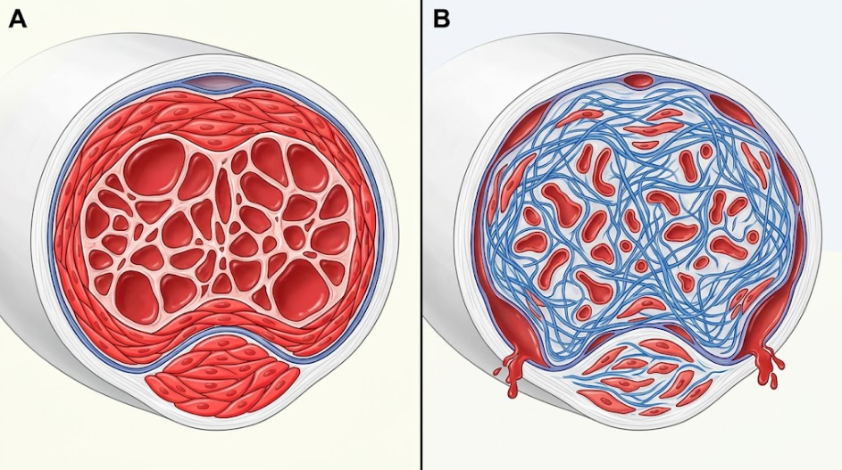 Figure 1. Biomechanical and microarchitectural properties of healthy and fibrotic cavernosal tissue in corporal veno-occlusive dysfunction (CVOD). Panel A depicts the healthy state where the sinusoids are compliant and contractile smooth muscle bundles (red staining) are abundant, allowing effective compression of the subtunical venules against the rigid tunica albuginea during erection. Panel B shows the pathological condition of CVOD. It shows the atrophy of the cavernosal smooth muscle and the progressive deposition of collagen (stained blue) leading to rigid, non-compliant sinusoids, uncompressed venules and persistent venous leakage.
Apoptosis (programmed cell death) of CSMCs initiates and drives the progressive decline in the cavernosal smooth muscle: collagen ratio.(Ferrini et al., 2009) Various systemic and local etiological factors such as age, diabetic hyperglycemia, chronic microvascular ischemia, and pelvic nerve injuries secondary to radical prostatectomy trigger this cellular depletion.(Hu et al., 2019) Despite the different initial triggers, these different etiologies converge on the common molecular pathway of chronic intracellular hypoxia, mitochondrial impairment and excessive oxidative stress in the cavernosal microenvironment.(Xiao, Guo, Zeng, Wu, & Jin, 2026) Regular erections provide high-oxygenated blood that keeps the cavernosal tissue healthy and maintains smooth muscle integrity. But when neuropathic or vascular diseases affect erection, the cavernosal tissue becomes chronically ischemic and hypoxic.(Akkus et al., 1997)
Concerning the molecular mechanism, this hypoxic condition leads to excessive production of reactive oxygen species (ROS) in the mitochondria of CSMCs, which exceeds the antioxidant systems. This oxidative stress causes peroxidation of membrane lipids and opening of mitochondrial permeability transition pore (mPTP) in the outer mitochondrial membrane.(Clavijo, Miner, & Rajfer, 2014) The opening of the mPTP causes the collapse of the mitochondrial membrane potential and the release of pro-apoptotic proteins, such as Cytochrome c, from the mitochondrial intermembrane space into the cytosol. Once in the cytosol, cytochrome c binds to the adaptor protein apoptotic protease activating factor-1 (Apaf-1) in the presence of dATP to form a heptameric complex called the apoptosome. This complex also recruits and activates the initiator protease Caspase 9. Active Caspase-9 then cleaves and activates the downstream effector protease Caspase-3. This ability of caspase-3 to cleave important structural proteins, trigger DNA fragmentation and induce cell shrinkage, chromatin condensation and cell death makes it the final executor of cell death.(Grünewald & Beal, 1999)
Animal experiments have helped to clarify the time course of these histopathological events. In rat models of bilateral cavernosal nerve injury (nerve resection or crush induced neuropraxia), damage to the cavernosal nerve results in rapid denervation induced apoptosis of the subtunical smooth muscle. Quantitative analysis showed a dramatic peak in apoptosis of CSMCs at only 3 days postinjury, as assessed by TUNEL staining and Western blot analysis of cleaved Caspase-3.(Ferrini et al., 2009) This early wave of programmed cell death results in a significant loss of smooth muscle mass, reflected in a 40% reduction in alpha-smooth muscle actin (α-SMA) expression by day 7. Interestingly, this severe histological damage occurs prior to the functional onset of CVOD, which can be detected by dynamic cavernosometry as early as 30 to 45 days following the initial injury.(Ferrini et al., 2009) This time separation underscores a fundamental clinical principle: microscopic cellular injury and smooth muscle loss are antecedent to and predictive of macroscopic functional failure. Also, a critical “loss threshold” of ~15% of the cavernosal smooth muscle mass has been defined by clinical and theoretical models.(Valente et al., 2003; Vernet et al., 2002) The penile hemodynamic system can compensate for small, sub-clinical decreases in smooth muscle volume, but when the cumulative loss of functional CSMCs exceeds this 15% threshold, the remaining muscle mass is simply not physically capable of generating the active compliance and tissue expansion needed to compress the venules, and symptomatic venous leakage and CVOD are unavoidable.(Lin, Bella, Lue, & Lin, 2006)
A profound change in the cellular identity of the remaining CSMCs also drives the progressive loss of functional cavernosal smooth muscle. Smooth muscle cells are phenotypically plastic to a much greater extent than are skeletal or cardiac muscle cells.(Chen et al., 2024) In response to alterations in their local microenvironment such as chronic hypoxia, mechanical stretch, inflammatory cytokines, hyperglycemic injury or hyperlipidemia, CSMCs may undergo a phenotypic switch from a functional, contractile state to a proliferative, synthetic state.(Wei et al., 2012) In the normal state, the overwhelming majority of CSMCs display the contractile phenotype. This phenotype is characterized by a highly organized contractile apparatus, low rate of cell proliferation and low secretion of extracellular matrix.(Wei et al., 2012) Contractile CSMCs express high levels of contractility-associated marker proteins such as alpha-smooth muscle actin (α-SMA), calponin-1, desmin and smooth muscle 22-alpha (SM22α) that are essential for the regulation of the active tone and relaxation compliance of the cavernosal sinusoids.(Wei et al., 2012)
However, local environmental stressors drive the phenotypic transition to the synthetic, proliferative state under pathological conditions.(H. B. Zhang et al., 2017) Phenotypic switch is defined by a strong downregulation of markers associated with contractility (e.g., calponin-1, α-SMA, desmin) and a concomitant upregulation of proteins associated with the synthetic state, in particular Osteopontin (OPN) and Vimentin. In line with this molecular shift, immunohistochemical analyzes of cavernosal tissue from diabetic, hyperlipidemic or nerve-injured animal models show a significant reduction of α-SMA and calponin-1 expression and an over-expression of osteopontin and vimentin.(Qian et al., 2020) The synthetic-phenotype CSMCs lose their physiological contractility and compliant relaxation ability. Instead, they become migratory, highly proliferative and actively produce and secrete large amounts of extracellular matrix proteins, especially collagen, into the intercellular space accelerating cavernosal fibrosis and rigidifying the trabecular walls.(El-Sakka & Yassin, 2010; Qian et al., 2020)
Several molecular signaling pathways orchestrate this phenotypic modulation. Chronic hyperglycemia and insulin resistance in T2DMED models disrupt cellular glucose metabolism, resulting in a dramatic loss of contractile proteins and a major phenotypic switch.(Malavige & Levy, 2009) Recent studies have demonstrated that the peripheral administration of the hormone Nesfatin-1 ameliorates diabetic erectile dysfunction by upregulating contractile markers and downregulating proliferative markers. Such protection is mediated by activation of the PI3K/Akt/mTOR signaling pathway, which maintains the contractile phenotype and blocks the synthetic transition.(Chew, Earle, Stuckey, Jamrozik, & Keogh, 2000) Likewise, in hyperlipidemia-induced ED models (e.g. high-cholesterol-fed rabbits), chronic lipid accumulation induces endothelial dysfunction, transforming growth factor-beta 1 (TGF-β1) overexpression, and an increase in phospho-myosin light chain 20 (p-MLC20) expression, which drive the synthetic phenotypic switch and extracellular matrix accumulation.(Hu et al., 2019) Conversely, anti-hypoxic pharmacological agents, such as salidroside (isolated from Rhodiola rosea), have been shown to counteract hypoxia-induced phenotypic switching by maintaining normal levels of α-SMA and desmin and suppressing vimentin and collagen expression in vitro and in vivo.(X. Zhang et al., 2017)
To understand the intracellular signaling that supports this phenotypic transition and the subsequent apoptotic cascade, we explore the molecular events occurring within the CSMC. Pathological stimuli (such as chronic hypoxia, high glucose or hyperlipidemia) act on the cell to induce two parallel, destructive pathways (as shown in Figure 2). On one hand, mitochondrial oxidative stress opens the mitochondrial permeability transition pore (mPTP), releasing Cytochrome c into the cytosol, which activates Caspase-9 and Caspase-3, leading to apoptosis and cell loss.(Wei et al., 2012) In contrast, these stressors promote the TGF-β1/Smad signaling pathway to transcriptionally upregulate synthetic proteins (Osteopontin and Vimentin) and downregulate contractility-associated proteins (α-SMA and Calponin-1). This phenotypic switch results in cells changing from their normal contractile state to synthetic cells that cannot relax and actively secrete excessive collagen into the extracellular matrix, thus aggravating cavernosal fibrosis.(Wei et al., 2012)
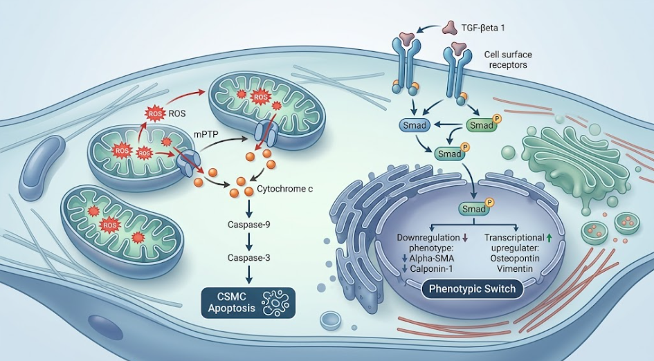
Figure 2. Intracellular Signaling Pathways Regulating Cavernosal Smooth Muscle Cell Apoptosis and Contractile to Synthetic Phenotypic Modulation. Schematic representation of two molecular cascades triggered by pathological stressors (hypoxia, hyperglycemia, hyperlipidemia). The left cascade depicts the apoptotic pathway mediated by mitochondrial oxidative stress leading to the opening of the mitochondrial permeability transition pore (mPTP), release of Cytochrome c and activation of Caspase-3. The right cascade describes the phenotypic transition driven by TGF-β1/Smad and HIF-1α pathways leading to downregulation of contractile markers (α-SMA and Calponin-1) and upregulation of synthetic markers (Osteopontin, Vimentin and Type I Collagen secretion).
Apoptosis of smooth muscle cells and their phenotypic switching result in profound structural remodeling of the cavernosal extracellular matrix (ECM). The ECM in normal erectile tissue is a highly organized, dynamic scaffolding that provides structural support while allowing for large volumetric expansion. The main structural components of this matrix are collagen fibers (mainly Type I, Type III and Type IV) and elastic fibers.(Raviv et al., 1997) Type I collagen consists of thick, rigid and tightly packed bundles of fibrils that provide high tensile strength, but low elastic compliance. Type III collagen is found in distensible, elastic tissues. It forms a fine, flexible network that permits high compliance, distensibility and elastic recoil.(Wespes et al., 1997) Type IV collagen is primarily found in the basement membrane of endothelial and vascular structures, supporting the microvascular integrity. In healthy conditions, these types of collagens are found in a balanced ratio that is adequate for the physical demands of tumescence and detumescence of the penis, such that the cavernosal trabeculae and the surrounding tunica albuginea can stretch up to 150% of their resting length during erection before becoming rigid.(El-Sakka & Yassin, 2010)
But in CVOD sustained apoptosis of CSMCs and hyper-synthetic activity of phenotypic-switched smooth muscle cells drive pathological fibroelastic remodeling. There is a dramatic increase in the total collagen content of both corpora cavernosa and tunica albuginea with a reduction or loss of functional elastic fibers.(Brock, Hsu, Nunes, von Heyden, & Lue, 1997) This progressive fibrosis is mediated by profibrotic cytokines, especially Transforming Growth Factor-beta 1 (TGF-β1) which is upregulated in hypoxic, ischemic or hyperglycemic conditions. TGF-β1 stimulates the intracellular Smad signaling pathway (i.e., promotes Smad2 and Smad3 phosphorylation) and directly stimulates collagen gene transcription, resulting in excessive accumulation of extracellular collagen.(Moreland et al., 1995; Nolazco et al., 2008)
Importantly, this pathological remodeling is not simply a quantitative increase in collagen deposition, but a qualitative change in collagen subtype composition. In models of nerve injury (BCNC) and hyperlipidemia, investigators have shown a dramatic increase in the Collagen Type I to Type III ratio (sometimes greater than 43% increase over healthy controls).(Qian et al., 2020) This transition suggests a preference for depositing the stiff, non-compliant Collagen Type I over the compliant, elastic Collagen Type III. At the same time, Collagen Type IV expression in the trabecular basement membrane is significantly reduced, indicating a disruption of the microvascular basement membrane. The substitution of compliant Type III collagen with rigid Type I collagen decreases the elasticity and compliance of the cavernosal trabeculae and the tunica albuginea.(Raviv et al., 1997) The excess rigid connective tissue forms a physical, noncompliant barrier that stiffens the trabecular walls and prevents the sinusoids from expanding and taking up blood at low pressures. In addition, loss of elasticity of the tunica albuginea results in structural subluxation and floppiness of the outer fibrous sheath. Instead of a hard, unyielding surface, the tunica is abnormally distended out of shape when blood enters the corpora. Thus, the subtunical venules are not compressed, and blood leaks continuously into the systemic vasculature, thereby preventing the development of rigid erections.(Grünewald & Beal, 1999)
After severe tissue injury, apoptosis and fibrotic remodeling, the erectile tissue initiates endogenous cellular defense mechanisms to maintain homeostasis. The most prominent cytoprotective pathway is the transcriptional induction of inducible nitric oxide synthase (iNOS, also called NOS II) in the corpus cavernosum.(Ferrini et al., 2009) iNOS is induced by tissue injury and generates sustained high levels of NO as opposed to nNOS and eNOS that generate transient levels of NO for physiological signaling. In animal models of cavernosal nerve resection (BCNR), nNOS expression decreases rapidly and is absent by day 30, whereas iNOS expression is induced as early as day 3 and steadily increases, reaching a peak at day 30 and maintaining high levels.(Ferrini et al., 2009; Wang et al., 2025)
This induction of iNOS and the subsequent continuous intracellular NO release act as an endogenous anti-fibrotic shield. Nitric oxide down-regulates pro-apoptotic signaling, quenches reactive oxygen species (ROS) and inhibits the transcription of profibrotic genes via the cGMP pathway retarding progressive collagen deposition. Studies have shown that the administration of iNOS inhibitors (e.g. L-NIL) significantly worsens cavernosal fibrosis, supporting a cytoprotective role for iNOS. However, the endogenous response is ultimately insufficient to fully overcome the aggressive factors induced by denervation. This state of cellular collapse is known as “corporal dystrophy”. CVOD and fibrosis still progress in untreated models despite iNOS upregulation.(Ferrini et al., 2017; Ferrini et al., 2009)
Thus, current research is focused on therapeutic interventions that enhance these rescue pathways to prevent or reverse the change in the smooth muscle-to-collagen ratio. A successful pharmacological approach to continuously administer Phosphodiesterase Type 5 inhibitors (PDE5i, e.g. sildenafil or tadalafil) has been developed.(El-Sakka & Yassin, 2010) The sustained use of PDE5i therapy prevents cGMP breakdown, thereby maintaining increased cGMP levels, which counteracts oxidative stress, inhibits TGF-β1, and prevents smooth muscle apoptosis.(Li et al., 2007) Studies have demonstrated that long-term therapy with PDE5i not only preserves smooth muscle, but also augments the replication of contractile smooth muscle cells, increasing the smooth muscle-to-collagen ratio.(Ferrini et al., 2009) Similarly, the daily use of a vacuum erectile device (VED) prevents penile shrinkage and CVOD by reducing hypoxia, preserving native collagen structures, and upregulating contractile calponin-1 and downregulating synthetic osteopontin.(Qian et al., 2020) Moreover, regenerative medicine approaches with intracavernosal injections of stem cells such as human umbilical cord mesenchymal stem cells (HUC-MSCs) or adipose-derived stem cells (ADSCs) stimulate endothelial and smooth muscle regeneration, reduce apoptosis (downregulating Caspase-3 and Bax and upregulating Bcl-2) and inhibit fibrosis, thus restoring tissue compliance and veno-occlusive function.(Liu et al., 2020)
This review synthesizes molecular, cellular and biomechanical data to establish the smooth muscle-to-collagen ratio in the corpora cavernosa as the defining cellular basis of penile veno-occlusive competence. The mechanical transition from flaccid to erect state requires perfect coordination of cellular compliance, tissue expandability and physical resistance. When this coordination is disrupted by pathological processes, the transition from a smooth muscle dominated tissue to a collagen dominated tissue directly leads to CVOD. This, along with the mechanical requirements of blood containment, means that any process that reduces the smooth muscle content below the critical threshold of about 15% will compromise the passive compression of the subtunical veins and venous leakage is bound to occur.(Ferrini et al., 2017)
In the past, to understand these cellular changes we had to perform invasive, painful, and clinically unacceptable penile biopsies which limited our ability to follow cavernosal tissue degeneration in real time. However, recent advances in diagnostic imaging now provide a new non-invasive approach to quantify these structural changes: two-dimensional shear-wave elastography (2-D SWE).(Hu et al., 2019) The technology uses acoustic radiation force to create shear waves in the targeted tissue and measures their propagation speed to calculate the tissue’s Young’s modulus (YM) or stiffness in kilopascals (kPa). 2-D SWE can be used to detect and quantify cavernosal fibrosis and smooth muscle loss in a non-invasive way due to the relatively high shear wave velocity of stiff collagenous connective tissue compared with compliant smooth muscle.(Hu et al., 2019)
The clinical and experimental studies have validated the use of 2-D SWE to assess cavernosal health in various age groups and pathological conditions. In healthy human cohorts, the Young’s modulus of the corpus cavernosum is strongly and positively correlated with chronological age (r = 0.949) and negatively correlated with erectile function as assessed by the International Index of Erectile Function-5 (IIEF-5) (r = -0.843).(Cheng et al., 2022) Histological correlations confirmed that this age-associated increase in tissue stiffness (YM) directly correlated with a progressive decrease in smooth muscle content (r = -0.738) and a reciprocal increase in collagen deposition (r = 0.732) leading to a significant decrease in the smooth muscle to collagen ratio (r = -0.720).(Cheng et al., 2022) In rabbit models of hyperlipidemia-induced erectile dysfunction, 2-D SWE scanning revealed a significant increase in cavernosal stiffness. The mean shear-wave quantitative values in hyperlipidemic rabbits increased to 24.53 ± 2.64 kPa, compared to 9.97 ± 0.54 kPa in normal controls.(Hu et al., 2019) This increased stiffness perfectly correlated with histology evidence of extracellular matrix accumulation, TGF-β1 overexpression and smooth muscle phenotypic switching. These results suggest that 2-D SWE represents an objective, non-invasive approach to assess cavernosal aging, fibrosis, and degeneration in a clinical setting, allowing early therapeutic intervention before irreversible “corporal dystrophy” is established.(Cheng et al., 2022; Hu et al., 2019)
Alteration in the ratio of cavernosal smooth muscle to collagen is the predominant cellular mechanism responsible for the development of Corporal Veno-Occlusive Dysfunction and penile structural failure. The slow loss of cavernosal smooth muscle cells through oxidative stress-induced apoptosis and the phenotypic switching of the remaining smooth muscle cells from a contractile to a synthetic phenotype, results in a pathological remodeling of the extracellular matrix. The compliant smooth muscle and elastic Type III collagen are replaced qualitatively and quantitatively by rigid, non-compliant Type I collagen which rigidifies the trabecular walls and compromises the elasticity of the tunica albuginea. Loss of compliance does not allow the sinusoidal spaces to expand enough to compress the subtunical venules. This leads to persistent uncontrolled venous leakage. Understanding these intricate molecular and cellular cascades highlights the limitations of managing erectile dysfunction with only transient, short-acting vasoactive drugs that do not address underlying structural tissue damage. Instead, it highlights the pressing clinical demand for early, preventive and regenerative interventions such as continuous PDE5 inhibitor therapy, vacuum rehabilitation and stem cell-based interventions that target these cellular mechanisms to maintain the smooth muscle-to-collagen ratio and prevent the progression of penile structural failure.
Akkus, E., Carrier, S., Baba, K., Hsu, G. L., Padma-Nathan, H., Nunes, L., & Lue, T. F. (1997). Structural alterations in the tunica albuginea of the penis: impact of Peyronie’s disease, ageing and impotence. Br J Urol, 79(1), 47-53. doi:10.1046/j.1464-410x.1997.26511.x
Brock, G., Hsu, G. L., Nunes, L., von Heyden, B., & Lue, T. F. (1997). The anatomy of the tunica albuginea in the normal penis and Peyronie’s disease. J Urol, 157(1), 276-281.
Chen, K., Huang, B., Feng, J., Hu, Z., Fan, S., Ren, S., . . . Li, G. (2024). Nesfatin-1 regulates the phenotype transition of cavernous smooth muscle cells by activating PI3K/AKT/mTOR signaling pathway to improve diabetic erectile dysfunction. PLOS ONE, 19(9), e0304485. doi:10.1371/journal.pone.0304485
Cheng, H., Liu, G. X., Wang, F., Wang, K., Ruan, L. T., & Yang, L. (2022). Quantitative assessment of the aging corpus cavernosum by shear wave elastography. Asian J Androl, 24(6), 628-632. doi:10.4103/aja2021119
Chew, K. K., Earle, C. M., Stuckey, B. G., Jamrozik, K., & Keogh, E. J. (2000). Erectile dysfunction in general medicine practice: prevalence and clinical correlates. Int J Impot Res, 12(1), 41-45. doi:10.1038/sj.ijir.3900457
Clavijo, R. I., Miner, M. M., & Rajfer, J. (2014). Erectile Dysfunction and Essential Hypertension: The Same Aging-related Disorder? Rev Urol, 16(4), 167-171.
El-Sakka, A. I., & Yassin, A. A. (2010). Amelioration of Penile Fibrosis: Myth or Reality. Journal of Andrology, 31(4), 324-335. doi:https://doi.org/10.2164/jandrol.109.008730
Ferrini, M. G., Gonzalez-Cadavid, N. F., & Rajfer, J. (2017). Aging related erectile dysfunction-potential mechanism to halt or delay its onset. Transl Androl Urol, 6(1), 20-27. doi:10.21037/tau.2016.11.18
Ferrini, M. G., Kovanecz, I., Sanchez, S., Umeh, C., Rajfer, J., & Gonzalez-Cadavid, N. F. (2009). Fibrosis and loss of smooth muscle in the corpora cavernosa precede corporal veno-occlusive dysfunction (CVOD) induced by experimental cavernosal nerve damage in the rat. J Sex Med, 6(2), 415-428. doi:10.1111/j.1743-6109.2008.01105.x
Grünewald, T., & Beal, M. F. (1999). NOS knockouts and neuroprotection. Nature Medicine, 5(12), 1354-1355. doi:10.1038/70918
Hu, J. L., Chen, H. X., Chen, H. R., Wu, Y., Sun, X. W., Li, Z., & Xing, J. F. (2019). Novel noninvasive quantification of penile corpus cavernosum lesions in hyperlipidemia-induced erectile dysfunction in rabbits by two-dimensional shear-wave elastography. Asian J Androl, 21(2), 143-149. doi:10.4103/aja.aja_78_18
Li, P., Oparil, S., Novak, L., Cao, X., Shi, W., Lucas, J., & Chen, Y. F. (2007). ANP signaling inhibits TGF-beta-induced Smad2 and Smad3 nuclear translocation and extracellular matrix expression in rat pulmonary arterial smooth muscle cells. J Appl Physiol (1985), 102(1), 390-398. doi:10.1152/japplphysiol.00468.2006
Lin, G., Bella, A. J., Lue, T. F., & Lin, C. S. (2006). Brain-derived neurotrophic factor (BDNF) acts primarily via the JAK/STAT pathway to promote neurite growth in the major pelvic ganglion of the rat: part 2. J Sex Med, 3(5), 821-829. doi:10.1111/j.1743-6109.2006.00292.x
Liu, M. C., Chang, M. L., Wang, Y. C., Chen, W. H., Wu, C. C., & Yeh, S. D. (2020). Revisiting the Regenerative Therapeutic Advances Towards Erectile Dysfunction. Cells, 9(5). doi:10.3390/cells9051250
Malavige, L. S., & Levy, J. C. (2009). Erectile dysfunction in diabetes mellitus. J Sex Med, 6(5), 1232-1247. doi:10.1111/j.1743-6109.2008.01168.x
Moreland, R. B., Traish, A., McMillin, M. A., Smith, B., Goldstein, I., & Saenz de Tejada, I. (1995). PGE1 suppresses the induction of collagen synthesis by transforming growth factor-beta 1 in human corpus cavernosum smooth muscle. J Urol, 153(3 Pt 1), 826-834.
Nolazco, G., Kovanecz, I., Vernet, D., Gelfand, R. A., Tsao, J., Ferrini, M. G., . . . Gonzalez-Cadavid, N. F. (2008). Effect of muscle-derived stem cells on the restoration of corpora cavernosa smooth muscle and erectile function in the aged rat. BJU Int, 101(9), 1156-1164. doi:10.1111/j.1464-410X.2008.07507.x
Qian, S. Q., Qin, F., Zhang, S., Yang, Y., Wei, Q., Wang, R., & Yuan, J. H. (2020). Vacuum therapy prevents corporeal veno-occlusive dysfunction and penile shrinkage in a cavernosal nerve injured rat model. Asian J Androl, 22(3), 274-279. doi:10.4103/aja.aja_57_19
Raviv, G., Vanegas, J. P., Petein, M., Schulman, C., Danguy, A., Kiss, R., & Wespes, E. (1997). Biochemical alterations of the tunica albuginea in impotence. J Urol, 158(5), 1778-1782. doi:10.1016/s0022-5347(01)64127-2
Sattar, A. A., Salpigides, G., Vanderhaeghen, J. J., Schulman, C. C., & Wespes, E. (1995). Cavernous oxygen tension and smooth muscle fibers: relation and function. J Urol, 154(5), 1736-1739.
Valente, E. G., Vernet, D., Ferrini, M. G., Qian, A., Rajfer, J., & Gonzalez-Cadavid, N. F. (2003). L-arginine and phosphodiesterase (PDE) inhibitors counteract fibrosis in the Peyronie’s fibrotic plaque and related fibroblast cultures. Nitric Oxide, 9(4), 229-244. doi:10.1016/j.niox.2003.12.002
Vernet, D., Ferrini, M. G., Valente, E. G., Magee, T. R., Bou-Gharios, G., Rajfer, J., & Gonzalez-Cadavid, N. F. (2002). Effect of nitric oxide on the differentiation of fibroblasts into myofibroblasts in the Peyronie’s fibrotic plaque and in its rat model. Nitric Oxide, 7(4), 262-276. doi:10.1016/s1089-8603(02)00124-6
Wang, W., Liu, Y., Zhou, Z.-H., Pang, K., Wang, J.-K., Huan, P.-F., . . . Han, C.-H. (2025). Effects of human umbilical cord-derived mesenchymal stem cell therapy for cavernous nerve injury-induced erectile dysfunction in the rat model. Asian Journal of Andrology, 27(4), 508-515. doi:10.4103/aja2024115
Wei, A. Y., He, S. H., Zhao, J. F., liu, Y., Liu, Y., Hu, Y. W., . . . Wu, Z. Y. (2012). Characterization of corpus cavernosum smooth muscle cell phenotype in diabetic rats with erectile dysfunction. Int J Impot Res, 24(5), 196-201. doi:10.1038/ijir.2012.16
Wespes, E., Sattar, A. A., Golzarian, J., Wery, D., Daoud, N., & Schulman, C. C. (1997). Corporeal veno-occlusive dysfunction: predominantly intracavernous muscular pathology. J Urol, 157(5), 1678-1680. doi:10.1016/s0022-5347(01)64833-x
Xiao, M., Guo, W., Zeng, R., Wu, S., & Jin, X. (2026). Improving erectile function in diabetic male mice by rescuing depalmitoylated FBP1 to reduce cavernosal lactate. Nature Communications, 17(1), 1740. doi:10.1038/s41467-026-68443-y
Zhang, H. B., Wang, Z. Q., Chen, F. Z., Ding, W., Liu, W. B., Chen, Z. R., . . . Wei, A. Y. (2017). Maintenance of the contractile phenotype in corpus cavernosum smooth muscle cells by Myocardin gene therapy ameliorates erectile dysfunction in bilateral cavernous nerve injury rats. Andrology, 5(4), 798-806. doi:10.1111/andr.12375
Zhang, X., Zhao, J. F., Zhao, F., Yan, J. F., Yang, F., Huang, X. J., . . . Lv, B. D. (2017). The Protective Effect of Salidroside on Hypoxia-Induced Corpus Cavernosum Smooth Muscle Cell Phenotypic Transformation. Evid Based Complement Alternat Med, 2017, 3530281. doi:10.1155/2017/3530281