Our Services
Others
- DFPP
- Shockwave
- IV Drip


“This comprehensive literature review elucidates the intricate molecular architecture of oligodendrocytes and the myelin sheath, detailing how the disruption of metabolic coupling and programmed cell death pathways within the axon-glial unit drives neurodegeneration in multiple sclerosis.”
Multiple sclerosis is traditionally recognized as an inflammatory demyelinating disease of the central nervous system, characterized by the progressive deterioration of myelin sheaths and the loss of mature oligodendrocytes. Early disease pathogenesis is driven by systemic and compartmentalized immune responses, while progressive neurological decline is fundamentally underpinned by the collapse of the axon-glial unit and subsequent neuroaxonal degeneration. The intricate anatomy and biochemistry of white matter of the central nervous system is reviewed with an emphasis on the specialized lipid and protein composition of compact myelin and its functional domains, the nodes of Ranvier, paranodes, and juxtaparanodes. We discuss the crucial, non-canonical roles of oligodendrocytes in metabolic and trophic support of axons via the activity-dependent lactate-pyruvate shuttle and the axo-myelinic synapse. Furthermore, the article extensively reviews the mechanisms involved in the regulation of oligodendrocyte injury and apoptosis as compared with classical apoptosis and receptor-interacting protein kinase–mediated necroptosis under oxidative and nitrosative stress conditions. Finally, we discuss the pathophysiological barriers to endogenous remyelination, in particular molecular inhibitors such as LINGO-1, Notch and Wnt signaling cascades in the hostile lesion microenvironment. Drawing on these biophysical and metabolic insights, this review outlines therapeutic approaches focused on stimulating myelin repair and maintaining axonal integrity, a paradigmatic shift in neurodegenerative research.
Keywords: Oligodendrocytes, Myelin Sheath, Multiple Sclerosis, Axon-Glial Unit, Metabolic Coupling, Necroptosis, Remyelination Barriers
The evolution of the central nervous system (CNS) in jawed vertebrates shows increasing complexity of cellular organization, one hallmark of which is the acquisition of myelin.(Zalc, Goujet, & Colman, 2008) Myelination is a highly regulated, bidirectional cellular interaction, which enables fast and energetically inexpensive conduction of electrical signals along axons by saltatory conduction.(Duncan, Simkins, & Emery, 2021) This specialized task is carried out only by oligodendrocytes in the central nervous system that undergo a strict lineage progression, migration and differentiation to build the myelin sheath.(Bokulic Panichi, Stanca, Dolciotti, & Bongioanni, 2025) For decades the scientific consensus has considered myelin as a static, inert insulating membrane, whose sole function is to speed up action potential propagation.(Zhang et al., 2025) However, modern neurobiology has redefined the myelin sheath as a highly dynamic, plastic and metabolically active organelle deeply embedded in the functional homeostasis of the neural network.(Stadelmann, Timmler, Barrantes-Freer, & Simons, 2019). Oligodendrocytes and the axons they ensheath are in a symbiotic relationship, termed the axon-glial unit, which actively controls electrical, ionic and metabolic homeostasis.(García-Domínguez, 2025)
In diseases like multiple sclerosis (MS) this delicate cellular relationship is systematically destroyed.(López-Muguruza & Matute, 2023) We have previously reported on the clinical spectrum of multiple sclerosis, where smoldering neurodegeneration is shown to be a persistent, compartmentalized driver of progressive neurological disability, independent of apparent, relapse-associated systemic inflammation. To understand the cellular and biophysical basis of this progressive decay, one must first zoom in on the micro-level architecture of the CNS white matter, and how the loss of oligodendrocyte-mediated support leads to axonal metabolic crises and degeneration. In multiple sclerosis, demyelination is not just the loss of a physical insulation, but the structural and energetic uncoupling of the neuron from its primary metabolic guardian.(Duncan et al., 2021)
This literature review offers an in-depth synthesis of the anatomy, molecular and metabolic architecture of white matter in the central nervous system. It details the particular lipidomic and proteomic composition of the myelin sheath and how the components come together to form specialized functional domains.(Oudejans, Luchicchi, Strijbis, Geurts, & van Dam, 2021; Podbielska, Banik, Kurowska, & Hogan, 2013) We discuss the biophysical mechanisms of metabolic coupling in the axon-glial unit, with a focus on the activity-dependent monocarboxylate shuttle and the physiology of the axo-myelinic synapse.(Oudejans et al., 2021) In addition, the current review is devoted to the specific programd cell death pathways, caspase-dependent apoptosis and receptor-interacting protein kinase (RIPK)-dependent necroptosis, that are responsible for oligodendrocyte death in active multiple sclerosis lesions. Finally, we discuss pathological signaling blocks that prevent oligodendrocyte progenitor cell (OPC) maturation and successful remyelination and novel therapeutic targets to restore myelin integrity and maintain long-term axonal survival.(García-Domínguez, 2025)
The integrity of the central nervous system white matter relies on the unique morphology and biosynthetic capacity of mature oligodendrocytes. Unlike peripheral nervous system Schwann cells that contact and myelinate only one segment of an individual axon, a single central nervous system oligodendrocyte can extend multiple processes to ensheath and insulate up to 40 to 60 individual axons. For this monumental task to be performed, one mature oligodendrocyte must synthesize, transport and organize enormous quantities of lipids and proteins, sustaining a membrane surface area estimated to be between 5,000 and 50,000 square micrometers per day.(Dawson, Polito, Levine, & Reynolds, 2003; Nishiyama, 2001)
The myelin membrane is biochemically very specialized, unlike other biological membranes. Cell membranes are usually a pretty even mix of proteins and lipids, but the myelin sheath is nearly all lipids, comprising somewhere between 70 and 85 percent of the dry weight of the sheath. The remainder, 15% to 30%, is protein.(Williamson & Lyons, 2018) This high lipid to protein ratio is required to obtain the high electrical resistance and low capacitance required for saltatory conduction. The lipidomic profile of myelin in the central nervous system consists of three major lipid classes, namely cholesterol, phospholipids and glycolipids. These lipids are present in a remarkably constant molar ratio of ~2:2:1. Cholesterol is a major physical stabilizer and a key player in myelin membrane growth allowing the exit of major membrane proteins from the endoplasmic reticulum and their delivery to the growing sheath. (Franklin & Ffrench-Constant, 2008) Phospholipids, such as phosphatidylcholine, phosphatidylethanolamine and plasmalogens, provide structural fluidity and membrane curvature.(López-Muguruza & Matute, 2023) Glycolipids are abundant in the white matter of the central nervous system, primarily as galactosylceramide (GalCer) and its sulfated derivative, sulfatide (sGalCer). These molecules have very long, saturated fatty acid tails, which favors tight hydrophobic packing and membrane compaction. These lipids cluster together with certain proteins creating highly ordered microdomains known as lipid rafts, which control the fluidity of the membrane and trafficking of proteins and signaling cascades across the membrane.(Bokulic Panichi et al., 2025)
Central nervous system myelin is composed of two major structural proteins, proteolipid protein (PLP) and myelin basic protein (MBP), which make up more than two-thirds of the total mass of myelin protein. Proteolipid protein is a hydrophobic, tetraspan transmembrane protein which makes up approximately 38% of central nervous system myelin protein.(Kim, An, Fan, & Park, 2021) The main role of proteolipid protein is to adhesive-stack and compact the extracellular leaflets of the spiraling myelin membrane. This preserves the physical stability of the multilamellar sheath and protects the underlying axon from environmental insults. Myelin basic protein is an intrinsically unstructured, positively charged extrinsic protein located on the cytoplasmic face of the myelin membrane and accounts for about 30% of the total protein mass.(Paez & Lyons, 2020) Electrostatic interactions between myelin basic protein and negatively charged headgroups of cytoplasmic lipids (e.g., phosphatidylserine) neutralize these charges, triggering a cohesive phase transition that zippers the cytoplasmic leaflets together to form the “major dense line” of compacted myelin. Quantitatively minor but functionally critical proteins include myelin associated glycoprotein (MAG), myelin oligodendrocyte glycoprotein (MOG) and 2’,3’-cyclic-nucleotide 3’-phosphodiesterase (CNPase). Myelin-associated glycoprotein is located only in the adaxonal or innermost layer of the myelin membrane and, therefore, directly contacts the axonal surface to modulate signaling. Myelin oligodendrocyte glycoprotein is located on the outermost surface and is thus an important target for autoimmune antibodies in multiple sclerosis. (Rosetti, Maggio, & Oliveira, 2008) CNPase is found in uncompacted cytoplasmic channels and interacts with the actin cytoskeleton to counteract the zippering forces of the myelin basic protein, thereby maintaining open cytosolic conduits that allow the distribution of metabolic substrates throughout the sheath.(Duncan et al., 2021)
The functional myelinated axon consists of four distinct anatomical domains: the nod of Ranvier, the paranode, the juxtaparanode and the internode. The node of Ranvier is a narrow unmyelinated gap of ~1 μm in which voltage-gated sodium channels (specifically NaV1.6) are highly clustered to allow inward sodium currents during action potential propagation.(Lajtha, Toth, Fujimoto, & Agrawal, 1977; LeBaron, Sanyal, & Jungalwala, 1981) The paranode, a critical site of physical attachment, flanks the node, and is where the terminal cytoplasmic loops of the myelin sheath are tightly bound to the axonal membrane. This physical seal is mediated by a highly specialized axo-glial junctional complex consisting of axonal contactin-associated protein (Caspr) and contactin-1, which bind in trans to glial neurofascin-155 (NF155).(Oudejans et al., 2021) This junction forms a tight diffusion barrier that separates the nodal sodium channels from juxtaparanodal potassium channels while maintaining tight triangular junctional clefts that allow diffusion of small molecules into the periaxonal space.(Mierzwa, Shroff, & Rosenbluth, 2010) The paranode is bordered by the juxtaparanode, which is rich in voltage-gated potassium channels (specifically Kv1.1 and Kv1.2) clustered by the Caspr2-contactin-2 complex to stabilize the axonal membrane potential and prevent abnormal electrical firing. The internode is the long-myelinated segment between these domains, where the axolemma is protected from the extracellular space by compact myelin, as seen in Figure 1 shown the structural architecture of the axon-glial unit.(Duncan et al., 2021)
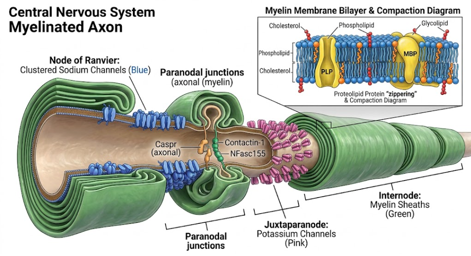
Figure 1. Structural domains of a Myelinated axon (central nervous system). Different regions of a myelinated axon in the central nervous system. The Node of Ranvier (middle) with clustered sodium channels (blue), flanked by the Paranode with specific cell-adhesion molecules and the Juxtaparanode with potassium channels (pink). The long internode region is shown enclosed by lipid-rich myelin sheaths (green). An inset displays a detailed view of the composition of the myelin membrane bilayer and the compaction driven by PLP and MBP proteins.
In its classic role, the myelin sheath is not only a physical and electrical insulator, but also a major metabolic conduit for the physical and energetic survival of the underlying axon. Axons are long, thin cell projections extending a long distance from the neuronal cell body, making active anterograde axonal transport of organelles, proteins and metabolites heavily dependent on continuous local ATP production.(Barres & Raff, 1999; Trapp, Nishiyama, Cheng, & Macklin, 1997) The compact myelin physically isolates the myelinated axolemma from the extracellular environment, and thus the axon is structurally limited in its ability to directly import glucose and nutrients from the surrounding capillaries. The oligodendrocyte and the myelin sheath solve this physical problem by forming a highly specialized metabolic partnership, the so-called oligodendrocyte-axon lactate shuttle.(Li & Sheng, 2023)
Compared to neurons that are almost entirely dependent on oxidative phosphorylation for ATP generation, myelinating oligodendrocytes maintain a surprisingly high rate of aerobic glycolysis. This preference for glycolysis is maintained even when oxygen is present, leading to the continuous production of pyruvate and its further transformation to lactate-by-lactate dehydrogenase. This metabolic configuration is very efficient, since glucose that is imported is processed within the oligodendrocyte cytosol, and the produced monocarboxylates (lactate and pyruvate) are actively transported across the inner, adaxonal myelin membrane, and into the narrow periaxonal space via glia-specific monocarboxylate transporter 1 (MCT1).(Simons & Nave, 2015) This lactate and pyruvate is rapidly and directly taken up by the axon into its axoplasm, which is densely populated with neuron-specific monocarboxylate transporter 2 (MCT2) on the internodal axolemma. Once inside the axon, these substrates are then transported into the axonal mitochondria, where they enter the tricarboxylic acid (TCA) cycle to fuel oxidative phosphorylation, producing the massive amounts of ATP necessary to power the Na+/K+-ATPase pumps and maintain axonal transport. This Figure 2 illustrates the spatial relationship of the metabolic coupling, which indicates that these monocarboxylate transporters are strategically positioned along the adaxonal myelinic channels, to favor direct and unobstructed metabolic transfer.(Tepavčević, 2021)
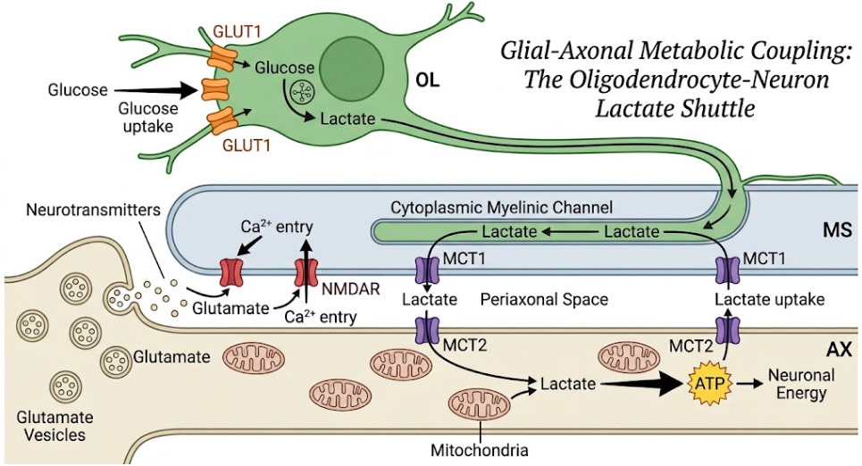
Figure 2. The Oligodendrocyte-Axon Lactate Shuttle. This figure shows the metabolic support system between oligodendrocytes (OL) and axons (AX). Oligodendrocytes take up glucose and metabolize it to lactate which they shuttle through the myelin sheath to the axon to support ATP production. Axonal release of glutamate that activates NMDA receptor on myelin sheath controls this energy transfer.
The activity of the oligodendrocyte-axon lactate shuttle is not static, but dynamically regulated by neuronal electrical activity through a specialized communication point, the axo-myelinic synapse (AMS).(Oudejans et al., 2021) When the action potential propagates along the internodal axolemma, voltage-gated calcium channels on the surface of the axon allow a local influx of calcium. The increased calcium results in an exocytotic release of glutamate from the axon into the periaxonal space. Ionotropic glutamate receptors (e.g., α-amino-3-hydroxy-5-methyl-4-isoxazolepropionic acid [AMPA] and N-methyl-D-aspartate [NMDA]) are present on the inner adaxonal membrane of the myelin sheath and are directly activated by glutamate released from the axon.(Philips & Rothstein, 2017) This activation of myelinic NMDA and AMPA receptors causes a localized calcium influx in the myelin compartment. This calcium surge provides a strong intracellular signal that triggers the rapid translocation of more glucose transporter 1 (GLUT1) molecules from intracellular vesicular pools to the outer cell membrane of the oligodendrocyte and the myelin sheath. Activity-dependent upregulation of glucose transporters facilitates the uptake of glucose from the vasculature, and adjacent astrocytes, to sustain glycolysis and export of lactate to meet the immediate, local energetic demands of the active axon. This bi-directional feed-back loop ensures that the axonal metabolic support is finely tuned to the electrical work load.(Fünfschilling et al., 2012)
In multiple sclerosis, the complex homeostatic and metabolic balance of the axon-glial unit is devastatingly disrupted. The main mechanism of demyelination is the loss of oligodendrocytes, which disrupts saltatory conduction and deprives the axon of its metabolic lifeline, leading to irreversible neuroaxonal degeneration and progressive clinical disability. The loss of oligodendrocytes in MS lesions is a complex, multi-factorial process that is driven by a hostile inflammatory microenvironment, severe oxidative and nitrosative stress and activation of distinct programmed cell death pathways.(Duncan et al., 2021; García-Domínguez, 2025)
Oligodendrocytes are especially susceptible to mitochondrial insults and oxidative stress due to the metabolic demands of synthesizing and maintaining the extensive myelin membrane. Under physiological conditions, oligodendrocytes depend on mitochondrial oxidative phosphorylation to fulfill the tremendous ATP demands of membrane synthesis, compaction and maintenance of ionic gradients through Na+/K+-ATPase pumps. In active lesions of multiple sclerosis, infiltrating inflammatory cells (including CD4+ and CD8+ T lymphocytes, B cells and activated macrophages/microglia) produce highly toxic levels of reactive oxygen species (ROS) and reactive nitrogen species (RNS). This oxidative storm results in severe lipid peroxidation, protein nitration, and mitochondrial DNA damage.(Miljković & Spasojević, 2013; Narine & Colognato, 2022)
In addition, multiple sclerosis tissues have severe defects in the mitochondrial respiratory chain, in particular, complex I (NADH: ubiquinone oxidoreductase) and complex IV (cytochrome c oxidase) which are highly sensitive to oxidative and nitrosative damage. The inhibition of these respiratory complexes leads to impairment of the proton gradient across the inner mitochondrial membrane, the mitochondrial membrane potential and the ATP synthesis via oxidative phosphorylation. This bioenergetic failure is especially catastrophic for oligodendrocytes, which cannot maintain membrane transport with sufficient ATP, resulting in intracellular osmotic imbalance and swelling that causes retraction of oligodendrocyte processes from the myelin sheath in a survival response that destabilizes compacted myelin and exposes the axon to toxic inflammatory mediators.(Jahn et al., 2020)
Oligodendrocyte death in multiple sclerosis is mediated by two genetically controlled pathways of programmed cell death, apoptosis and necroptosis. Apoptosis is a regulated cell death process characterized by cell shrinkage, chromatin condensation and caspase activation and is prominent in early-stage lesions of multiple sclerosis, particularly in type III lesions characterized by primary oligodendrogliopathy. The extrinsic apoptotic pathway is triggered via the interaction of pro-inflammatory cytokines such as tumor necrosis factor-alpha (TNF-α) or Fas ligand (FasL) with death receptors (TNFR1 and Fas, respectively) that are highly upregulated on oligodendrocytes within active lesions.(Prineas & Parratt, 2012) The binding recruits Fas-associated death domain (FADD) and procaspase-8 to form the death-inducing signaling complex (DISC) leading to the activation of initiator caspase-8 and downstream executioner caspases, mainly caspase-3 and caspase-9. Or CD8+ cytotoxic T lymphocytes directly attack oligodendrocytes that display major histocompatibility complex class I (MHC-I) molecules, releasing perforin and granzyme B to induce caspase dependent apoptosis.(Denic, Wootla, & Rodriguez, 2013) The classical complement pathway can also induce apoptosis; autoantibodies binding to myelin-specific antigens (e.g. MBP or MOG) trigger the complement cascade, resulting in the assembly of the membrane attack complex (MAC). MAC insertion into the oligodendrocyte membrane leads to a large influx of extracellular calcium (Ca2+) and sodium (Na+) ions and a rapid efflux of potassium (K+), resulting in mitochondrial outer membrane permeabilization (MOMP), osmotic imbalance and apoptotic caspase activation.(Caprariello, Mangla, Miller, & Selkirk, 2012)
In contrast, necroptosis is a type of regulated, lytic cell death that occurs under highly inflammatory conditions, especially when classical caspase activity is blocked or overwhelmed, as for example in the presence of high concentrations of nitric oxide, which strongly inhibit caspase-3. Necroptosis is a highly pro-inflammatory cell death pathway due to its lytic nature that ruptures the plasma membrane, releasing highly immunogenic damage-associated molecular patterns (DAMPs) into the extracellular space, perpetuating chronic neuroinflammation. As in the Figure 3 shown cell death mechanisms, necroptosis is mediated by a specific molecular signaling pathway.(Yuan, Amin, & Ofengeim, 2019) When TNFR1 is activated by TNF-α in the absence or blockade of caspase-8 activity, receptor-interacting protein kinase 1 (RIPK1) and receptor-interacting protein kinase 3 (RIPK3) autophosphorylation and associate to form a functional amyloid-like signaling complex, called the necrosome. The active necrosome then recruits and phosphorylates mixed lineage kinase domain-like protein (MLKL). Upon phosphorylation, MLKL oligomerizes and translocates directly to the plasma membrane where it inserts into the lipid bilayer, disrupting membrane integrity and causing rapid, lytic cell rupture and necrotic death. Detection of phosphorylated MLKL in active MS lesions is evidence for active necroptosis in oligodendrocyte death.(Zhou et al., 2024)
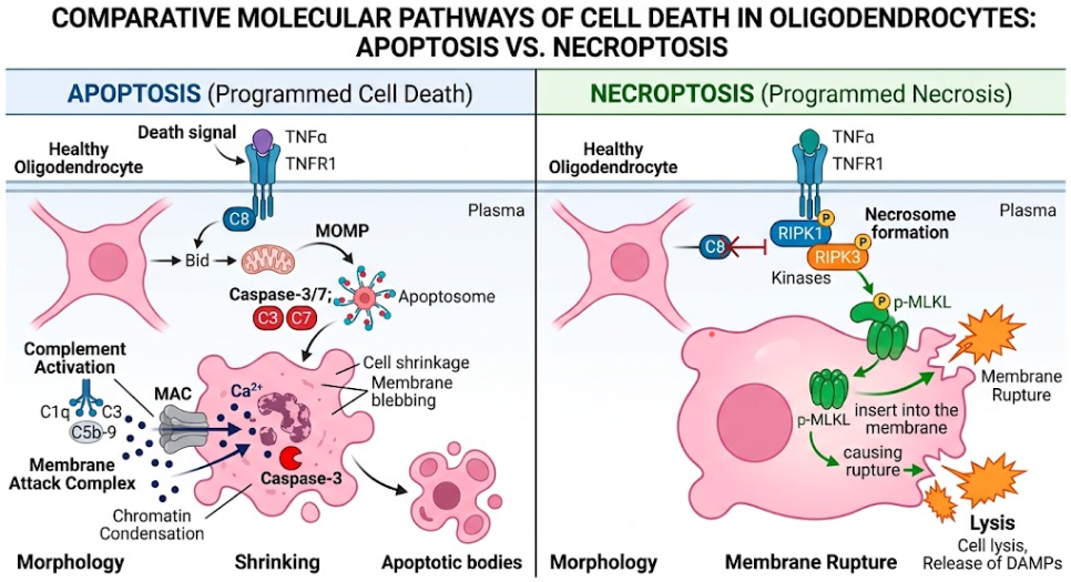
Figure 3. Distinct Molecular Pathways of Oligodendrocyte Cell Death: Apoptosis vs. Necroptosis. Schematic representation of the mechanisms of apoptosis (left; cell shrinkage and caspase activation) and necroptosis (right; rupture of the membrane and swelling via MLKL).
The central nervous system has an endogenous capacity for repair called endogenous remyelination following inflammatory demyelination. This regenerative process is mediated by a ubiquitous population of adult stem cells, the oligodendrocyte progenitor cells (OPCs) which make up approximately 5% to 10% of the total cell population in the adult central nervous system. Local OPCs react to demyelinating injury via inflammatory and chemotactic signals to induce rapid proliferation and migration to the denuded lesion site.(Chamberlain, Nanescu, Psachoulia, & Huang, 2016) These recruitment of OPCs to the lesion must then be followed by a series of tightly regulated differentiation steps so that they become mature, myelinating oligodendrocytes, capable of extending processes, wrapping denuded axons and restoring saltatory conduction and metabolic support.(Glezer, Lapointe, & Rivest, 2006)
In the chronic stages of multiple sclerosis, however, this regenerative process fails frequently and systematically, leading to permanent demyelination, progressive axonal metabolic starvation and irreversible axonal loss. Although OPCs are often found in large numbers in chronic lesions, they are arrested in an immature, pre-myelinating state and fail to undergo terminal differentiation into mature, myelinating cells. This maturation arrest is driven by an intricate interplay of cell-intrinsic inhibitory pathways and a hostile extrinsic extracellular microenvironment.(Yang et al., 2022)
There are several highly conserved cell-intrinsic signaling pathways that serve as powerful biological “brakes” to prevent OPC differentiation and inhibit remyelination. The most prominent of these is the LINGO-1 (leucine-rich repeat and immunoglobulin-like domain-containing Nogo receptor-interacting protein 1) pathway. LINGO-1 is a transmembrane glycoprotein expressed on both central nervous system neurons and oligodendrocytes. When myelination or remyelination is initiated, LINGO-1 associates with Nogo receptor 1 (NgR1) and p75 (or its functional homolog TROY) to form a tripartite receptor complex that binds myelin-associated inhibitory factors (such as MAG, MOG, and Nogo-A) present in the lesion debris.(Mi, 2008) The activation of this complex trigger downstream activation of the RhoA (Rho GTPase) pathway, which actively collapses the actin cytoskeleton, repels OPC processes, and potently blocks OPC differentiation, myelin gene expression, and myelin sheath formation.(Goldschmidt & McGinley, 2021)
Another major blockade of maturation is canonical Notch signaling. The canonical Notch signaling is activated by axonal ligands (such as Jagged1) binding to Notch1 receptors on the surface of OPCs. This pathway inhibits the formation of myelin sheaths and timely differentiation and maintains the cells in a proliferative progenitor state. In addition, an important obstacle is the dysregulation of the canonical Wnt/β-catenin signaling pathway. Transient activation of Wnt is required for early lineage specification, but sustained high level Wnt tone in demyelinated lesions actively prevents OPC maturation and timely myelination, locking OPCs in an immature state. Class 3 Semaphorins (Sema3A specifically) and Semaphorin 4D (Sema4D/CD100) are also critical inhibitory cues acting on OPCs by binding plexin and neuropilin receptors inducing process collapse and inhibiting recruitment and differentiation.(Chamberlain et al., 2016)
The chronic multiple sclerosis lesion has a hostile extrinsic microenvironment which further contributes to the arrest of maturation. Dense extracellular matrix components, such as chondroitin sulfate proteoglycans (CSPGs) and tenascin-C, deposited in the glial scar form a physical and chemical barrier that actively inhibits OPC migration into the lesion core. Moreover, the persistent presence of non-phagocytosed myelin debris containing highly inhibitory proteins, such as MAG, MOG and Nogo-A, directly maintains activation of LINGO-1 and RhoA signaling in recruited OPCs, thus maintaining the differentiation arrest.(Podbielska et al., 2013) A timely clearance of this debris is critical to successful remyelination, and necessitates a phenotypic switch in macrophages and microglia from a classically activated, pro-inflammatory (CAM/M1-like) phenotype to an alternatively activated, pro-regenerative (AAM/M2-like) phenotype. Alternatively activated macrophages actively phagocytose myelin debris and secrete key pro-remyelinating growth factors, such as insulin-like growth factor 1 (IGF-1), platelet-derived growth factor (PDGF) and neuregulin-1 (Nrg-1), which promote OPC differentiation, survival and myelination.(Thornton & Hughes, 2020)
The balance of these positive and negative molecular cues will determine whether a demyelinated axon will be functionally repaired or abandoned to progressive degeneration, as summarized in the Figure 4 of modulators of myelin repair.(Chamberlain et al., 2016)
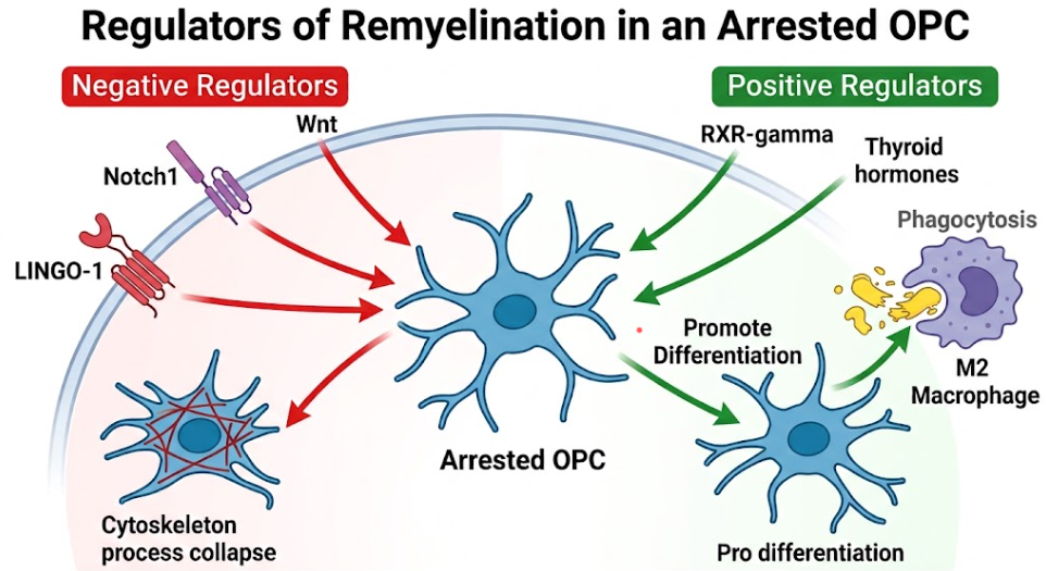
Figure 4. Molecular Regulation of Remyelination in OPCs. This diagram compares the impact of negative regulators (LINGO-1, Notch1, Wnt), which cause cytoskeleton collapse and halt remyelination, against positive regulators (RXR-gamma, thyroid hormones) and M2 macrophage phagocytosis, which promote cell differentiation for repair.
The structural and metabolic findings summarized in this review emphasize a major paradigm change in the pathophysiology of multiple sclerosis. For decades, multiple sclerosis was conceptualized as an immunological disorder of the white matter, and therapeutic development was focused almost exclusively on systemic immune suppression to reduce the frequency and severity of clinical relapses. However, as shown by recent neuroglial studies, the progressive neurological degeneration is essentially driven by a chronic bioenergetic and structural crisis of the axon-glial unit. Immunomodulatory therapies are very effective in preventing the infiltration of autoreactive immune cells across the blood–brain barrier in early, relapsing–remitting phases of the disease, but are largely ineffective in stopping the compartmentalized, smoldering neurodegeneration that characterizes progressive multiple sclerosis. (García-Domínguez, 2025)
Critical to this smoldering decay is the uncoupling of metabolic support within the axon-glial unit. Demyelinated axons lose the highly localized delivery of ATP and monocarboxylates provided by the oligodendrocyte-axon lactate shuttle. For electrical conduction along the naked axolemma to be maintained, a demyelinated neuron must undergo a massive structural reorganization, up-regulating and dispersing voltage-gated sodium channels (specifically NaV1.6) along the entire length of the denuded segment.(Duncan et al., 2021) This structural change is an exponential increase in the metabolic and ATP demands of the axon. The continuous extrusion of sodium ions by the Na+/K+-ATPase pump requires massive energy expenditure. Simultaneously, the demyelination process causes dramatic mitochondrial damage in a localized manner. This is evidenced by impaired complex I and IV activity, impaired membrane potential and failing ATP synthesis.(García-Domínguez, 2025; López-Muguruza & Matute, 2023)
An increase in energetic workload together with a reduced bioenergetic capacity lead to an intra-axonal energy crisis. Under the same conditions, the Na+/K+-ATPase pumps fail and intracellular sodium accumulates, resulting in reversal of the Na+/Ca2+ exchanger and a toxic intra-axonal calcium overload. This calcium surge activates calcium-dependent proteases such as calpains, which systematically dismantle the axonal cytoskeleton, resulting in axonal swelling, focal transection and permanent, irreversible neuroaxonal loss. So, demyelination is not only a structural deficiency but an energetic catastrophe that inevitably leads to axonal death if timely myelin repair is not installed.(Duncan et al., 2021)
There is thus a strong clinical need to go beyond pure immune suppression to neuroregenerative and bioenergetic therapies that directly target oligodendrocyte survival and foster endogenous remyelination. A major frontier for therapy is to induce oligodendrocyte resilience to oxidative stress. Oligodendrocytes have limited antioxidant defense mechanisms and are therefore particularly susceptible to mitochondrial damage mediated by ROS and RNS. Thus, the administration of anti-oxidative pathway modulators and vitagene network activators (e.g., L-carnitine or sirtuin activators) represents a promising approach to avoid bioenergetic collapse and maintain compact myelin stability.(Kim et al., 2021)
Furthermore, it is necessary to actively eliminate the pathophysiological barriers that inhibit OPC maturation to restore the arrested remyelination process. For example, generation of humanized monoclonal antibodies against the LINGO-1 pathway, such as Opicinumab, has shown promising efficacy in pre-clinical models to induce timely OPC differentiation and myelin formation. Initial clinical trials have produced mixed results, but the therapeutic window and particular patient subpopulations are still under active investigation.(López-Muguruza & Matute, 2023)
Also exciting candidates for regeneration are drugs that activate positive regulatory pathways. RXR-γ agonists and muscarinic acetylcholine receptor (mAChR) antagonists like clemastine have been reported to enhance OPC differentiation and myelin sheath thickness and length in demyelination models.(Stadelmann et al., 2019) The combination of these pro-remyelinating agents with CNS-selective thyroid hormone analogs (e.g., sobetirome) or metabolic boosters that increase lipid synthesis and modulate glial mitochondrial bioenergetics, could provide the metabolic and structural support needed to halt progressive degeneration. The future of multiple sclerosis therapy will at last be an integrative, multi-pronged therapeutic approach that simultaneously suppresses compartmentalized inflammation, protects oligodendrocytes from bioenergetic collapse, and actively stimulates endogenous remyelination.(García-Domínguez, 2025)
To summarize, oligodendrocytes and myelin sheath are the structural and metabolic basis of the architecture of white matter in the central nervous system and are central to the pathogenesis and resolution of multiple sclerosis. The switch from healthy, activity-dependent metabolic coupling to the severe bioenergetic failure and programmed lytic cell death observed in multiple sclerosis active lesions highlights the cellular interdependence within the axon-glial unit. Demyelination is a major pathological event that strips the axon of essential monocarboxylate support, initiating a devastating cascade of secondary axonal energy failure, toxic calcium overload, and permanent neurodegeneration. While the adult central nervous system maintains an endogenous capacity for repair via OPC recruitment, the chronic multiple sclerosis microenvironment presents formidable cell-intrinsic and extrinsic barriers—including LINGO-1, Notch, and Wnt signaling blockades, and dense extracellular matrix deposition—that arrest regeneration.
Overcoming this maturation arrest and protecting mature oligodendrocytes from bioenergetic collapse represent the primary frontiers of modern neurobiology. By integrating precise mechanistic insights into lipid metabolism, metabolic coupling, programmed cell death pathways, and remyelination barriers, future therapeutic strategies can transition beyond classical immunomodulation to active, long-term neuroregeneration, preserving axonal integrity and ultimately restoring neurological function in patients with multiple sclerosis.
Barres, B. A., & Raff, M. C. (1999). Axonal control of oligodendrocyte development. J Cell Biol, 147(6), 1123-1128. doi:10.1083/jcb.147.6.1123
Bokulic Panichi, L., Stanca, S., Dolciotti, C., & Bongioanni, P. (2025). The Role of Oligodendrocytes in Neurodegenerative Diseases: Unwrapping the Layers. Int J Mol Sci, 26(10). doi:10.3390/ijms26104623
Caprariello, A. V., Mangla, S., Miller, R. H., & Selkirk, S. M. (2012). Apoptosis of oligodendrocytes in the central nervous system results in rapid focal demyelination. Ann Neurol, 72(3), 395-405. doi:10.1002/ana.23606
Chamberlain, K. A., Nanescu, S. E., Psachoulia, K., & Huang, J. K. (2016). Oligodendrocyte regeneration: Its significance in myelin replacement and neuroprotection in multiple sclerosis. Neuropharmacology, 110(Pt B), 633-643. doi:10.1016/j.neuropharm.2015.10.010
Dawson, M. R., Polito, A., Levine, J. M., & Reynolds, R. (2003). NG2-expressing glial progenitor cells: an abundant and widespread population of cycling cells in the adult rat CNS. Mol Cell Neurosci, 24(2), 476-488. doi:10.1016/s1044-7431(03)00210-0
Denic, A., Wootla, B., & Rodriguez, M. (2013). CD8(+) T cells in multiple sclerosis. Expert Opin Ther Targets, 17(9), 1053-1066. doi:10.1517/14728222.2013.815726
Duncan, G. J., Simkins, T. J., & Emery, B. (2021). Neuron-Oligodendrocyte Interactions in the Structure and Integrity of Axons. Front Cell Dev Biol, 9, 653101. doi:10.3389/fcell.2021.653101
Franklin, R. J., & Ffrench-Constant, C. (2008). Remyelination in the CNS: from biology to therapy. Nat Rev Neurosci, 9(11), 839-855. doi:10.1038/nrn2480
Fünfschilling, U., Supplie, L. M., Mahad, D., Boretius, S., Saab, A. S., Edgar, J., . . . Nave, K. A. (2012). Glycolytic oligodendrocytes maintain myelin and long-term axonal integrity. Nature, 485(7399), 517-521. doi:10.1038/nature11007
García-Domínguez, M. (2025). White Matter in Crisis: Oligodendrocytes and the Pathophysiology of Multiple Sclerosis. Cells, 14(18). doi:10.3390/cells14181408
Glezer, I., Lapointe, A., & Rivest, S. (2006). Innate immunity triggers oligodendrocyte progenitor reactivity and confines damages to brain injuries. Faseb j, 20(6), 750-752. doi:10.1096/fj.05-5234fje
Goldschmidt, C., & McGinley, M. P. (2021). Advances in the Treatment of Multiple Sclerosis. Neurol Clin, 39(1), 21-33. doi:10.1016/j.ncl.2020.09.002
Jahn, O., Siems, S. B., Kusch, K., Hesse, D., Jung, R. B., Liepold, T., . . . Werner, H. B. (2020). The CNS Myelin Proteome: Deep Profile and Persistence After Post-mortem Delay. Front Cell Neurosci, 14, 239. doi:10.3389/fncel.2020.00239
Kim, D., An, H., Fan, C., & Park, Y. (2021). Identifying oligodendrocyte enhancers governing Plp1 expression. Hum Mol Genet, 30(23), 2225-2239. doi:10.1093/hmg/ddab184
Lajtha, A., Toth, J., Fujimoto, K., & Agrawal, H. C. (1977). Turnover of myelin proteins in mouse brain in vivo. Biochem J, 164(2), 323-329. doi:10.1042/bj1640323
LeBaron, F. N., Sanyal, S., & Jungalwala, F. B. (1981). Turnover rate of molecular species of sphingomyelin in rat brain. Neurochem Res, 6(10), 1081-1089. doi:10.1007/bf00964414
Li, S., & Sheng, Z. H. (2023). Oligodendrocyte-derived transcellular signaling regulates axonal energy metabolism. Curr Opin Neurobiol, 80, 102722. doi:10.1016/j.conb.2023.102722
López-Muguruza, E., & Matute, C. (2023). Alterations of Oligodendrocyte and Myelin Energy Metabolism in Multiple Sclerosis. Int J Mol Sci, 24(16). doi:10.3390/ijms241612912
Mi, S. (2008). Troy/Taj and its role in CNS axon regeneration. Cytokine Growth Factor Rev, 19(3-4), 245-251. doi:10.1016/j.cytogfr.2008.04.007
Mierzwa, A., Shroff, S., & Rosenbluth, J. (2010). Permeability of the paranodal junction of myelinated nerve fibers. J Neurosci, 30(47), 15962-15968. doi:10.1523/jneurosci.4047-10.2010
Miljković, D., & Spasojević, I. (2013). Multiple sclerosis: molecular mechanisms and therapeutic opportunities. Antioxid Redox Signal, 19(18), 2286-2334. doi:10.1089/ars.2012.5068
Narine, M., & Colognato, H. (2022). Current Insights Into Oligodendrocyte Metabolism and Its Power to Sculpt the Myelin Landscape. Front Cell Neurosci, 16, 892968. doi:10.3389/fncel.2022.892968
Nishiyama, A. (2001). NG2 cells in the brain: a novel glial cell population. Hum Cell, 14(1), 77-82.
Oudejans, E., Luchicchi, A., Strijbis, E. M. M., Geurts, J. J. G., & van Dam, A. M. (2021). Is MS affecting the CNS only? Lessons from clinic to myelin pathophysiology. Neurol Neuroimmunol Neuroinflamm, 8(1). doi:10.1212/nxi.0000000000000914
Paez, P. M., & Lyons, D. A. (2020). Calcium Signaling in the Oligodendrocyte Lineage: Regulators and Consequences. Annu Rev Neurosci, 43, 163-186. doi:10.1146/annurev-neuro-100719-093305
Philips, T., & Rothstein, J. D. (2017). Oligodendroglia: metabolic supporters of neurons. J Clin Invest, 127(9), 3271-3280. doi:10.1172/jci90610
Podbielska, M., Banik, N. L., Kurowska, E., & Hogan, E. L. (2013). Myelin recovery in multiple sclerosis: the challenge of remyelination. Brain Sci, 3(3), 1282-1324. doi:10.3390/brainsci3031282
Prineas, J. W., & Parratt, J. D. (2012). Oligodendrocytes and the early multiple sclerosis lesion. Ann Neurol, 72(1), 18-31. doi:10.1002/ana.23634
Rosetti, C. M., Maggio, B., & Oliveira, R. G. (2008). The self-organization of lipids and proteins of myelin at the membrane interface. Molecular factors underlying the microheterogeneity of domain segregation. Biochim Biophys Acta, 1778(7-8), 1665-1675. doi:10.1016/j.bbamem.2008.02.007
Simons, M., & Nave, K. A. (2015). Oligodendrocytes: Myelination and Axonal Support. Cold Spring Harb Perspect Biol, 8(1), a020479. doi:10.1101/cshperspect.a020479
Stadelmann, C., Timmler, S., Barrantes-Freer, A., & Simons, M. (2019). Myelin in the Central Nervous System: Structure, Function, and Pathology. Physiological Reviews, 99(3), 1381-1431. doi:10.1152/physrev.00031.2018
Tepavčević, V. (2021). Oligodendroglial Energy Metabolism and (re)Myelination. Life (Basel), 11(3). doi:10.3390/life11030238
Thornton, M. A., & Hughes, E. G. (2020). Neuron-oligodendroglia interactions: Activity-dependent regulation of cellular signaling. Neurosci Lett, 727, 134916. doi:10.1016/j.neulet.2020.134916
Trapp, B. D., Nishiyama, A., Cheng, D., & Macklin, W. (1997). Differentiation and death of premyelinating oligodendrocytes in developing rodent brain. J Cell Biol, 137(2), 459-468. doi:10.1083/jcb.137.2.459
Williamson, J. M., & Lyons, D. A. (2018). Myelin Dynamics Throughout Life: An Ever-Changing Landscape? Front Cell Neurosci, 12, 424. doi:10.3389/fncel.2018.00424
Yang, J., Ran, M., Li, H., Lin, Y., Ma, K., Yang, Y., . . . Yang, S. (2022). New insight into neurological degeneration: Inflammatory cytokines and blood-brain barrier. Front Mol Neurosci, 15, 1013933. doi:10.3389/fnmol.2022.1013933
Yuan, J., Amin, P., & Ofengeim, D. (2019). Necroptosis and RIPK1-mediated neuroinflammation in CNS diseases. Nat Rev Neurosci, 20(1), 19-33. doi:10.1038/s41583-018-0093-1
Zalc, B., Goujet, D., & Colman, D. (2008). The origin of the myelination program in vertebrates. Curr Biol, 18(12), R511-512. doi:10.1016/j.cub.2008.04.010
Zhang, N., Yi, R., Zhong, F., Lu, Y., Chen, W., Ke, Z., . . . Li, W. (2025). Oligodendrocytes and myelination: pioneering new frontiers in cognitive neuroscience. Front Neurosci, 19, 1618468. doi:10.3389/fnins.2025.1618468
Zhou, Y., Xiang, Y., Liu, S., Li, C., Dong, J., Kong, X., . . . Zhang, L. (2024). RIPK3 signaling and its role in regulated cell death and diseases. Cell Death Discov, 10(1), 200. doi:10.1038/s41420-024-01957-w