Our Services
Others
- DFPP
- Shockwave
- IV Drip


“This comprehensive review describes the continuum of clinical and pathologic features of multiple sclerosis highlighting the complex cellular and molecular alterations that take place as patients transition from the initial inflammatory attack to a silent phase of progressive neurodegeneration.”
Multiple sclerosis (MS) is a chronic, heterogeneous, autoimmune and neurodegenerative disorder of the central nervous system (CNS) characterized by demyelination, synaptic dysfunction, axonal injury, and progressive brain atrophy. In clinical practice, MS has been classified into fixed clinical phenotypes (e.g. Relapsing-Remitting MS [RRMS], Secondary Progressive MS [SPMS]) based on relapses and disability accumulation. However, a major paradigm shift has occurred: MS is increasingly recognized as a continuum of clinical presentation, where diffuse neuroinflammation and neurodegeneration coexist and interact from the onset of disease. This review represents a comprehensive analysis of this clinical continuum, directly addressing the transition from CIS to relapsing remitting and then to the steady accumulation of disability of Secondary Progressive MS.
The cellular and molecular mechanisms of systemic adaptive immune-mediated blood-brain barrier destruction, compartmentalized CNS-restricted inflammation, meningeal B-cell follicles, smoldering slowly expanding lesions (SELs) with paramagnetic rims, and chronic axonal mitochondrial dysfunction are detailed. The review also discusses the clinical challenges of the transition phase, underdiagnosis of SPMS in global registries and the predictive potential of emerging fluid biomarkers (including serum neurofilament light chain [sNfL] and glial fibrillary acidic protein [GFAP]) and digital instruments. Finally, we discuss the changing therapeutic landscape with clinical management moving away from traditional peripherally acting anti-inflammatory drugs to highly CNS-penetrant neuroprotective and remyelinating interventions that change the disease course and improve patient outcomes.
Multiple sclerosis (MS) is a major cause of non-traumatic neurologic disability among young adults in the United States and Europe. It typically affects people between the ages of 20 and 40 years and has a striking female predominance.(Tullman, 2013) Multiple sclerosis is characterized by autoimmune-mediated demyelination, axonal degeneration, gliosis and progressive loss of brain tissue. Clinical presentation can vary widely and include motor, sensory, visual and cognitive deficits.(Compston & Coles, 2008) Traditionally, MS has been defined into rigid, static clinical subtypes based on relapses and gradual, continuous worsening.(Uawithya et al., 2026) This phenotypic classification, first proposed in 1996 and updated in 2013, outlined boundaries between Relapsing-Remitting MS (RRMS), Primary Progressive MS (PPMS) and Secondary Progressive MS (SPMS).(Klineova & Lublin, 2018)
During the past decade, neuroimmunology has undergone a major paradigm shift. The traditional distinction between “relapsing” and “progressive” forms of MS has been challenged by current clinical, pathological, and neuroradiological research. These phenotypes do not describe separate diseases, but rather coexist on a common, continuous clinical spectrum.(Cross, Trotter, & Lyons, 2001) The pathological processes that underlie progressive disability, such as diffuse neuroaxonal loss, reactive astrogliosis and gray matter atrophy, are now known to be active from the very beginning of the disease course, even in the early, clinically stable stages of RRMS.(Dhib-Jalbut, 2002; Sie, Korn, & Mitsdoerffer, 2014)
Understanding this clinical continuum is key to optimizing patient care, particularly during the critical transition from relapsing-remitting to secondary progression. Historically, this transition has been associated with significant diagnostic delays, clinical uncertainty and lack of specific biological or radiological markers.(Klineova & Lublin, 2018) This comprehensive literature review discusses the continuum in multiple sclerosis, from the first clinical presentation to progressive disability. We bridge the gap between neurobiological research and clinical practice by explaining the cellular and molecular mechanisms in detail, in an easy-to-understand language for healthcare professionals, patients and their families. We also discuss the clinical realities of diagnosis, underdiagnosis in European registries, the emerging role of molecular and digital biomarkers, and the paradigm shift in the therapeutic landscape toward highly CNS-penetrant, neuroprotective and remyelinating interventions.
The clinical course of MS is very heterogeneous. The presentation of symptoms, the frequency of relapses and the rate of disability accumulation differ significantly among individuals.(Compston & Coles, 2008) Nevertheless, the revised 2013 clinical descriptors outline several important landmark phases along the disease continuum.(Klineova & Lublin, 2018)
Clinically Isolated Syndrome is the initial acute or subacute attack of neurologic symptoms that are consistent with inflammatory demyelination of the central nervous system. To be formally classified as CIS, the episode must have an acute or subacute onset of symptoms, which evolve over days to weeks, last for at least 24 hours, and occur in the absolute absence of fever, active infection, or systemic metabolic derangement.(Klineova & Lublin, 2018; Madineni, S V, & Bhuma, 2023) CIS usually appears as a monofocal neurological deficit, such as optic neuritis (with unilateral vision loss and painful eye movement), a brainstem-cerebellar syndrome (with double vision, vertigo or ataxia) or partial transverse myelitis (with sensory disturbances or motor weakness in the limbs). CIS may have multifocal presentation with simultaneous involvement of multiple anatomical locations within CNS.(Tullman, 2013)
The major clinical challenge in the management of CIS is to determine the individual risk of conversion to clinically definite multiple sclerosis (CDMS). The most powerful and reliable predictor of future clinical conversion is the presence of asymptomatic silent demyelinating T2 white matter lesions on baseline brain MRI, as shown in robust long-term prospective studies. These silent, pre-existing demyelinating lesions are found in about 50% to 70% of people presenting with CIS. For these individuals, the risk of conversion to CDMS is extraordinarily high, with observational studies reporting conversion rates of up to 80% in a 20-year follow-up period. In contrast, patients with a completely normal brain MRI at onset have a remarkably low risk of conversion estimated to be only 4% to 23%.(Klineova & Lublin, 2018)
A pathological cerebrospinal fluid (CSF) profile, such as an increased IgG index or the presence of oligoclonal bands (OCBs), is also a very sensitive biological marker of persistent, compartmentalized intrathecal inflammation. The presence of CSF-restricted OCBs has been shown to increase the risk of clinical conversion by 2 times even in patients with a completely normal brain MRI at baseline, increasing the risk from 4% to about 23%. Under the revised 2017 McDonald criteria, CSF-restricted oligoclonal bands can now substitute for dissemination in time, permitting an immediate definitive diagnosis of MS at the onset of the first clinical attack, thus allowing for earlier initiation of disease-modifying therapies.(Compston & Coles, 2008; Klineova & Lublin, 2018)
Relapsing-Remitting MS is diagnosed in approximately 85% of people after the first clinical event.(Andravizou et al., 2019; Antel, Antel, Caramanos, Arnold, & Kuhlmann, 2012) It is characterized by alternating episodes of acute neurological deterioration (relapse) and subsequent periods of relative clinical stability (remission) without the emergence of new symptoms.(Klineova & Lublin, 2018) Relapses are discrete, active inflammatory attacks on central nervous system myelin sheaths that last for days to several weeks and then fade gradually. Remyelination is very efficient early in the disease and clinical recovery is frequently complete. However, subsequent relapses often result in residual cumulative deficits. This incomplete recovery leads to a stepwise, staircase-like accumulation of permanent physical impairment over time as in the Figure 1.(Klineova & Lublin, 2018)
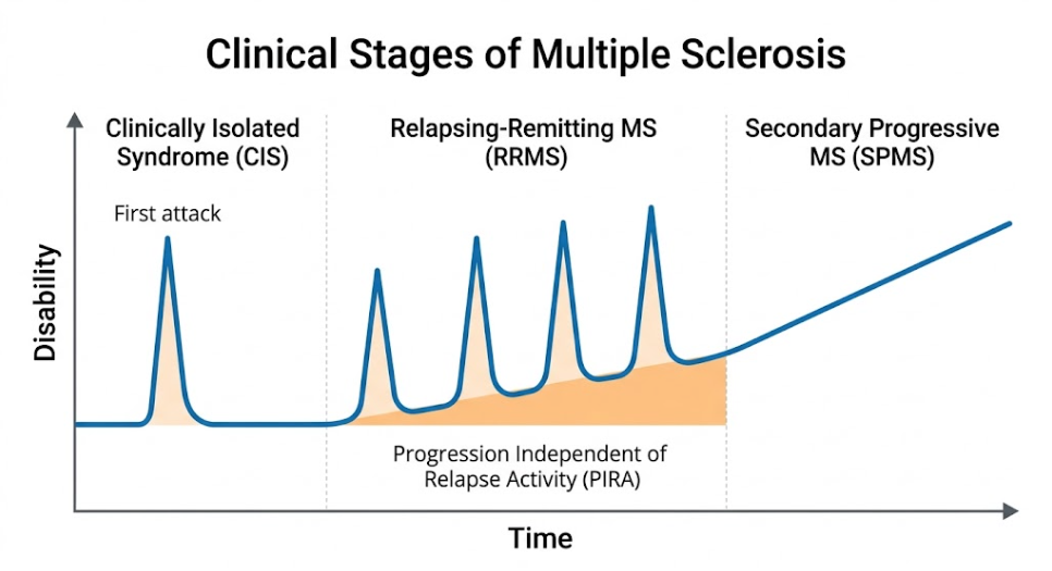
Figure 1: Spectrum and clinical continuum of multiple sclerosis. A timeline of MS disability progression over time, from the first attack (CIS) and relapsing-remitting phases (RRMS) with underlying progression (PIRA), to continuous, steady increase in disability (SPMS).
The traditional view was that disability accumulation in RRMS was entirely relapse-driven. This concept is called Relapse-Associated Worsening (RAW).(Uawithya et al., 2026) In this context, progressive disability was seen as the cumulation of partial repairs of tissues following acute inflammatory episodes. However, this assumption has been disproven by recent clinical trial databases and registries that have identified a parallel, background process called Progression Independent of Relapse Activity (PIRA).(Kappos et al., 2020; Uawithya et al., 2026) PIRA means progressive, documented accrual of neurological disability in the complete absence of recent clinical relapses.(Kappos et al., 2020)
Registry data over the long term have shown that PIRA starts very early in the RRMS phase and accounts for up to half of the total disability accumulation in relapsing patients and up to 89.1% of events in patients treated with highly effective therapies who achieve complete suppression of acute clinical relapses. This implies that a slow degenerative process, a “smoldering” one, is in place from the very beginning of the disease, silently driving progression behind the mask of acute inflammatory relapses.(Ciubotaru et al., 2024; Lublin et al., 2022; Pozzilli et al., 2023)
Most patients with RRMS who do not receive treatment will progress to Secondary Progressive Multiple Sclerosis. Median conversion time ~19-20 years after RRMS onset.(Klineova & Lublin, 2018) SPMS is defined by a progressive build-up of neurological disability and a gradual decline in physical and cognitive function independent of clinical relapses. Some SPMS patients may still have superimposed clinical relapses (active SPMS) and some may develop a non-active progressive phase with complete cessation of clinical relapse activity.(Uawithya et al., 2026)
The transition from RRMS to SPMS is an important milestone in the disease course, and is associated with a substantial physical and psychological burden for patients and caregivers.(Caseby, Woodhouse, Montgomery, Kroes, & Duddy, 2022) As the disease progresses, daily tasks become more and more difficult and often lead to decreased employment, loss of functional independence, and social isolation.(Caseby et al., 2022) SPMS transition is clinically very important, but is a huge clinical challenge to diagnose, as there are no definitive imaging or biological markers to demarcate when a patient has transitioned. Therefore, SPMS is typically a retrospective diagnosis, with 3 to 12 months of documented gradual progression of disability in the clinic required, resulting in a lengthy transition period marked by a diagnostic uncertainty lasting an average of 2.9 to 3.3 years.(Klineova & Lublin, 2018)
Approximately 10 to 15% of people are diagnosed with Primary Progressive MS. This form of MS is characterized by a slow and steady decline of neurological function from the onset of the disease, without any initial relapses or remissions.(Krieger & Sumowski, 2018) Although PPMS is clinically defined as the absence of a first relapsing phase, pathological, genetic and imaging data suggest that PPMS and SPMS are different aspects of the same progressive MS spectrum. In fact, natural history studies have demonstrated that following the onset of the progressive phase, disability accumulates at a strikingly similar rate in both PPMS and SPMS, irrespective of past relapses.(Klineova & Lublin, 2018) Moreover, the finding that up to 10% of patients with Radiologically Isolated Syndrome (RIS) can directly convert to a PPMS course suggests that the absence of a clinically evident relapsing phase in PPMS may be simply due to clinically silent, subclinical demyelinating lesions in the early stages of the disease.(Madineni et al., 2023)
To understand the evolution of the relapsing-remitting to a secondary progressive course of MS we have to focus on the changing underlying pathophysiological processes. Conversely, SPMS is characterized by chronic neurodegeneration and localized innate immunity, while RRMS is characterized by acute focal inflammation and predominantly systemic adaptive immunity.(Cree et al., 2021; Frischer et al., 2009) As shown in Figure 2.
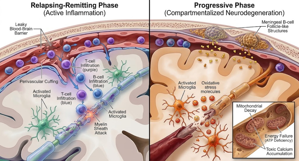
Figure 2. Two mechanisms of MS pathophysiology. The left panel shows the Relapsing-Remitting phase in which a leaky blood-brain barrier allows immune cells to actively invade the brain and attack nerve myelin. The right panel illustrates the Progressive phase characterized by compartmentalized neurodegeneration, where trapped meningeal inflammation, chronic oxidative stress and mitochondrial energy failure result in irreversible nerve damage.
The systemic adaptive immune response is the major driver of damage in the early relapsing phase of MS. Autoreactive CD4+ T helper cells (Th1 and Th17) are primed in the periphery to unknown myelin antigens. Th1 cells release proinflammatory cytokines such as interleukin (IL)-1 and interferon-gamma (IFN-γ), and Th17 cells release IL-17.(Klineova & Lublin, 2018) These cytokines up-regulate adhesion molecules and induce secretion of matrix metalloproteinases that digest the extracellular matrix of the vascular endothelial wall systematically.(Weiner, 2008) This destructive process results in disruption of the blood-brain barrier (BBB) and enables autoreactive T cells, B cells and macrophages to migrate from the bloodstream into the central nervous system parenchyma.(Compston & Coles, 2008) In the CNS these immune cells initiate a cascade of focal inflammatory attacks that result in demyelination and acute axonal injury. In the early phase of the disease, anti-inflammatory Th2 cells migrating into the CNS support tissue repair and may thus explain the clinical remissions in RRMS.(Tullman, 2013)
As the disease advances to SPMS, the inflammatory response undergoes a dramatic change.(Cree et al., 2021) The blood brain barrier often heals or closes and thus the active recruitment of systemic immune cells into the CNS wanes. Rather, the inflammatory process becomes localized within the CNS, persisting behind an intact or repaired BBB.(Cree et al., 2021) This localized inflammation is driven by resident microglia and astrocytes as well as by immune cells that migrated into the CNS in the early stages of disease and became trapped.(Klineova & Lublin, 2018)
Progressive pathology in MS is characterized by the formation of meningeal lymphoid follicle-like structures or tertiary lymphoid structures (TLS) . They are dense clusters of B lymphocytes, plasma cells and follicular dendritic cells in the meninges, especially around subpial cortical lesions.(Cree et al., 2021) These meningeal follicles release soluble toxic factors such as cytokines and antibodies, which diffuse into the cerebral cortex causing widespread subpial and cortical demyelination, microglial activation and progressive gray matter atrophy.(Klineova & Lublin, 2018)
Relapsing-remitting MS features the development of new focal white matter lesions, whereas secondary progressive MS is characterized as “smoldering MS”—the slow and steady growth of pre-existing, chronic active lesions (slowly expanding lesions, or SELs).(Mahad, Trapp, & Lassmann, 2015) In SELs an inactive, hypocellular necrotic core is surrounded by a highly active rim of activated microglia and macrophages. Susceptibility-weighted imaging (SWI) or quantitative susceptibility mapping (QSM) in MRI visualizes paramagnetic rim lesions (PRLs) at the lesion rim, which directly depicts the accumulation of iron-laden activated microglia and macrophages at the lesion edges.(Pozzilli et al., 2023) Iron-loaded microglia generate reactive oxygen species and nitric oxide, resulting in chronic demyelination and progressive axonal destruction over time, independent of infiltration of peripheral immune cells.(Uawithya et al., 2026)
Hence, in SPMS, mitochondrial dysfunction and persistent energy deficit in demyelinated axons promote axonal degeneration.(Albelo-Martínez & Rizvi, 2025) Axons are usually insulated by myelin sheaths that allow saltatory conduction and protect axons from metabolic stress. Permanent demyelination induces substantial reorganization and upregulation of voltage-gated sodium channels (NaV1.6) along the entire length of the demyelinated axolemma to preserve nerve conduction. This structural change increases the energy (adenosine triphosphate or ATP) demands of the axon.(Klineova & Lublin, 2018)
But the demyelinating process also induces simultaneous and severe mitochondrial dysfunction in the axon, with damaged mitochondrial DNA, impaired respiratory chain complexes and failing ATP production.(Albelo-Martínez & Rizvi, 2025) The failure of ATP production along with the increased energy demand results in the failure of the energy-dependent sodium-potassium ATPase pump. So, sodium builds up inside the cell and the sodium-calcium exchanger runs in reverse. This results in a toxic influx and accumulation of intracellular calcium which activates calcium dependent proteases and lipases that systematically digest the axonal cytoskeleton resulting in permanent axonal transection and neurodegeneration.(Klineova & Lublin, 2018)
Transition from RRMS to SPMS is a significant clinical bottleneck, with delays in diagnosis resulting in missed therapeutic opportunities. Healthcare professionals experience a high level of diagnostic hesitancy when officially diagnosing a patient with SPMS.(Uawithya et al., 2026) A large share of this caution is related to reimbursement policies and regulatory structures. In many countries, disease-modifying therapies (DMTs) are licensed and reimbursed primarily for RRMS and a formal diagnosis of SPMS can lead to patients losing access to their current effective treatments.(Caseby et al., 2022; Cree et al., 2021) This is to prevent this, as clinicians often continue to treat progressive patients with a “RRMS” label, continuing treatment with HETs even after recognition of insidious progression. This pragmatism emphasizes a unique clinical reality in which administrative and financial guidelines directly affect disease staging and clinical decision-making.(Caseby et al., 2022)
In addition, epidemiological studies using European MS registries (e.g. Italian, Swedish and German registries) have demonstrated that SPMS is systematically underdiagnosed in clinical practice.(Forsberg et al., 2023) Large cohorts of patients when analyzed using objective data-driven algorithms show a substantially increased proportion of SPMS patients. For example, a decision tree classifier using only a single EDSS score and the age of a patient raises the estimated proportion of SPMS in registries from a clinical baseline of ~17.7% to ~28.1%.(Forsberg et al., 2023) The decision tree classifier is a very sensitive and objective tool that could be used for the harmonization of classifications between clinical registries. The MSBase algorithm is similarly a good framework, as it includes a three-strata progression magnitude, requiring a confirmed increase in EDSS for at least 3 months and an EDSS score of 4.0 or more and pyramidal functional system score of 2.0 or more.(Forsberg et al., 2023)
A landmark Italian register study of 9,958 patients over 30 years showed decreasing rates of SPMS conversion over time, partially explained by earlier treatment initiation and therapeutic coverage.(Zanghì et al., 2025) In multivariable modeling a 10% increase in coverage was associated with 19% decreased risk of conversion to SPMS (hazard ratio 0.89, 95% confidence interval 0.87 to 0.90).(Zanghì et al., 2025) Qualitative data from the Italian ManTra project also showed that as many as 43% of patients in transition were not aware of their SPMS diagnosis , highlighting a major gap in patient-physician communication and a long time in which patients are left to deal with worsening symptoms without support.(Giovannetti et al., 2020; Ziemssen et al., 2023)
To alleviate these diagnostic delays the scientific community has adopted highly sensitive multimodal monitoring strategies. Digital biomarkers and patient reported outcome measures (PROMs) are increasingly added to clinician administered tools.(Ziemssen et al., 2023) Digital devices such as the smartphone-based FLOODLIGHT app allow passive remote monitoring of motor symptoms and active cognitive screening, capturing subtle changes in a patient’s daily life that are often missed during routine clinical visits. Cognitive function, not well characterized by the ambulation-heavy Expanded Disability Status Scale (EDSS), is now being increasingly quantified with the Symbol Digit Modalities Test (SDMT) or digital applications such as CogEval to detect early progressive cognitive decline.(Uawithya et al., 2026)
Fluid biomarkers appear to be another promising candidate for early detection of progression beside digital instruments. sNfL is a promising marker of active axonal injury. Elevated sNfL levels have been detected with highly sensitive SIMOA assays that correlate with annual progression of EDSS, future brain atrophy and cognitive decline.(Ciubotaru et al., 2024) Serum GFAP is also emerging as a marker of reactive astrogliosis and CSF sCD27 and sCD40L are emerging as markers of compartmentalized neuroinflammation and future progression.(Uawithya et al., 2026)
The therapeutic landscape of SPMS is finally changing from traditional anti-inflammatory drugs to neuroprotective and central-nervous-system-penetrant interventions.(Uawithya et al., 2026) Targeting peripherally-driven inflammation with ocrelizumab (anti-CD20 monoclonal antibody) and S1P modulators (such as siponimod and fingolimod) has been successful, but limited in efficacy in non-active progressive MS where neurodegeneration is the predominant driver of disability.(Albelo-Martínez & Rizvi, 2025; Dimitriou, Meuth, Martinez-Lapiscina, Albrecht, & Menge, 2023) Compartmentalized CNS inflammation is being directly targeted in clinical trials using small-molecule inhibitors of Bruton’s tyrosine kinase (BTK). Others include tolebrutinib, fenebrutinib and orelabrutinib. BTK inhibitors are highly able to cross the blood-brain barrier and directly inhibit activated microglia and B-cell follicles in the CNS parenchyma, which may slow smoldering MS and PIRA. Other novel approaches such as frexalimab (blocking CD40-CD40L pathway), CAR-T cell therapies targeting CD19/BCMA and remyelinating agents like clemastine are promising strategies to increase myelin repair and maintain functional reserve in patients who are entering the progressive phase of this complex disease.(Albelo-Martínez & Rizvi, 2025; Uawithya et al., 2026)
The clinical spectrum of MS is a complex and continuous continuum connecting early relapsing-remitting neuroinflammation to progressive compartmentalized neurodegeneration. CIS is the first step in this direction and RRMS is characterized by overt clinical relapses, the emergence of SPMS draws attention to the background neurodegeneration that often goes unnoticed clinically until a substantial degree of disability has been reached. MS is progressive from the very beginning, as shown by pathological processes such as systemic adaptive immune attacks, BBB breakdown, compartmentalized meningeal inflammation, slowly expanding chronic active lesions, and mitochondrial dysfunction.
The historical division of relapsing and progressive MS is slowly being replaced by a united spectrum model. Despite the huge challenge of making the transition diagnosis due to administrative policies and clinical uncertainty, the combination of advanced quantitative neuroimaging, digital remote monitoring and molecular biomarkers such as sNfL and GFAP provides a pathway toward earlier and more accurate detection of progressive disease. Ultimately, bridging these diagnostic and therapeutic gaps in the next decade is paramount. With therapies that can penetrate the CNS, target-specific interventions and remyelinating strategies, clinicians can hope to intervene in the early “window of opportunity” to change the course of the disease and to improve long-term outcomes and quality of life for those living with MS.
Albelo-Martínez, M., & Rizvi, S. (2025). Progressive multiple sclerosis: Evaluating current therapies and exploring future treatment strategies. Neurotherapeutics, 22(4), e00601. doi:10.1016/j.neurot.2025.e00601
Andravizou, A., Dardiotis, E., Artemiadis, A., Sokratous, M., Siokas, V., Tsouris, Z., . . . Hadjigeorgiou, G. M. (2019). Brain atrophy in multiple sclerosis: mechanisms, clinical relevance and treatment options. Auto Immun Highlights, 10(1), 7. doi:10.1186/s13317-019-0117-5
Antel, J., Antel, S., Caramanos, Z., Arnold, D. L., & Kuhlmann, T. (2012). Primary progressive multiple sclerosis: part of the MS disease spectrum or separate disease entity? Acta Neuropathol, 123(5), 627-638. doi:10.1007/s00401-012-0953-0
Caseby, S. C. L., Woodhouse, F. A., Montgomery, S. M., Kroes, M. A., & Duddy, M. E. (2022). Transition to secondary progressive multiple sclerosis: The consequences for patients and healthcare systems, a healthcare professional survey. Health Sci Rep, 5(1), e474. doi:10.1002/hsr2.474
Ciubotaru, A., Grosu, C., Alexa, D., Covali, R., Maștaleru, A., Leon, M. M., . . . Ignat, E. B. (2024). The Faces of “Too Late”-A Surprisingly Progressive Cohort of “Stable” Relapsing Remitting Multiple Sclerosis Patients. Medicina (Kaunas), 60(9). doi:10.3390/medicina60091401
Compston, A., & Coles, A. (2008). Multiple sclerosis. Lancet, 372(9648), 1502-1517. doi:10.1016/s0140-6736(08)61620-7
Cree, B. A. C., Arnold, D. L., Chataway, J., Chitnis, T., Fox, R. J., Pozo Ramajo, A., . . . Lassmann, H. (2021). Secondary Progressive Multiple Sclerosis: New Insights. Neurology, 97(8), 378-388. doi:10.1212/wnl.0000000000012323
Cross, A. H., Trotter, J. L., & Lyons, J. (2001). B cells and antibodies in CNS demyelinating disease. J Neuroimmunol, 112(1-2), 1-14. doi:10.1016/s0165-5728(00)00409-4
Dhib-Jalbut, S. (2002). Mechanisms of action of interferons and glatiramer acetate in multiple sclerosis. Neurology, 58(8 Suppl 4), S3-9. doi:10.1212/wnl.58.8_suppl_4.s3
Dimitriou, N. G., Meuth, S. G., Martinez-Lapiscina, E. H., Albrecht, P., & Menge, T. (2023). Treatment of Patients with Multiple Sclerosis Transitioning Between Relapsing and Progressive Disease. CNS Drugs, 37(1), 69-92. doi:10.1007/s40263-022-00977-3
Forsberg, L., Spelman, T., Klyve, P., Manouchehrinia, A., Ramanujam, R., Mouresan, E., . . . Hillert, J. (2023). Proportion and characteristics of secondary progressive multiple sclerosis in five European registries using objective classifiers. Mult Scler J Exp Transl Clin, 9(1), 20552173231153557. doi:10.1177/20552173231153557
Frischer, J. M., Bramow, S., Dal-Bianco, A., Lucchinetti, C. F., Rauschka, H., Schmidbauer, M., . . . Lassmann, H. (2009). The relation between inflammation and neurodegeneration in multiple sclerosis brains. Brain, 132(Pt 5), 1175-1189. doi:10.1093/brain/awp070
Giovannetti, A. M., Pietrolongo, E., Borreani, C., Giordano, A., Schiffmann, I., Barabasch, A., . . . Solari, A. (2020). Conversion to secondary progressive multiple sclerosis: Multistakeholder experiences and needs in Italy. PLoS One, 15(2), e0228587. doi:10.1371/journal.pone.0228587
Kappos, L., Wolinsky, J. S., Giovannoni, G., Arnold, D. L., Wang, Q., Bernasconi, C., . . . Hauser, S. L. (2020). Contribution of Relapse-Independent Progression vs Relapse-Associated Worsening to Overall Confirmed Disability Accumulation in Typical Relapsing Multiple Sclerosis in a Pooled Analysis of 2 Randomized Clinical Trials. JAMA Neurol, 77(9), 1132-1140. doi:10.1001/jamaneurol.2020.1568
Klineova, S., & Lublin, F. D. (2018). Clinical Course of Multiple Sclerosis. Cold Spring Harb Perspect Med, 8(9). doi:10.1101/cshperspect.a028928
Krieger, S. C., & Sumowski, J. (2018). New Insights into Multiple Sclerosis Clinical Course from the Topographical Model and Functional Reserve. Neurol Clin, 36(1), 13-25. doi:10.1016/j.ncl.2017.08.003
Lublin, F. D., Häring, D. A., Ganjgahi, H., Ocampo, A., Hatami, F., Čuklina, J., . . . Bermel, R. A. (2022). How patients with multiple sclerosis acquire disability. Brain, 145(9), 3147-3161. doi:10.1093/brain/awac016
Madineni, K. U. C., S V, N. P., & Bhuma, V. (2023). The Clinical Spectrum of Multiple Sclerosis in a Tertiary Care Hospital.
Mahad, D. H., Trapp, B. D., & Lassmann, H. (2015). Pathological mechanisms in progressive multiple sclerosis. Lancet Neurol, 14(2), 183-193. doi:10.1016/s1474-4422(14)70256-x
Pozzilli, C., Pugliatti, M., Vermersch, P., Grigoriadis, N., Alkhawajah, M., Airas, L., & Oreja-Guevara, C. (2023). Diagnosis and treatment of progressive multiple sclerosis: A position paper. Eur J Neurol, 30(1), 9-21. doi:10.1111/ene.15593
Sie, C., Korn, T., & Mitsdoerffer, M. (2014). Th17 cells in central nervous system autoimmunity. Exp Neurol, 262 Pt A, 18-27. doi:10.1016/j.expneurol.2014.03.009
Tullman, M. J. (2013). Overview of the epidemiology, diagnosis, and disease progression associated with multiple sclerosis. Am J Manag Care, 19(2 Suppl), S15-20.
Uawithya, E., Mytych, J. S., Muwenda, I., Reidy, M., Khan, M., & Mao-Draayer, Y. (2026). Challenges and Innovation for Diagnosing and Treatment of Secondary Progressive Multiple Sclerosis. International Journal of Molecular Sciences, 27(10), 4558.
Weiner, H. L. (2008). A shift from adaptive to innate immunity: a potential mechanism of disease progression in multiple sclerosis. J Neurol, 255 Suppl 1, 3-11. doi:10.1007/s00415-008-1002-8
Zanghì, A., Copetti, M., Avolio, C., Paolicelli, D., Romeo, M. A. L., Patti, F., . . . D’Amico, E. (2025). Multiple sclerosis from onset to secondary progression: a 30-year Italian register study. J Neurol Neurosurg Psychiatry, 96(11), 1061-1069. doi:10.1136/jnnp-2025-335958
Ziemssen, T., Bhan, V., Chataway, J., Chitnis, T., Campbell Cree, B. A., Havrdova, E. K., . . . Hach, T. (2023). Secondary Progressive Multiple Sclerosis: A Review of Clinical Characteristics, Definition, Prognostic Tools, and Disease-Modifying Therapies. Neurol Neuroimmunol Neuroinflamm, 10(1). doi:10.1212/nxi.0000000000200064