Our Services
Others
- DFPP
- Shockwave
- IV Drip


“This review reveals the silent crisis within the inner ear, showing how unprotected mitochondrial DNA mutations and disastrous free radical storms imprison outer hair cells in a vicious cycle of self-destruction, silencing our hearing forever.”
Worldwide, hearing loss is the most prevalent sensory impairment and has profound impacts on cognitive health and quality of life. A major feature of acquired and hereditary sensorineural hearing loss (SNHL) is the progressive degeneration of the sensory epithelium of the mammalian cochlea. The OHCs of these cells serve as mechanical amplifiers of the ear, converting acoustic vibrations into electrical signals. OHCs are postmitotic, non-regenerating, and have a high metabolic demand and are thus particularly vulnerable to stress. In this review, we study the pathophysiology of OHC death in hearing loss, with special interest in mitochondrial DNA (mtDNA) mutations and reactive oxygen species (ROS) overload. We follow the trail of the unprotected mitochondrial genome being prone to mutations that compromise oxidative phosphorylation (OXPHOS). This impairment leads to a vicious cycle of electron leakage, lipid peroxidation and collapse of membrane fluidity. Finally, the accumulated insults activate the intrinsic apoptotic pathway via Bax/Bak, leading to caspase-dependent cell death of OHCs. We also discuss paradigms in single-cell transcriptomics, including somatic ‘cryptic’ mutations, and explore the therapeutic potential of mitochondria-targeted antioxidants for cochlear health preservation.
The mammalian cochlea is a biophysical engineering marvel, capable of discriminating and amplifying sounds over a large range of frequencies. This task is divided in the organ of Corti between inner hair cells (IHCs) that transmit auditory signals to the brain and outer hair cells (OHCs) that provide active mechanical amplification of traveling sound waves.(Lin et al., 2024) As sound vibrations move thru the cochlea, they cause the stereocilia on the apical surfaces of OHCs to be displaced. This mechanical shearing opens transduction ion channels to generate a receptor current that drives OHC electromotility, a physical amplification of the vibration of the basilar membrane.(Lin et al., 2024)
This electromechanical amplification requires continuous, rapid mechanical work and, therefore, the metabolic demand within the OHCs is very high. To meet this demand, OHCs are packed with mitochondria, especially along their lateral walls and under their nuclei. Mitochondria are thus the primary energy providers for hearing. However, such a high metabolic activity also implies that OHCs are exposed to byproducts of aerobic respiration all the time.(Miura et al., 2022; Wijnker, Sequeira, Kuster, & Velden, 2019) Unlike most other cell types, mammalian hair cells are post-mitotic, and do not regenerate once they are lost. Thus, loss of hair cells leads to permanent hearing impairment.(Esteves & Caria, 2026; Inês Silva, Malitckii, Santos, & Vilaça, 2023)
Prolonged exposure to high environmental sounds, ototoxic drugs or natural aging can cause overload of this system, mostly affecting OHCs located at the basal turn of the cochlea, tuned to high frequency sounds. Free radical damage and metabolic stress are inherently more damaging for basal turn OHCs. This differential vulnerability is why most progressive hearing loss starts with loss of high-frequency sensitivity. Understanding the molecular basis of this susceptibility is important for developing preventive strategies.(Uchida, Sugiura, Nakashima, Ando, & Shimokata, 2012)
Mitochondria contain their own genome (mtDNA), which is a circular molecule that is inherited maternally. It contains 37 genes, among which are 13 polypeptides that are essential components of the respiratory chain and 2 rRNAs and 22 tRNAs that are necessary for mitochondrial translation.(Wallace, 2005) Unlike nuclear DNA, mtDNA lacks the protective histone proteins and robust repair pathways found in the nucleus. mtDNA is located very near the electron transport chain (ETC), the main site of free radical production in the cell, and is therefore under continuous attack from reactive oxygen species (ROS). Environmental factors cause mtDNA to mutate as much as 10,000 times faster than nuclear DNA.(Pfeiffer et al., 2003; Wijnker et al., 2019)
mtDNA mutations result in defective synthesis of respiratory chain subunits, impairing oxidative phosphorylation and metabolic homeostasis. Hereditary hearing loss is associated with several mutations in mtDNA that lead to severe auditory phenotypes. For example, the MT-RNR1 gene codes for the 12S ribosomal RNA and the m.1555A>G mutation is a classic example.(X. Li et al., 2004) This mutation modifies the spatial structure of the mitochondrial ribosome, thus making it more similar to the bacterial ribosome and increasing the susceptibility of the host to aminoglycoside-induced ototoxicity. Point mutations in mitochondrial tRNA genes are also very common in deafness syndromes. For instance, mutations A7445G, 7472insC, and G8363A in the MT-TS1 gene coding for tRNA^Ser(UCN) influence the rate of tRNA precursor processing, reducing the steady-state levels of tRNA and stopping mitochondrial protein synthesis.(Lin et al., 2024; Xu & Yang, 2025)
A typical example is the heteroplasmic A3243G mutation of the tRNA^Leu(UUR) gene causing maternally inherited diabetes and deafness (MIDD) and MELAS. The A3243G mutation alters crucial taurine modifications within the anticodon loop of the tRNA, leading to a defective Complex I ND6 subunit. The C3388A change is in the ND1 subunit of Complex I. The T12201C mutation in the tRNA^His gene affects histidyl-tRNA metabolism and severely impacts respiratory chain complexes leading to bioenergetic collapse.(Esteves & Caria, 2026; Tanaka et al., 2004)
Mutations in mtDNA are not only inherited but also somatic mtDNA mutations increase in the cochlea with aging.(Van Campen, Murphy, Franks, Mathias, & Toraason, 2002) Such nuclear-encoded mitochondrial maintenance proteins are defective, and thus accelerate this somatic decay.(Kudryavtseva et al., 2016) Mutations or deficiencies in the mitochondrial DNA polymerase gamma (POLG), which is responsible for the fidelity of mtDNA replication, result in a dramatic increase in the accumulation of somatic mtDNA point mutations and deletions. This results in early loss of hair cells and increased age-related hearing loss.(P. Li et al., 2023)
mtDNA mutations, when affecting the structural integrity of the subunits of the respiratory chain, cause a destabilization of the electrochemical gradient across the inner mitochondrial membrane.(Inês Silva et al., 2023; Swerdlow et al., 2017) Inside the mitochondrial electron transport chain, there are seven distinct sites of superoxide production with the highest capacity of electron leakage at Complex I and Complex III. Under normal circumstances electrons flow smoothly from electron donors to oxygen.(Esteves & Caria, 2026) But in a mutated or dysfunctional state, electrons are transferred prematurely to molecular oxygen, resulting in the superoxide radical anion (O2•-).(Inês Silva et al., 2023; Wijnker et al., 2019)
The superoxide is converted to hydrogen peroxide (H2O2) by the endogenous superoxide dismutase (e.g. cytoplasmic SOD1, mitochondrial matrix SOD2).(Umeda et al., 2005) The Fenton chemistry is catalyzed by iron and other transition metals to decompose hydrogen peroxide into the highly reactive hydroxyl radical (•OH) that attacks all nearby macromolecules such as lipids, proteins and DNA. That creates a vicious circle feeding on itself.(Miura et al., 2022) mtDNA mutations cause OXPHOS dysfunction and electron leakage, leading to ROS overload. These free radicals then attack the mtDNA that is close by and unshielded, causing more mutations and deletions, which cause more severe respiratory chain dysfunction and ROS production.(Pei et al., 2016)
Such ROS excess has disastrous physical effects on the structural and mechanical properties of OHCs. ROS directly react with the polyunsaturated fatty acids in the cell and mitochondrial membranes initiating a lipid peroxidation cascade which produces toxic end-products like malondialdehyde (MDA) and 4-hydroxynonenal (4-HNE).(Yamasoba et al., 2013) These lipid peroxides damage the membrane integrity and reduce plasma membrane fluidity considerably. Plasma membrane fluidity is key to the lateral wall micromechanics of OHCs, allowing for fast contraction and extension during sound amplification. Lipid peroxidation decreases the fluidity of membranes, which impairs OHC electromotility, resulting in a direct loss of cochlear sensitivity.(Liu et al., 2012)
As illustrated in the Figure 1. depicting the cellular cascade of mitochondrial oxidative stress (see below Figure 1.), the ROS overload leads to damage to adjacent structures such as the stria vascularis and spiral ganglion neurons. Basal OHCs are particularly prone to this oxidative stress because they have intrinsically lower levels of endogenous antioxidants than their apical counterparts, i.e. they are easily overwhelmed during metabolic stress.(Van Campen et al., 2002)
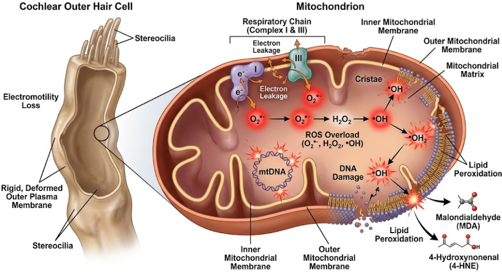
Figure 1. The Outer Hair Cell Mitochondrial Oxidative Stress Loop. This diagram depicts the metabolic cascade of OHCs under stress. It points to the mitochondria as the main site of ROS production. When compromised (by mtDNA mutations or environmental toxins) electrons leak from Complex I and Complex III to produce superoxide (O2•-), which is converted to hydrogen peroxide (H2O2) and then to hydroxyl radicals (•OH). Such radicals cause lipid peroxidation of the cell membrane, resulting in the formation of MDA and 4-HNE, which directly decrease membrane fluidity and damage OHC electromotility.
When the ROS accumulation and mitochondrial damage exceed the ability of the cell to deal with it, the OHC switches from metabolic dysfunction to an active program of self-destruction. This programmed cell death is mediated mainly by the intrinsic or mitochondrial-dependent apoptotic pathway. The loss of mitochondrial outer membrane integrity is the key initiator for this pathway.(Fu et al., 2021; Inês Silva et al., 2023)
Oxidative stress induces damage to the inner mitochondrial membrane, leading to the collapse of the mitochondrial membrane potential (ΔΨm) and the opening of the mitochondrial permeability transition pore (mPTP).(Nishigaki et al., 2007) This allows the release of pro-apoptotic factors from the mitochondrial intermembrane space into the cytosol. Cytochrome c is the key of these factors. In the cytosol, Cytochrome c in dATP-dependently binds to the adapter protein Apaf-1 and procaspase-9 to form a wheel-like multi-protein complex, called the apoptosome.(Inês Silva et al., 2023) The apoptosome is an activating platform that cleaves and activates the initiator caspase-9, which cleaves and activates the downstream executioner caspases-3 and -7. These active caspases cleave structural proteins and activate endonucleases resulting in DNA fragmentation, cytoskeletal breakdown and cell shrinkage.(Fujimoto & Yamasoba, 2014)
This apoptotic cascade is tightly controlled by the Bcl-2 family of proteins, the gatekeepers of mitochondrial membrane permeability. The Anti-apoptotic members (e.g., Bcl-2) act to maintain the integrity of the membrane, whereas the pro-apoptotic members (e.g., Bax and Bak) promote pore formation.(Johnson, Y Zheng, Bykhovskaya, Spirina, & Fischel-Ghodsian, 2001) Antiapoptotic Bcl-2 is downregulated and proapoptotic Bax and Bak are activated with constant excess ROS. Bax and Bak oligomerize on the mitochondrial outer membrane to form macromolecular pores that directly promote Cytochrome c release.(Xie et al., 2022)
The cell also undergoes characteristic morphological changes such as nuclear condensation and stereociliary disarray (shown in the apoptotic execution schematic, see Figure 2. below) resulting in cell death without damage to the surrounding tissues.(Teraoka, Hato, Inufusa, & You, 2024) Since OHCs are not regenerative, this apoptotic cascade causes a permanent silencing of the active amplifiers of the cochlea.(Esteves & Caria, 2026; Meng et al., 2025)
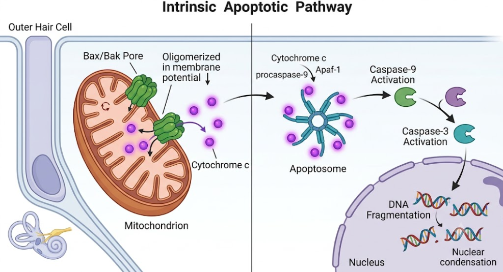
Figure 2. The Outer Hair Cell Caspase-Dependent Apoptotic Pathway. This schematic illustrates the molecular steps of intrinsic apoptosis. ROS-induced damage results in oligomerization of Bax and Bak and permeabilization of the outer mitochondrial membrane with collapse of the membrane potential (ΔΨm). This causes the release of Cytochrome c, which binds Apaf-1 and procaspase-9 to form the apoptosome. Activated caspase-9 cleavage of procaspase-3 to active caspase-3 results in translocation to the nucleus to execute DNA fragmentation and chromatin condensation.
Mutations in mtDNA maternally inherited have been associated with several specific syndromes of congenital deafness. However, recent advances in single-cell multi-omics have created a paradigm shift in understanding age-related cochlear degeneration.(Chen et al., 2026; Domínguez-de-la-Cruz et al., 2020) In tissue aggregate sequencing, somatic mutations specific to single cells, called “cryptic” mutations, are entirely hidden, as they are averaged out in bulk samples. Yet, single-cell mtDNA datasets from post-mitotic tissues illustrate that these cryptic mutations accumulate steadily over the lifespan, accounting for the vast majority of mtDNA variation in advanced age and reaching functionally significant levels of heteroplasmy that align with species-specific mid-to-late life.(Domínguez-de-la-Cruz et al., 2020; Pérez-Amado et al., 2020)
The accumulation of cryptic mtDNA mutations in OHCs correlates with the hallmarks of aging, most notably loss of proteostasis, ER stress and chronic inflammation. Cryptic mutations interrupt translation of mitochondrial encoded respiratory proteins resulting in the accumulation of misfolded or unfolded mitochondrial proteins. This results in the unfolded protein response (UPR) and ER stress with activation of chaperones and stress-response pathways (e.g. the ATF4 and p53 pathways).(Chen et al., 2026) Importantly, studies have demonstrated that dietary manipulations like calorie restriction can greatly slow the rate of accumulation of these cryptic mtDNA mutations, and delay the onset of age-related hearing loss. Calorie restriction maintains the quality control of mitochondria by maintaining the balance of mitochondrial fusion/fission and promoting mitophagy which selectively removes damaged mitochondria before they can trigger apoptosis.(Nishigaki et al., 2007)
These mechanistic insights have opened exciting therapeutic avenues targeting mitochondrial homeostasis and antioxidant defenses in a restorative fashion. Clinical trials with conventional food-derived antioxidants, including vitamins C and E, have given equivocal results, primarily due to their poor bioavailability and inability to cross the blood-labyrinth barrier to reach mitochondria.(Steinhubl, 2008) To overcome this barrier scientists have developed mitochondria-targeted antioxidants such as MitoQ and SkQR1. These compounds comprise an antioxidant moiety covalently conjugated to a lipophilic triphenylphosphonium (TPP) cation. The positive charge of TPP endows high lipophilicity, which leads to accumulation of the antioxidant several hundred-fold directly in the inner mitochondrial membrane due to the high negative potential of the mitochondrial matrix, where it scavenges ROS at their primary site of generation, protecting OHCs from apoptotic death.(Green et al., 2025; Inês Silva et al., 2023)
Also, natural compounds that activate the nuclear factor erythroid 2-related factor 2 (Nrf2) pathway may be useful for improving the intrinsic protective mechanisms of the cochlea.(Collino et al., 2006) Nrf2 is the master transcriptional regulator of cell homeostasis binding to antioxidant response elements (ARE) located in the promoter regions of genes encoding cytoprotective enzymes such as heme oxygenase-1 (HO-1), superoxide dismutases (SODs) and glutathione peroxidase (GPx).(Someya & Prolla, 2010) For example, the major bioactive constituent of Panax notoginseng, Notoginsenoside R1 (NGR1), can protect auditory cells and cochlear explants from cisplatin ototoxicity. NGR1 increased the expression of HO-1 at mRNA and protein levels, reduced the generation of ROS, cleaved caspase-3 and prevented loss of OHC stereociliary bundles.(Benkafadar et al., 2019) The protective effects of NGR1 are abolished by the pharmacological inhibition of HO-1 with zinc protoporphyrin IX (ZNPPIX), indicating the relevance of the HO-1 pathway for the protection of auditory cells.(Inês Silva et al., 2023; Van Campen et al., 2002)
To prevent and treat sensorineural hearing loss, we must move beyond reactive management to precision, mechanism-based interventions. Modern therapies aim to target the unshielded mitochondrial genome, prevent ROS-mediated lipid peroxidation, and actively inhibit the Bax/Bak-mediated apoptotic pathway, so as to preserve the whispering powerhouses of the inner ear and maintain OHC electromotility, ultimately protecting human hearing across the lifespan.
Benkafadar, N., François, F., Affortit, C., Casas, F., Ceccato, J. C., Menardo, J., . . . Wang, J. (2019). ROS-Induced Activation of DNA Damage Responses Drives Senescence-Like State in Postmitotic Cochlear Cells: Implication for Hearing Preservation. Mol Neurobiol, 56(8), 5950-5969. doi:10.1007/s12035-019-1493-6
Chen, Y., Shi, H., Xiao, M., Pan, H., Yu, X., Zhu, Y., . . . Shi, S. (2026). Mitochondrial DNA mutations and intercellular mitochondrial transfer in cancer: mechanisms, biological effects, and clinical potential. Biomark Res, 14(1), 29. doi:10.1186/s40364-026-00902-6
Collino, M., Aragno, M., Mastrocola, R., Gallicchio, M., Rosa, A. C., Dianzani, C., . . . Fantozzi, R. (2006). Modulation of the oxidative stress and inflammatory response by PPAR-gamma agonists in the hippocampus of rats exposed to cerebral ischemia/reperfusion. Eur J Pharmacol, 530(1-2), 70-80. doi:10.1016/j.ejphar.2005.11.049
Domínguez-de-la-Cruz, E., Muñoz, M. L., Pérez-Muñoz, A., García-Hernández, N., Moctezuma-Meza, C., & Hinojosa-Cruz, J. C. (2020). Reduced mitochondrial DNA copy number is associated with the haplogroup, and some clinical features of breast cancer in Mexican patients. Gene, 761, 145047. doi:10.1016/j.gene.2020.145047
Esteves, F., & Caria, H. (2026). Genetic and Environmental Factors Shaping Hearing Loss: Xenobiotics, Mechanisms and Translational Perspectives. J Xenobiot, 16(1). doi:10.3390/jox16010027
Fu, X., Wan, P., Li, P., Wang, J., Guo, S., Zhang, Y., . . . Chai, R. (2021). Mechanism and Prevention of Ototoxicity Induced by Aminoglycosides. Frontiers in Cellular Neuroscience, Volume 15 – 2021. doi:10.3389/fncel.2021.692762
Fujimoto, C., & Yamasoba, T. (2014). Oxidative stresses and mitochondrial dysfunction in age-related hearing loss. Oxid Med Cell Longev, 2014, 582849. doi:10.1155/2014/582849
Green, A. P., Klimm, F., Marshall, A. S., Leetmaa, R., Aryaman, J., Gómez-Durán, A., . . . Jones, N. S. (2025). Cryptic mitochondrial DNA mutations coincide with mid-late life and are pathophysiologically informative in single cells across tissues and species. Nat Commun, 16(1), 2250. doi:10.1038/s41467-025-57286-8
Inês Silva, M., Malitckii, E., Santos, T. G., & Vilaça, P. (2023). Review of conventional and advanced non-destructive testing techniques for detection and characterization of small-scale defects. Progress in Materials Science, 138, 101155. doi:https://doi.org/10.1016/j.pmatsci.2023.101155
Johnson, K. R., Y Zheng, Q., Bykhovskaya, Y., Spirina, O., & Fischel-Ghodsian, N. (2001). A nuclear-mitochondrial DNA interaction affecting hearing impairment in mice. Nature Genetics, 27(2), 191-194. doi:10.1038/84831
Kudryavtseva, A. V., Krasnov, G. S., Dmitriev, A. A., Alekseev, B. Y., Kardymon, O. L., Sadritdinova, A. F., . . . Snezhkina, A. V. (2016). Mitochondrial dysfunction and oxidative stress in aging and cancer. Oncotarget, 7(29), 44879-44905. doi:10.18632/oncotarget.9821
Li, P., Li, S., Wang, L., Li, H., Wang, Y., Liu, H., . . . Zhang, Y. (2023). Mitochondrial dysfunction in hearing loss: Oxidative stress, autophagy and NLRP3 inflammasome. Frontiers in Cell and Developmental Biology, Volume 11 – 2023. doi:10.3389/fcell.2023.1119773
Li, X., Fischel-Ghodsian, N., Schwartz, F., Yan, Q., Friedman, R. A., & Guan, M. X. (2004). Biochemical characterization of the mitochondrial tRNASer(UCN) T7511C mutation associated with nonsyndromic deafness. Nucleic Acids Res, 32(3), 867-877. doi:10.1093/nar/gkh226
Lin, Y. C., Ho, Y. J., Lin, Y. Y., Liao, A. H., Kuo, C. Y., Chen, H. K., . . . Shih, C. P. (2024). Notoginsenoside R1 Attenuates Cisplatin-Induced Ototoxicity by Inducing Heme Oxygenase-1 Expression and Suppressing Oxidative Stress. Int J Mol Sci, 25(21). doi:10.3390/ijms252111444
Liu, L., Feng, D., Chen, G., Chen, M., Zheng, Q., Song, P., . . . Chen, Q. (2012). Mitochondrial outer-membrane protein FUNDC1 mediates hypoxia-induced mitophagy in mammalian cells. Nat Cell Biol, 14(2), 177-185. doi:10.1038/ncb2422
Meng, L., Liu, S., Luo, J., Tu, Y., Li, T., Li, P., . . . Shi, L. (2025). Oxidative stress and reactive oxygen species in otorhinolaryngological diseases: insights from pathophysiology to targeted antioxidant therapies. Redox Rep, 30(1), 2458942. doi:10.1080/13510002.2025.2458942
Miura, S., Sasaki, A., Kasai, S., Sugawara, T., Maeda, Y., Goto, S., . . . Matsubara, A. (2022). Association of mitochondrial DNA haplogroup and hearing impairment with aging in Japanese general population of the Iwaki Health Promotion Project. J Hum Genet, 67(6), 369-375. doi:10.1038/s10038-022-01011-6
Nishigaki, Y., Yamada, Y., Fuku, N., Matsuo, H., Segawa, T., Watanabe, S., . . . Tanaka, M. (2007). Mitochondrial haplogroup N9b is protective against myocardial infarction in Japanese males. Hum Genet, 120(6), 827-836. doi:10.1007/s00439-006-0269-z
Pei, H., Yang, Y., Zhao, H., Li, X., Yang, D., Li, D., & Yang, Y. (2016). The Role of Mitochondrial Functional Proteins in ROS Production in Ischemic Heart Diseases. Oxid Med Cell Longev, 2016, 5470457. doi:10.1155/2016/5470457
Pérez-Amado, C. J., Tovar, H., Gómez-Romero, L., Beltrán-Anaya, F. O., Bautista-Piña, V., Dominguez-Reyes, C., . . . Jiménez-Morales, S. (2020). Mitochondrial DNA Mutation Analysis in Breast Cancer: Shifting From Germline Heteroplasmy Toward Homoplasmy in Tumors. Front Oncol, 10, 572954. doi:10.3389/fonc.2020.572954
Pfeiffer, K., Gohil, V., Stuart, R. A., Hunte, C., Brandt, U., Greenberg, M. L., & Schägger, H. (2003). Cardiolipin stabilizes respiratory chain supercomplexes. J Biol Chem, 278(52), 52873-52880. doi:10.1074/jbc.M308366200
Someya, S., & Prolla, T. A. (2010). Mitochondrial oxidative damage and apoptosis in age-related hearing loss. Mech Ageing Dev, 131(7-8), 480-486. doi:10.1016/j.mad.2010.04.006
Steinhubl, S. R. (2008). Why have antioxidants failed in clinical trials? Am J Cardiol, 101(10a), 14d-19d. doi:10.1016/j.amjcard.2008.02.003
Swerdlow, R. H., Koppel, S., Weidling, I., Hayley, C., Ji, Y., & Wilkins, H. M. (2017). Mitochondria, Cybrids, Aging, and Alzheimer’s Disease. Prog Mol Biol Transl Sci, 146, 259-302. doi:10.1016/bs.pmbts.2016.12.017
Tanaka, M., Cabrera, V. M., González, A. M., Larruga, J. M., Takeyasu, T., Fuku, N., . . . Shimodaira, H. (2004). Mitochondrial genome variation in eastern Asia and the peopling of Japan. Genome Res, 14(10a), 1832-1850. doi:10.1101/gr.2286304
Teraoka, M., Hato, N., Inufusa, H., & You, F. (2024). Role of Oxidative Stress in Sensorineural Hearing Loss. Int J Mol Sci, 25(8), 4146.
Uchida, Y., Sugiura, S., Nakashima, T., Ando, F., & Shimokata, H. (2012). [Estimates of the size of the hearing-impaired elderly population in Japan and 10-year incidence of hearing loss by age, based on data from the National Institute for Longevity Sciences-Longitudinal Study of Aging (NILS-LSA)]. Nihon Ronen Igakkai Zasshi, 49(2), 222-227. doi:10.3143/geriatrics.49.222
Umeda, N., Suzuki, T., Yukawa, M., Ohya, Y., Shindo, H., Watanabe, K., & Suzuki, T. (2005). Mitochondria-specific RNA-modifying enzymes responsible for the biosynthesis of the wobble base in mitochondrial tRNAs. Implications for the molecular pathogenesis of human mitochondrial diseases. J Biol Chem, 280(2), 1613-1624. doi:10.1074/jbc.M409306200
Van Campen, L. E., Murphy, W. J., Franks, J. R., Mathias, P. I., & Toraason, M. A. (2002). Oxidative DNA damage is associated with intense noise exposure in the rat. Hear Res, 164(1-2), 29-38. doi:10.1016/s0378-5955(01)00391-4
Wallace, D. C. (2005). A mitochondrial paradigm of metabolic and degenerative diseases, aging, and cancer: a dawn for evolutionary medicine. Annu Rev Genet, 39, 359-407. doi:10.1146/annurev.genet.39.110304.095751
Wijnker, P. J. M., Sequeira, V., Kuster, D. W. D., & Velden, J. V. (2019). Hypertrophic Cardiomyopathy: A Vicious Cycle Triggered by Sarcomere Mutations and Secondary Disease Hits. Antioxid Redox Signal, 31(4), 318-358. doi:10.1089/ars.2017.7236
Xie, C., Zhuang, X.-X., Niu, Z., Ai, R., Lautrup, S., Zheng, S., . . . Fang, E. F. (2022). Amelioration of Alzheimer’s disease pathology by mitophagy inducers identified via machine learning and a cross-species workflow. Nature Biomedical Engineering, 6(1), 76-93. doi:10.1038/s41551-021-00819-5
Xu, S., & Yang, N. (2025). The Role and Research Progress of Mitochondria in Sensorineural Hearing Loss. Molecular Neurobiology, 62(6), 6913-6921. doi:10.1007/s12035-024-04470-4
Yamasoba, T., Lin, F. R., Someya, S., Kashio, A., Sakamoto, T., & Kondo, K. (2013). Current concepts in age-related hearing loss: epidemiology and mechanistic pathways. Hear Res, 303, 30-38. doi:10.1016/j.heares.2013.01.021