Our Services
Others
- DFPP
- Shockwave
- IV Drip


“This review of the literature elucidates the mechanism by which the structural degradation of PRG4/lubricin results in failure of boundary lubrication, converting forces of joint articulation into damaging surface shear stresses that cause superficial zone chondrocyte apoptosis and progressive cartilage atrophy in osteoarthritis.”
Osteoarthritis (OA) is a complex whole-joint disease with progressive articular cartilage degeneration, subchondral bone remodeling and chronic synovial inflammation. Osteoarthritis has traditionally been regarded as a mechanical phenomenon of wear and tear. Modern biomechanical research demonstrates that osteoarthritis is a consequence of a catastrophic breakdown of the joint tribology and interfacial protective mechanisms. A critical event in the early stages of osteoarthritis is the loss of boundary lubrication at the articular surface, primarily mediated by Proteoglycan 4 (PRG4), also known as lubricin or superficial zone protein. PRG4 is a specialized mucinous glycoprotein that is produced by the superficial zone chondrocytes and synovial fibroblasts. In healthy articulation, PRG4 forms a dense, hydrated macromolecular brush layer, the lamina splendens, which reduces solid-to-solid contact during high compressive loads and low sliding velocities. Pro-inflammatory cytokines, reactive oxygen species and proteolytic enzymes cleave PRG4, while its biosynthesis is downregulated in osteoarthritic conditions. This degradation ruptures the boundary lubricating film and leads to abrupt increase of surface coefficient of friction. When the superficial top zone of cartilage is deprived of friction-reducing coverage, it becomes susceptible to excessive unbuffered shear forces during joint movement. These stresses induce local shear stresses that cause mechanical deformation, mitochondrial dysfunction, depletion of the pericellular matrix, and widespread apoptosis of superficial zone chondrocytes via caspase-3 activation. Loss of these essential cells impairs the maintenance of the extracellular matrix, leading to fibrillation of the superficial zone, delamination and progressive cartilage atrophy. Moreover, loss of functional PRG4 disrupts key anti-inflammatory signaling pathways mediated by CD44 and Toll-like receptors (TLR2/4) and creates a vicious mechano-inflammatory feedback loop that accelerates joint degradation. This review aims to review the current literature to outline the molecular and biomechanical mechanisms linking PRG4 degradation, boundary lubrication failure and superficial zone atrophy in osteoarthritis.
Articular cartilage offers an extremely efficient low-friction bearing surface that enables smooth pain-free articulation across synovial joints for a human life span (Li et al., 2026). The physiological function of this tissue depends on a delicate balance between fluid phase load bearing and boundary mode surface lubrication (McNary, Athanasiou, & Reddi, 2012). Surprisingly, coefficient of friction values measured in healthy diarthrodial joints are often in the range of 0.001 to 0.02 under physiological loading conditions. However, when joint homeostasis fails due to aging, mechanical trauma or metabolic dysfunction, this exceptional tribological operation deteriorates, precipitating the onset and progression of OA(Li et al., 2026).
Osteoarthritis is the most common degenerative joint disease in the world and affects hundreds of millions of people around the world with a significant clinical challenge. From a pathological point of view, initial osteoarthritic changes develop predominantly in the articular surface in the superficial zone(W. Chen, Ye, Zhang, Xie, & Xu, 2025). The superficial zone is the most superficial and thinnest layer of articular cartilage, with chondrocytes that are flattened parallel to the joint surface and densely woven type II collagen fibrils. The specialized zone is the main mechanical barrier to the tensile and shear forces generated during articulation and at the same time the factory for the synthesis of the essential boundary lubricants(Saito, 2022; Waller et al., 2013).
Proteoglycan 4 (PRG4) is a multifunctional mucinous glycoprotein encoded by the PRG4 gene on human chromosome 1q25 and is essential for maintenance of articular surface integrity(Rhee et al., 2005). PRG4 is also called lubricin or superficial zone protein (SZP) and is uniquely secreted into the synovial fluid and onto the cartilage tidemark by superficial zone chondrocytes and synovial type B fibroblasts(Waller et al., 2013). PRG4 acts as the primary boundary lubricant under high axial loads and low sliding speeds, kinematic regimes where interstitial fluid pressurization subsides, acting as a protective molecular shield preventing direct contact of opposing surface asperities(Waller et al., 2013).
Boundary lubrication does not occur when PRG4 is transcriptionally downregulated or enzymatically degraded. Such a tribological failure causes sudden frictional spikes all over the cartilage surface exposing the superficial chondrocytes to extreme unbuffered kinetic shear stresses(Waller et al., 2013). Chronic friction induces stress responses in the cells, depletion of their energy resources and programd cell death (apoptosis) in the superficial layer. Superficial zone chondrocytes die causing loss of structural integrity of the superficial collagen-proteoglycan meshwork and subsequent cartilage fibrillation, surface delamination and rapid superficial zone atrophy(Coles et al., 2010; Rhee et al., 2005).
This literature review provides an in-depth analysis of the mechanistic cascade between PRG4 degradation, boundary lubrication failure and superficial zone atrophy in osteoarthritis. This review provides a profound, integrated understanding of the role of boundary layer breakdown in the pathogenesis of structural cartilage destruction through an exploration of the molecular frameworks, tribological regimes, mechanotransduction cascades, and cellular signaling networks involved.
To appreciate the devastating impact of PRG4 degradation in osteoarthritis, it is first necessary to consider the unique molecular architecture and physiological functions of intact lubricin(Fu et al., 2025). PRG4 is a large, highly conserved glycoprotein of ∼1,404 amino acids and an estimated molecular weight of 220–460 kDa depending on species-specific posttranslational glycosylation profiles. The entire protein has a typical “bottle-brush” polymer structure with three distinct domain regions: a cysteine-rich N-terminal domain, a central mucin-like core domain and a globular C-terminal domain(Lin & Klein, 2022).
The N-terminus includes two Somatomedin B-like (SMB) motifs, a heparin binding site and a covalently attached chondroitin sulfate side chain. Important disulfide bonds in the N-terminus allow PRG4 molecules to self-assemble into higher order homodimers and multimers. Multimeric forms of PRG4 showed dramatically superior boundary lubricating efficiency to monomeric species. The central core domain encoded by exon 6 of the PRG4 gene is the largest part of the molecule and is composed of long tandem repetitions of amino acids (KEPAPTTT and XXTTTX sequences). This central domain is heavily O-glycosylated with negative sialic acid and core-1 galactosyl (β-1,3) N-acetylgalactosamine (Gal-GalNAc) oligosaccharides that constitute more than 50% of the total molecular mass(Jay, Harris, & Cha, 2001). These hydrophilic negatively charged sugar chains stick out from the polypeptide backbone, creating a stiff hydrated steric barrier. Finally, the C-terminal region possesses a Hemopexin-like (PEX) domain which is structurally homologous to vitronectin and matrix metalloproteinases, mediating specific interactions with extracellular matrix components(Li et al., 2026; Rhee et al., 2005).
In healthy joints, PRG4 functions via an intricate ‘bristle-cap’ mechanism. The electrostatics and hydrophobicity of the amphiphilic globular N- and C-terminal “caps” allow binding to collagen type II fibers, fibronectin and surface-active phospholipids on the lamina splendens, the outermost layer of cartilage. Meanwhile the central negatively charged mucin-like “bristles” protrude out into the intra-articular space(Flowers et al., 2017). When two opposing cartilage surfaces are brought together under high compressive loads, the opposing mucin domains which are hydrated, generate strong steric and electrostatic repulsive forces. The hydrated brush layer is capable of trapping water molecules to form a thin, robust liquid film that prevents direct solid-to-solid interaction between microscopic cartilage asperities(Yakubov, McColl, Bongaerts, & Ramsden, 2009).
As discussed in the previous review of fluid-phase load support, over 90% of the joint loads during dynamic sliding are supported by the rheology of synovial fluid and biphasic cartilage pressurization. However, in stationary loading, directional change, or slow sliding motion, interstitial fluid escapes from the contact region leading to interstitial fluid pressure decay(McNary et al., 2012). In these particular kinematic conditions, the joint articulation enters completely into the boundary lubrication regime. In here, the molecular brush layer of PRG4 is the only mechanical protection against wear(Waller et al., 2013).
As shown in Figure 1, the structural organization of PRG4 on the healthy cartilage surface is a continuous protective boundary film. This hydrated molecular architecture effectively disperses tangential sliding forces, maintains low friction, and protects the underlying superficial zone chondrocytes from direct mechanical trauma(Waller et al., 2013).
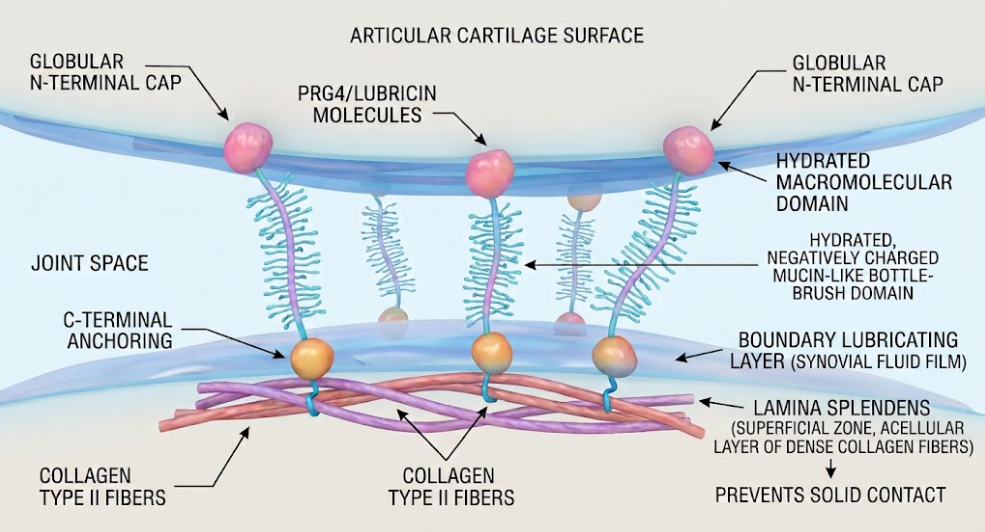
Figure 1. Molecular Mechanism for Lubrication at Cartilage Boundaries. Molecular-level schematic of the organization of PRG4 (Lubricin) and collagen type II on the articular surface. PRG4 that is anchored forms a hydrated boundary lubricating layer to prevent solid contact between opposing surfaces.
In osteoarthritis, a combination of transcriptional repression, altered glycosylation and aggressive enzymatic cleavage severely impair the structural integrity and concentration of PRG4 in the joint environment. While transient compensatory increases in PRG4 mRNA transcription may exist in some joint compartments in early or post-traumatic stages of osteoarthritis, the functional pool of active, surface-adsorbed PRG4 protein decreases precipitously with increasing disease severity(Antunes et al., 2020).
The inflammatory mediators in the osteoarthritic intra-articular microenvironment are primarily responsible for the pathological degradation of PRG4. Pro-inflammatory cytokines, such as Interleukin-1 beta (IL-1β) and Tumor Necrosis Factor-alpha (TNF-α), decrease PRG4 gene expression in superficial zone chondrocytes and synoviocytes through activated Nuclear Factor-kappa B (NF-κB) signaling pathways(Young et al., 2006). Simultaneously, they stimulate the overexpression of catabolic enzymes, such as matrix metalloproteinases (MMPs), aggrecanases (ADAMTS-4 and ADAMTS-5), cathepsins B and G, and neutrophil elastase(L. Chen, Zhang, & Liu, 2024).
Enzymatic proteolysis directly cleaves the functional domains of PRG4. Cleavage of either the N-terminal or C-terminal globular domains abrogates the anchoring mechanisms that tether PRG4 to the cartilage surface. Unanchored lubricin molecules are quickly washed out to the synovial fluid bulk or washed out through the lymphatic system leaving the lamina splendens denuded(Coles et al., 2010). In addition, the proteolytic digestion of the central mucin core results in the degradation of the tandem repeat sequences, thus removing the hydrophilic sugar chains and eliminating the steric repulsion capacity of the lubricant(Boushehri et al., 2024).
Moreover, oxidative stress in the osteoarthritic joints contributes significantly to PRG4 dysfunction in addition to direct cleavage of protein. An example of reactive nitrogen species (RNS) generated by activated chondrocytes is peroxynitrite which induces oxidative modifications and cleavage of PRG4 polypeptide chains(Watkins & Reesink, 2020). Oxidative stress also modified the glycosylation machinery of superficial chondrocytes leading to abnormal sialylation or truncation of O-linked oligosaccharide chains. These modified glycan structures decrease the water-binding ability of PRG4 such that it is unable to form a stable hydration layer(Fu et al., 2025; Han et al., 2024).
Another emerging mechanism of PRG4 inactivation is related to abnormal ionic conditions in osteoarthritic synovial fluid. The research shows that cartilage demineralization and subchondral bone remodeling increases dissolved calcium ion concentrations in osteoarthritic joint fluid. The elevated calcium ions interact with the negatively charged sialic acid and sulfate groups of PRG4, causing conformational collapsing of the mucin domain and suppressing its boundary lubricating performance(Han et al., 2024).
The function of PRG4 is not redundant with other components of synovial fluid. HMW-HA provides excellent bulk fluid viscosity and shear-thinning properties, but HA alone is missing the specific surface anchoring domains needed to provide boundary lubrication in the absence of PRG4. Enzymatic degradation and consumption of surface-bound PRG4 result in an irreversible collapse of the boundary lubrication regime(Sakata et al., 2015; Waller et al., 2013).
The main tribological effect of PRG4 degradation is a large and significant increase in the coefficient of friction across the entire articular surface. Surface friction values of PRG4-deficient cartilage were documented to be two to five times higher than those of healthy, lubricated controls in experimental studies using atomic force microscopy, pendulum testing devices, and explant cartilage-on-cartilage bearing systems. The opposing cartilage asperities are in direct contact with each other during joint movement, with no cushioning boundary lubricant layer(Waller et al., 2013).
This increase in friction fundamentally alters the mechanical state of stress within the tissue. The tangential sliding forces are dissipated smoothly across the surface fluid film for physiological boundary lubrication. Surface sliding leads to a high kinetic friction when boundary lubrication fails, transmitting strong shear stress (τ) directly to the underlying extracellular matrix. Biomechanical strain analyzes reveal that the shear strain induced by friction is not uniformly distributed across the cartilage depth, but is highly concentrated in the most superficial zone and the immediate sub-superficial transitional region(Waller et al., 2013).
Chondrocytes in the superficial zone are uniquely sensitive to mechanical shear strain. These cells are susceptible to rapid membrane deformation, excessive influx of calcium ions through mechanosensitive ion channels (e.g., TRPV4 and Piezo1) and severe intracellular mechanical trauma under excessive shear stress caused by friction. This mechanical overstimulation results in acute mitochondrial dysregulation characterized by loss of mitochondrial membrane potential, increased production of mitochondrial reactive oxygen species and release of cytochrome c into the cytosol(Li et al., 2026; Watkins & Reesink, 2020).
Release of cytochrome c activates the intrinsic apoptotic pathway, which in turn activates the executioner enzyme, mainly caspase-3. Activated caspase-3 cleaves key cellular proteins resulting in nuclear chromatin condensation, DNA fragmentation (detected by TUNEL staining), and cell shrinkage. In vitro and ex vivo cartilage bearing studies demonstrate a linear correlation between the static coefficient of friction and the percentage of apoptotic chondrocytes localized specifically in the superficial zone. Supplementing lubricin back into the system decreases friction and significantly suppresses chondrocyte apoptosis(Larson, Zhang, Badger, & Jay, 2017; Waller et al., 2013).
The cascade from PRG4 loss to superficial zone atrophy follows a clear mechanobiological path as in the Figure 2. Boundary lubrication failure increases friction and produces large shear stress on the surface. This shear stress causes superficial zone chondrocyte apoptosis leading to matrix degradation, collagen fibrillation and ultimate structural atrophy(Waller et al., 2013).
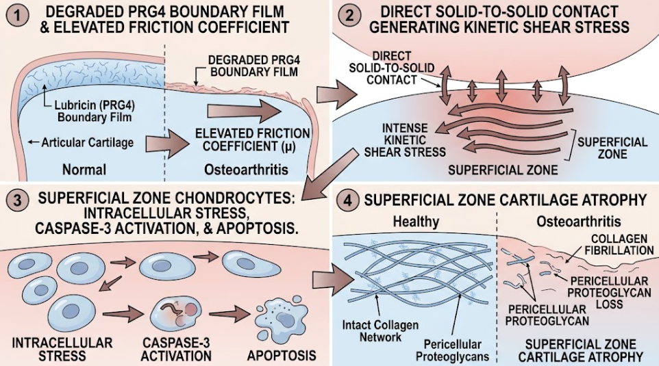
Figure 2. Biomechanical Cascade of Osteoarthritis Atrophy. Schematic overview of the transition from boundary lubrication failure to cartilage degeneration in OA. (1) PRG4 degradation causes increase in friction coefficient, (2) leading to solid-to-solid contact and high superficial zone kinetic shear stress. (3) The resulting intracellular stress activates caspase-3 and leads to chondrocyte apoptosis, (4) resulting in matrix degradation and superficial zone atrophy.
Loss of superficial zone chondrocytes has severe long-term consequences for homeostasis of articular cartilage. Superficial chondrocytes are non-dividing, highly specialized cells that synthesize collagen type II, PRG4 and clusterin and maintain the tight, tangential collagen meshwork that prevents tissue swelling(Saito, 2022). Further, recent lineage-tracing studies have identified a subpopulation of PRG4-expressing superficial zone cells as resident articular cartilage progenitor cells (ACPCs). These progenitor cells are multi-lineage differentiating and belong to the intrinsic cellular reservoir for maintenance and repair of cartilage(Watkins & Reesink, 2020).
Frictional shear stress causes selective destruction of superficial zone chondrocytes and progenitor populations, which compromises the ability of cartilage to repair surface micro-damage or to replenish degraded lubricin molecules(Coles et al., 2010). Without resident cells to maintain the extracellular matrix, the superficial collagen network breaks down by mechanical fatigue. Type II collagen fibrils are denatured, broken and fibrillated. At the same time, the pericellular proteoglycans (e.g. aggrecan) are leached out into the joint space(Waller et al., 2013).
Structural integrity of the topmost layer deteriorates with increasing collagen fibrillation and loss of pericellular matrix. The superficial zone gradually delaminates and erodes leading to measurable thinning and complete structural atrophy of the superficial zone(Rhee et al., 2005). This thinning of the superficial zone exposes the underlying intermediate and deep zones of cartilage to unbuffered mechanical loads, which accelerates the progression to full-thickness erosion of cartilage, subchondral bone sclerosis, and end-stage osteoarthritis(Waller et al., 2013).
PRG4 functions as a classic biomechanical boundary lubricant to reduce friction and also acts as a major biological signaling guardian in the joint. Emerging evidence suggests that PRG4 directly attenuates pro-inflammatory cascades, inhibits abnormal cell proliferation and alters immune cell phenotypes. Thus, PRG4 degradation in osteoarthritis is not just a matter of physical friction but also disinhibits major pro-inflammatory signaling networks simultaneously, creating a destructive mechano-inflammatory feedback loop(Alquraini et al., 2015).
CD44 cell surface receptor is one major biological pathway controlled by PRG4. CD44 is a transmembrane glycoprotein, highly expressed by chondrocytes, synoviocytes and intra-articular macrophages. Matrix degradation in osteoarthritic joints results in the generation of high concentrations of low-molecular-weight hyaluronic acid (LMW-HA) fragments. LMW-HA binding to CD44 leads to pro-inflammatory signaling pathways, resulting in NF-κB nuclear translocation and up-regulation of catabolic cytokines (IL-1β, IL-6, TNF-α) and matrix-degrading enzymes (MMP-1, MMP-3, MMP-13)(Fu et al., 2025; Li et al., 2026).
Physiological PRG4 binds CD44 with high affinity through its central and terminal regions. PRG4 competitively inhibits LMW-HA binding by binding to CD44 receptors, thereby preventing subsequent NF-κB activation and inflammatory cytokine production. If PRG4 is enzymatically degraded or absent, CD44 receptors are fully available for LMW-HA fragments, resulting in uncontrolled NF-κB activation and inflammatory amplification(Fu et al., 2025).
PRG4 also interacts directly with Toll-like receptors such as TLR2, TLR4 and TLR5. The primary pattern recognition receptors recognizing damage-associated molecular patterns (DAMPs) released from degraded cartilage matrix, such as fibronectin fragments, S100 proteins and hyaluronan fragments, are Toll-like receptors. Binding of DAMP to TLR2/4 recruits the adaptor protein MyD88, leading to downstream activation of the IκB kinase (IKK) complex, phosphorylation of IκBα, and nuclear translocation of NF-κB(Fu et al., 2025; Li et al., 2026).
PRG4 is an endogenous natural antagonist of TLR2 and TLR4. PRG4 directly binds the extracellular domains of TLR2 and TLR4, preventing DAMP-induced receptor dimerization and MyD88 recruitment. Without PRG4, TLR2 and TLR4 signaling is left unchecked, which causes synovial macrophages to adopt a pro-inflammatory M1 phenotype and also stimulates proliferation of synoviocytes, synovial hyperplasia, and capsular fibrosis(Rhee et al., 2005).
PRG4 acts as a dual biomechanical and immunomodulatory barrier, as shown in Figure 3. PRG4 reduces boundary layer friction on a mechanical level and protects against shear stress. At the molecular scale, PRG4 binds to CD44 and TLR2/4 receptors so that it can inhibit NF-κB signaling, prevent synovial inflammation and inhibit synovial cell hyperproliferation (Alquraini et al., 2017).
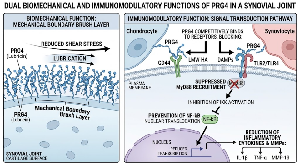
Figure 3. PRG4 has two protective mechanisms, mechanical and biological, within the synovial joint. PRG4 reduces shear stress at the cartilage surface as a boundary lubricant (Left). PRG4 competes for binding to cell surface receptors (CD44, TLRs) and inhibits pro-inflammatory ligands (LMW-HA, DAMPs), thus inhibiting the NF-κB signaling cascade and decreasing the production of inflammatory cytokines and MMPs (Right).
Moreover, PRG4 expression is tightly regulated by mechanotransduction and growth factor pathways via Transforming Growth Factor-beta (TGF-β) and specialized transcription factors. Under physiological conditions, moderate fluid flow shear stress (FSS) and TGF-β1 signaling activate cAMP response element-binding protein (CREB) and its specific co-factor Creb5 that bind to proximal promoter regulatory elements (E1 and E2) of the PRG4 gene and maintain high lubricin expression(Saito, 2022).
Pro-inflammatory cytokines dysregulate TGF-β/Creb5 signaling in osteoarthritis, driving TGF-β receptor expression toward the catabolic ALK1/Smad1/5/8 pathway away from the protective ALK5/Smad2/3 pathway. This signaling switch downregulates Creb5 and suppresses transcription of PRG4(Young et al., 2006). Loss of PRG4 increases boundary friction, inducing pathological mechanical stress that further promotes release of inflammatory cytokines, matrix degradation and receptor-mediated catabolism. This self-enhancing cycle of molecular inflammation being driven by mechanical friction and molecular inflammation destroying boundary lubrication speeds up superficial zone atrophy and joint destruction(Li et al., 2026).
The evidence collected in this literature review demonstrates that osteoarthritis can no longer be considered merely as a structural deterioration of the deeper extracellular matrix with age. Rather, the onset and early development of osteoarthritis are fundamentally at the articular surface, driven by the breakdown of boundary lubrication and the resultant atrophy of the superficial zone(W. Chen et al., 2025).
This pathological sequence is initiated by the key molecule, PRG4/lubricin. Full-length PRG4 has a uniquely evolved domain architecture that combines mechanical surface tethering, steric hydration lubrication and anti-inflammatory receptor signaling. The loss of PRG4 destabilizes the entire diarthrodial joint, as demonstrated in multiple experimental, genetic, and clinical models including Prg4 knockout mice, CACP syndrome patients and post-traumatic meniscectomy models(Li et al., 2026; Waller et al., 2013).
If boundary lubrication fails, the coefficient of friction will spike. The main biomechanical insight from the literature is that high friction at the surface directly results in concentrated shear stress in the superficial zone. Chondrocytes of the superficial zone are adapted for tangential orientation but are not able to cope with sustained kinetic shear stress. This caspase-3 mediated apoptosis therefore removes the very cells that synthesize PRG4 and maintain the superficial collagen framework. This loss of cells directly leads to fibrillation, delamination and atrophy of the superficial zone(Waller et al., 2013).
Crucially, the loss of PRG4 links biomechanical dysfunction with chronic aseptic inflammation. PRG4 deficiency releases the inhibitory control on CD44 and TLR2/4 receptors, allowing DAMPs and LMW-HA fragments to induce unrestrained NF-kB activation, which leads to synoviocyte hyperproliferation, M1 macrophage polarization and inflammatory cytokine secretion. This mechano-inflammatory coupling can account for the observation that simple mechanical unloading of the joint fails to halt osteoarthritis progression after surface boundary failure has occurred(Li et al., 2026).
These results identify PRG4 as an ideal target for disease-modifying osteoarthritis drugs (DMOADs) from a therapeutic perspective. Standard viscosupplementation using hyaluronic acid injections only offers a temporary fluid phase cushioning and fails to restore proper boundary lubrication as HA lacks cartilage anchoring domains(McNary et al., 2012). In contrast, tribosupplementation with full-length recombinant human PRG4 (rhPRG4), PRG4-mimetic biomaterials or gene therapy vectors capable of restoring endogenous PRG4 expression is very promising. Re-installation of an intact boundary lubricating film reduces surface friction, prevents superficial chondrocyte apoptosis, and re-engages CD44 and TLR2/4 receptor blockade to extinguish intra-articular inflammation(Waller et al., 2013).
However, major translational hurdles remain. The delivered recombinant lubricants must be capable of anchoring to an already damaged, fibrillated cartilage surface in a protease-rich environment. Future work should focus on improving biomimetic delivery systems, engineering of protease-resistant PRG4 variants and identification of early tribological biomarkers that can detect boundary lubrication failure before irreversible superficial zone atrophy.
To summarize, the failure of boundary lubrication and breakdown of PRG4/lubricin is a defining pathological mechanism in the initiation and progression of osteoarthritis. PRG4 is essential for joint homeostasis and serves as a physical friction-reducing and biological regulator of intra-articular inflammation. When PRG4 is enzymatically cleaved or transcriptionally repressed by pro-inflammatory cytokines, boundary lubrication is lost and there is a surge in surface friction. This increased friction exposes the most superficial zone of the cartilage to extreme kinetic shear stresses, leading to caspase-3-mediated apoptosis of superficial zone chondrocytes. These cells are essential to the matrix, and their loss results in matrix depletion, surface fibrillation, and superficial zone atrophy. Concurrently, PRG4 loss relieves inhibition of CD44 and TLR2/4 signaling to form a self-amplifying mechano-inflammatory feedback loop. Novel PRG4-targeted biological and biomimetic therapies for restoration of boundary lubrication have tremendous therapeutic potential to arrest superficial zone atrophy and preserve long-term joint function.
Alquraini, A., Garguilo, S., D’Souza, G., Zhang, L. X., Schmidt, T. A., Jay, G. D., & Elsaid, K. A. (2015). The interaction of lubricin/proteoglycan 4 (PRG4) with toll-like receptors 2 and 4: an anti-inflammatory role of PRG4 in synovial fluid. Arthritis Res Ther, 17, 353. doi:10.1186/s13075-015-0877-x
Alquraini, A., Jamal, M., Zhang, L., Schmidt, T., Jay, G. D., & Elsaid, K. A. (2017). The autocrine role of proteoglycan-4 (PRG4) in modulating osteoarthritic synoviocyte proliferation and expression of matrix degrading enzymes. Arthritis Res Ther, 19(1), 89. doi:10.1186/s13075-017-1301-5
Antunes, B. P., Vainieri, M. L., Alini, M., Monsonego-Ornan, E., Grad, S., & Yayon, A. (2020). Enhanced chondrogenic phenotype of primary bovine articular chondrocytes in Fibrin-Hyaluronan hydrogel by multi-axial mechanical loading and FGF18. Acta Biomater, 105, 170-179. doi:10.1016/j.actbio.2020.01.032
Boushehri, S., Holey, H., Brosz, M., Gumbsch, P., Pastewka, L., Aponte-Santamaría, C., & Gräter, F. (2024). O-glycans Expand Lubricin and Attenuate Its Viscosity and Shear Thinning. Biomacromolecules, 25(7), 3893-3908. doi:10.1021/acs.biomac.3c01348
Chen, L., Zhang, Z., & Liu, X. (2024). Role and Mechanism of Mechanical Load in the Homeostasis of the Subchondral Bone in Knee Osteoarthritis: A Comprehensive Review. J Inflamm Res, 17, 9359-9378. doi:10.2147/jir.S492415
Chen, W., Ye, Q., Zhang, M., Xie, R., & Xu, C. (2025). Lubrication for Osteoarthritis: From Single-Function to Multifunctional Lubricants. Int J Mol Sci, 26(5). doi:10.3390/ijms26051856
Coles, J. M., Zhang, L., Blum, J. J., Warman, M. L., Jay, G. D., Guilak, F., & Zauscher, S. (2010). Loss of cartilage structure, stiffness, and frictional properties in mice lacking PRG4. Arthritis Rheum, 62(6), 1666-1674. doi:10.1002/art.27436
Flowers, S. A., Zieba, A., Örnros, J., Jin, C., Rolfson, O., Björkman, L. I., . . . Karlsson, N. G. (2017). Lubricin binds cartilage proteins, cartilage oligomeric matrix protein, fibronectin and collagen II at the cartilage surface. Sci Rep, 7(1), 13149. doi:10.1038/s41598-017-13558-y
Fu, P. J., Zheng, S. Y., Luo, Y., Ren, Z. Q., Li, Z. H., Wang, Y. P., & Lu, B. B. (2025). Prg4 and Osteoarthritis: Functions, Regulatory Factors, and Treatment Strategies. Biomedicines, 13(3). doi:10.3390/biomedicines13030693
Han, M., Russo, M. J., Desroches, P. E., Silva, S. M., Quigley, A. F., Kapsa, R. M. I., . . . Greene, G. W. (2024). Calcium ions have a detrimental impact on the boundary lubrication property of hyaluronic acid and lubricin (PRG-4) both alone and in combination. Colloids and Surfaces B: Biointerfaces, 234, 113741. doi:https://doi.org/10.1016/j.colsurfb.2023.113741
Jay, G. D., Harris, D. A., & Cha, C. J. (2001). Boundary lubrication by lubricin is mediated by O-linked beta(1-3)Gal-GalNAc oligosaccharides. Glycoconj J, 18(10), 807-815. doi:10.1023/a:1021159619373
Larson, K. M., Zhang, L., Badger, G. J., & Jay, G. D. (2017). Early genetic restoration of lubricin expression in transgenic mice mitigates chondrocyte peroxynitrite release and caspase-3 activation. Osteoarthritis Cartilage, 25(9), 1488-1495. doi:10.1016/j.joca.2017.05.012
Li, H., Yi, G., Zhou, D., Zhou, J., Cui, Z., Zhang, H., & Chen, Z. (2026). Lubricin in osteoarthritis: functions and therapeutic prospects. Front Immunol, 17, 1790804. doi:10.3389/fimmu.2026.1790804
Lin, W., & Klein, J. (2022). Hydration Lubrication in Biomedical Applications: From Cartilage to Hydrogels. Acc Mater Res, 3(2), 213-223. doi:10.1021/accountsmr.1c00219
McNary, S. M., Athanasiou, K. A., & Reddi, A. H. (2012). Engineering lubrication in articular cartilage. Tissue Eng Part B Rev, 18(2), 88-100. doi:10.1089/ten.TEB.2011.0394
Rhee, D. K., Marcelino, J., Baker, M., Gong, Y., Smits, P., Lefebvre, V., . . . Carpten, J. D. (2005). The secreted glycoprotein lubricin protects cartilage surfaces and inhibits synovial cell overgrowth. J Clin Invest, 115(3), 622-631. doi:10.1172/jci22263
Saito, T. (2022). The superficial zone of articular cartilage. Inflamm Regen, 42(1), 14. doi:10.1186/s41232-022-00202-0
Sakata, R., McNary, S. M., Miyatake, K., Lee, C. A., Van den Bogaerde, J. M., Marder, R. A., & Reddi, A. H. (2015). Stimulation of the superficial zone protein and lubrication in the articular cartilage by human platelet-rich plasma. Am J Sports Med, 43(6), 1467-1473. doi:10.1177/0363546515575023
Waller, K. A., Zhang, L. X., Elsaid, K. A., Fleming, B. C., Warman, M. L., & Jay, G. D. (2013). Role of lubricin and boundary lubrication in the prevention of chondrocyte apoptosis. Proc Natl Acad Sci U S A, 110(15), 5852-5857. doi:10.1073/pnas.1219289110
Watkins, A. R., & Reesink, H. L. (2020). Lubricin in experimental and naturally occurring osteoarthritis: a systematic review. Osteoarthritis Cartilage, 28(10), 1303-1315. doi:10.1016/j.joca.2020.05.009
Yakubov, G. E., McColl, J., Bongaerts, J. H., & Ramsden, J. J. (2009). Viscous boundary lubrication of hydrophobic surfaces by mucin. Langmuir, 25(4), 2313-2321. doi:10.1021/la8018666
Young, A. A., McLennan, S., Smith, M. M., Smith, S. M., Cake, M. A., Read, R. A., . . . Little, C. B. (2006). Proteoglycan 4 downregulation in a sheep meniscectomy model of early osteoarthritis. Arthritis Res Ther, 8(2), R41. doi:10.1186/ar1898