Our Services
Others
- DFPP
- Shockwave
- IV Drip


“This literature review summarizes the current scientific evidence on the role of the abnormal activation and sensitization of the trigeminovascular system, through vessel-to-neuron signaling and neuroimmune cascades, in the pathophysiology and chronification of pain of Chronic Migraine.”
Chronic migraine is a highly disabling neurological disorder characterized by recurrent episodes of severe headache and persistent sensory hypersensitivity. The pain of migraine is due to dysfunction and hyper-excitability of the trigeminovascular system (TVS), an anatomical and functional network linking cranial blood vessels to sensory neurons of the trigeminal nerve. Activation of the perivascular dural nociceptors causes release of important vasoactive neuropeptides such as calcitonin gene-related peptide (CGRP) and pituitary adenylate cyclase-activating polypeptide (PACAP) resulting in sterile neurogenic inflammation, local vasodilation, and plasma protein extravasation. Prolonged perivascular signaling induces peripheral sensitization of primary afferent neurons in the trigeminal ganglion, converting the normal vascular pulsation into the characteristic throbbing of migraine. Repetitive centrally conducted nociceptive barrages lead to central sensitization of second-order neurons in the trigeminocervical complex and third-order neurons in the thalamus, with the clinical manifestation of cephalic and extracephalic cutaneous allodynia. Moreover, recent literature suggests the importance of neuroimmune interactions within the meninges, trigeminal ganglion and central nervous system, mediated by satellite glia, microglial activation and damage-associated molecular patterns such as High-Mobility Group Box 1 (HMGB1), in perpetuating neural hyperexcitability and transitioning episodic migraine into a chronic state. In this literature review, we provide an in-depth analysis of the molecular, cellular and circuit mechanisms underlying TVS activation, sensitization and disease chronification.
Migraine is one of the leading causes of neurological disability worldwide. It affects more than 1 billion people and poses an immense personal and socioeconomic burden. Migraine is a particularly debilitating evolution of the disorder, the chronic form being clinically defined as ≥15 headache days per month for at least 3 months(Krivoshein et al., 2026; Suzuki, Suzuki, Shiina, Kobayashi, & Hirata, 2022). Historically, theories of migraine pathophysiology have oscillated between purely vascular models, in which the pain is due solely to dilation of the cerebral blood vessels and neuronal models in which cortical or brainstem hyperexcitability is emphasized.These views have been integrated into a neurovascular model of modern neuroscience with trigeminovascular system (TVS) at its center(Christensen, Ashina, & Ashina, 2025; Erdener & Dalkara, 2014).
The TVS is the principal nociceptive network of the intracranial pain-sensitive structures, including the dura mater, pial arteries, large cerebral vessels and dural venous sinuses. The primary sensory neurons of the trigeminal ganglion innervate the TVS that transmits mechanical, chemical and inflammatory signals from the cranial vasculature to the central pain processing circuits in the brainstem, thalamus and cerebral cortex(Noseda & Burstein, 2013; Zhang, Simoes, Guo, & Cao, 2024). Acute activation of TVS leads to self-limited headache attacks, while sustained and repetitive activation leads to progressive physiologic changes involving peripheral sensitization of dural afferents and central sensitization of dorsal horn and thalamic neurons (Bernstein & Burstein, 2012; Christensen et al., 2025).
There is increasing evidence that vessel-neuron communication and neuroimmune interactions are key drivers of this sensitized state. Chemical mediators released by vascular smooth muscle cells, endothelial cells and activated immune cells continuously modulate neuronal excitability resulting in self-amplifying feedback loops to maintain pain signals(Zhang et al., 2024). Understanding these cellular and molecular changes in detail is the key to understanding how episodic migraine attacks turn into chronic migraine. This review integrates current literature on the anatomical substrate of the TVS, the molecular mediators of neurogenic inflammation, the transition from peripheral to central sensitization, and the neuroimmune cascades underlying migraine chronification.
The anatomical configuration of the TVS is the structural basis for the perception of cranial pain. The trigeminal ganglion is the location of cell bodies of the primary sensory neurons of the TVS. These pseudounipolar neurons send peripheral axons via the ophthalmic division (V1) of the trigeminal nerve, and to a lesser extent the maxillary (V2) and mandibular (V3) branches, to densely innervate the meninges, middle meningeal artery, superior sagittal sinus, and pial vasculature(Burstein, Blake, Schain, & Perry, 2017). The dorsal root ganglia of the upper cervical spinal nerves (C1-C3) also innervate the dura of the posterior cranial fossa. These perivascular sensory nerve fibers are predominantly made up of unmyelinated C-fibers and thinly myelinated Aδ-fibers and express specialized mechanosensitive and chemosensitive ion channels(Olesen, Burstein, Ashina, & Tfelt-Hansen, 2009).
The central axons of the trigeminal ganglion neurons project to the brainstem and synapse with second order nociceptive neurons in the trigeminocervical complex (TCC) which includes the trigeminal nucleus caudalis (TNC) and dorsal horn of the upper cervical spinal cord. Converging input to the TCC derives from both the intracranial meninges and the extracranial periorbital facial skin(Burstein, Yamamura, Malick, & Strassman, 1998; Ferrari et al., 2022). The ascending axons from the TCC cross the midline and ascend through the quintothalamic tract to the third-order neurons in the thalamus such as the ventral posteromedial (VPM), posterior (Po) and lateral posterior (LP) thalamic nuclei. Thalamic trigeminovascular neurons then project to a number of cortical domains including primary and secondary somatosensory, insular, visual, auditory and motor cortexes, encoding the sensory-discriminative, affective and cognitive dimensions of migraine pain and associated sensory sensitivities(Noseda & Burstein, 2013).
As shown in the Figure 1 shows the structural organization of the trigeminovascular system, illustrating the ascending pain pathway from perivascular meningeal afferents via the trigeminal ganglion, trigeminocervical complex and thalamic nuclei to various cortical areas.
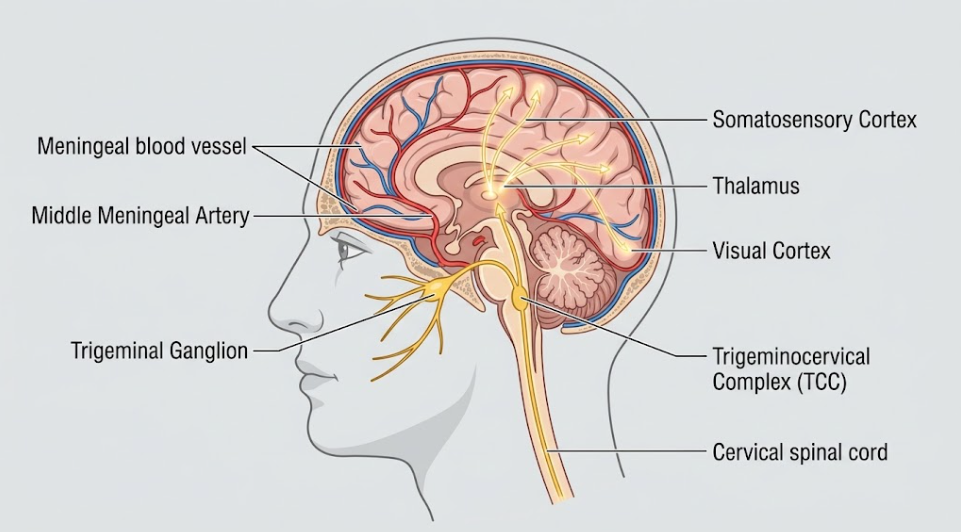
Figure 1. The Anatomical Pathways of the Trigeminovascular Systemin Migraine. The diagram illustrates trigeminal ganglion innervation of meningeal blood vessels, signal transmission to trigeminocervical complex (TCC) and ascending pathways to the thalamus. Further signal transmission to the somatosensory and visual cortexes highlights the pathways proposed in migraine pathophysiology and central sensitization.
In recent literature, the “vessel-to-neuron” hypothesis has been put forward as the primary trigger mechanism for the activation of TVS. Intracranial blood vessels are not passive responders to neural activity; structural and chemical changes in vascular smooth muscle cells (VSMCs) can directly depolarize perivascular nociceptors in their vicinity(Della Pietra et al., 2024). In clinical provocation studies, pharmacological agents that induce vasodilation (e.g. nitric oxide donors, adenosine triphosphate (ATP)-sensitive potassium () channel openers, and large-conductance calcium-activated potassium () channel openers) reliably trigger migraine attacks. Opening of VSMC and channels causes efflux of intracellular potassium into the tightly confined perivascular space around meningeal afferents(Krishnamoorthy-Natarajan & Koide, 2016). This leads to the build-up of extracellular potassium ions, and reduces the resting membrane potential of perivascular nerve endings leading to membrane depolarization and generation of action potentials. Also, the mechanical stretching of arterial wall during vasodilation leads to the deformation of the plasma membrane of nearby nociceptors and activation of mechanosensitive ion channels (e.g., Piezo1/2 and Two-Pore Domain Potassium () channels that transduce mechanical vascular stress into nociceptive electrical signals(Della Pietra et al., 2024).
Molecular Mechanisms of Peripheral Sensitization: Neuropeptides, Inflammatory Loops and Ion Channels
Activation of perivascular dural afferents leads to local release of vasoactive neuropeptides from their peripheral terminals, giving rise to a cascade referred to as neurogenic inflammation. The main neuropeptides involved in this process are calcitonin gene-related peptide (CGRP), pituitary adenylate cyclase-activating polypeptide (PACAP) and substance P(Ashina et al., 2019). CGRP is a 37 amino acid peptide synthesized in abundance by small- and medium-sized neurons of the trigeminal ganglion. Upon release into the dural microenvironment CGRP binds to canonical Calcitonin Receptor-Like Receptor / Receptor Activity-Modifying Protein 1 (CLR/RAMP1) complexes and AMY1 receptors expressed on vascular smooth muscle cells, endothelial cells and dural mast cells(Song et al., 2026). Activation of these receptors leads to stimulation of adenylate cyclase and subsequent increase of intracellular cyclic adenosine monophosphate (cAMP), which produces potent and long-lasting vasodilation of meningeal arteries and dural mast cell degranulation(Song et al., 2026).
The molecular neuroimmune interactions within the dura mater are shown in Figure 2, and demonstrate how primary afferent terminals release neuropeptides to activate vascular cells, mast cells and satellite glia to create a self-sustaining inflammatory soup.
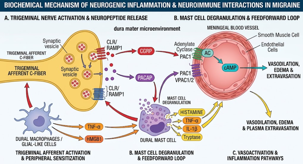
Figure 2. Neuroimmune interactions and neurogenic inflammation in the dural microenvironment in migraine. Schematic diagram of the biochemical mechanisms in migraine attack Activation of trigeminal afferent C-fibers leads to release of neuropeptides (CGRP and PACAP) inducing meningeal vasodilation and plasma extravasation through cAMP pathways. These neuropeptides also cause dural mast cell degranulation. Then, release of pro-inflammatory mediators (TNF-α, IL-1β, histamine, HMGB1, etc.) creates a feedforward loop that perpetuates peripheral nerve sensitization and ongoing neurogenic inflammation.
PACAP acts synergistically with CGRP through PAC1, VPAC1 and VPAC2 receptors to potentiate cAMP-dependent vasodilation, neurogenic inflammation and dural mast cell degranulation. Degranulating mast cells release a cocktail of pro-inflammatory and pro-nociceptive mediators including histamine, serotonin (5-HT), bradykinin, prostaglandin () and pro-inflammatory cytokines such as Tumor Necrosis Factor-alpha (TNF-α), Interleukin-1 beta (IL-1β) and Interleukin-6 (IL-6). This cocktail, often called “inflammatory soup” in the past, bathes the perivascular dural nociceptors and dramatically increases their excitability(Bernstein & Burstein, 2012; Syed, Koide, Braas, May, & Wellman, 2012).
A major molecular axis maintaining peripheral sensitization is the signaling pathway of High Mobility Group Box 1 (HMGB1) and Nuclear Factor kappa B (NF-κB). HMGB1 is an endogenous damage-associated molecular pattern (DAMP) that is released from trigeminal neurons under hypoxic or inflammatory stress. HMGB1 binds to Toll-Like Receptor 4 (TLR4) and Receptors for Advanced Glycation Endproducts (RAGE) on neighboring vascular endothelial cells and satellite glial cells, inducing NF-κB nuclear translocation(Song et al., 2026). Activated NF-κB increases endothelial expression and release of HMGB1 and inflammatory mediators that feedback on trigeminal sensory neurons to stimulate CALCA gene transcription and CGRP hyper-synthesis. This positive feedback loop between neurons and endothelium and neurons and glia leads to further increased CGRP release and neurovascular signaling in the TVS(Song et al., 2026).
In addition, dural nociceptors express a wide variety of Transient Receptor Potential (TRP) ion channels including TRPA1, TRPV1 and TRPM8. Environmental irritants, reactive oxygen species (ROS) and the products of lipid peroxidation formed during neurogenic inflammation activate TRPA1 and TRPV1 channels on C-fibers leading to calcium influx and vesicular exocytosis of more CGRP and substance P(Raggi et al., 2024).
The physiological consequences of this ongoing chemical exposure is peripheral sensitization. Peripheral sensitization means altered primary meningeal nociceptor response with lower activation threshold and increased responsiveness. Dural afferents that are not sensitized are silent at rest and respond only to high intensity mechanical force(Strassman, Raymond, & Burstein, 1996). These sensitized neurons, which are activated by inflammatory mediators, exhibit spontaneous firing and respond vigorously to low-intensity mechanical stimuli such as normal intravascular pressure pulses. Clinically, peripheral sensitization manifests as the pulsatile or throbbing quality of migraine headache and its characteristic worsening with routine activities that transiently increase intracranial pressure, such as coughing, bending over, or climbing stairs(Bernstein & Burstein, 2012; Zhang et al., 2024).
Peripheral sensitization explains the localized, throbbing headache and central sensitization explains the progression of the headache and the widespread sensory features of chronic migraine. Central sensitization is an increased excitability of nociceptive neurons in the central nervous system to their normal or subthreshold afferent input(Suzuki et al., 2022). Sensitized dural afferents fire tonically to release glutamate, CGRP and substance P into the TCC, overstimulating postsynaptic ionotropic glutamate receptors (AMPA and NMDA) on second order trigeminovascular neurons(Spekker, Nagy-Grócz, & Vécsei, 2023).
Prolonged activity removes the voltage dependent magnesium block from NMDA receptors, allowing intracellular calcium to enter the cell, activating intracellular protein kinases (such as p38 MAPK and ERK) and causing phosphorylation of synaptic receptors. This synaptic plasticity increases the gain of TCC cells(Suzuki et al., 2022). We show that central sensitization in the TCC leads to innocuous tactile stimuli applied to the face being processed as painful because second-order TCC neurons receive convergent sensory input from the cutaneous receptive fields of the periorbital facial skin and the intracranial dura mater. In clinical practice, this phenomenon is known as cephalic cutaneous allodynia. Patients with cephalic allodynia describe common nonpainful activities, such as combing hair, shaving, wearing eyeglasses, touching periorbital skin, or having shower water strike the face, as painful or distressing(Zhang et al., 2024).
If intense nociceptive input from the periphery is sustained, central sensitization spreads rostrally to involve third-order trigeminovascular neurons in the posterior and ventral posteromedial nuclei of the thalamus. Sensitized thalamic neurons expand the size of their receptive fields, and receive convergent inputs from the entire dorsal horn of the spinal cord(Burstein et al., 2010). In this way, thalamic sensitization turns localized head pain into extracephalic, whole-body cutaneous allodynia. At this advanced stage patients have cutaneous hypersensitivity over distant areas of the body, making it uncomfortable or painful to wear tight clothing, bracelets or socks or to lie under a blanket(Bernstein & Burstein, 2012).
In functional neuroimaging studies, allodynic migraineurs showed an increased blood-oxygen-level-dependent (BOLD) signal in the pulvinar and posterior thalamus in response to mechanical or thermal stimuli to the skin(Noseda & Burstein, 2013). The convergence of retinal ganglion cell projections onto light-sensitive, dura-sensitive thalamic neurons in the posterior thalamic nuclei also explains photophobia, the worsening of migraine pain by exposure to light. Thalamic processing is also altered in phonophobia and osmophobia. These features are a generalized failure of central sensory filtering and gating mechanisms(Noseda, Jakubowski, Kainz, Borsook, & Burstein, 2011).
In some individuals, especially those with migraine with aura, TVS activation is induced by Cortical Spreading Depression (CSD). CSD is a slowly propagating wave (2-6 mm/min) of near-total neuronal and glial depolarization, traveling across the cerebral cortex, followed by prolonged bioelectrical suppression(Charles & Baca, 2013). CSD (cortical spreading depression) results in a massive breakdown of cellular ion homeostasis, releasing high concentrations of extracellular potassium, glutamate, hydrogen ions, ATP and nitric oxide into the cortical parenchyma and subarachnoid space. CSD also leads to the opening of neuronal Pannexin-1 (Panx1) megachannels, triggering caspase-1 activation and release of pro-inflammatory molecules (IL-1β, HMGB1) which diffuse to the pial and dural nerve terminals. This parenchymal-to-meningeal cascade leads to neurogenic inflammation and activation of primary dural afferents with a physiological delay (15–30 min) that mimics the clinical latency between aura symptoms and headache onset(Noseda & Burstein, 2013).
Conversion from episodic to chronic migraine is characterized by persistent neuroinflammation and structural-functional remodeling of the TVS. Non-neuronal cells like trigeminal ganglion satellite glial cells, TCC microglia and astrocytes, and infiltrated immune cells are major drivers for disease chronification(Zhang et al., 2024).
In the trigeminal ganglion, the cell bodies of primary sensory neurons are completely enveloped by satellite glial cells (SGCs) to form functional neuron-glia units. Repeated TVS activation leads to the release of CGRP from neuronal cell bodies that bind to CGRP receptors on SGCs and induces the expression and release of nitric oxide synthase (NOS), nitric oxide, and pro-inflammatory cytokines (IL-1β, and TNF-α)(Morgan & Nkadimeng, 2025). SGCs are coupled to neighboring glia via gap junctions, and inflammatory signals can thus be transmitted throughout the ganglion, sensitizing neighboring, non-stimulated neurons. Concurrently, the chemokine CCL2 (Monocyte Chemoattractant Protein-1) is expressed and released by trigeminal ganglion neurons. CCL2 acts on the CCR2 receptors of infiltrating macrophages and T lymphocytes present in the ganglion and dura mater.These CCR2-expressing immune cells produce reactive oxygen species and carbonyl compounds that activate TRPA1 channels on dural afferents, leading to a prolonged state of peripheral sensitization that cannot be resolved by standard CGRP-blocking monotherapies(Zhang et al., 2024).
Central sensitization of migraine chronification by microglial purinergic receptors and inflammatory signaling pathways in the central neuroimmune signaling cascades of the trigeminocervical complex as illustrated in Figure 3.
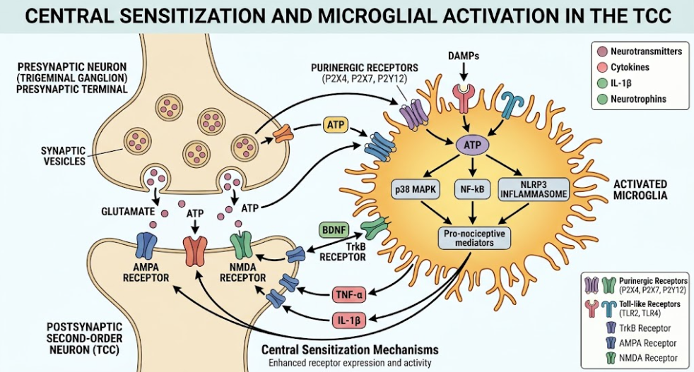
Figure 3. Mechanisms of microglia-neuron interaction in central sensitization of the trigeminocervical complex. Simplified schematic of synaptic signaling and microglial activation in the TCC. Presynaptic release of Glutamate and ATP activates postsynaptic neurons and nearby microglia. Microglial activation through Purinergic and Toll-like receptors activates intracellular pathways (p38 MAPK, NF-κB, NLRP3 inflammasome) to release pro-nociceptive mediators (BDNF, TNF-α, IL-1β) to enhance postsynaptic receptor activity and spread central sensitization.
Microglial activation in TCC is a feature of central nervous system pathophysiology of chronic migraine. Under conditions of recurrent, high frequency nociceptive input, such as modeled by repeated nitroglycerin or inflammatory soup exposures, sustained release of ATP from trigeminal central terminals activates purinergic P2X4, P2X7, and P2Y12 receptors on resident TCC microglia. Microglial purinergic signaling activates downstream p38 MAPK, RhoA/ROCK, NF-κB and NLRP3 inflammasome pathways(Zhang et al., 2024).
This intracellular cascade results in a robust production and release of brain derived neurotrophic factor (BDNF) and cytokines (TNF-α, IL-1β, IL-6). BDNF activates the neuronal TrkB receptors on second-order TCC neurons, resulting in down-regulation of the potassium-chloride co-transporter KCC2(Zhang et al., 2024). This causes a shift in the chloride equilibrium potential and abolishes GABAergic and glycinergic synaptic inhibition leading to TCC neuron hyperexcitability. Likewise, microglial TNF-α and IL-1β promote trafficking of AMPA and NMDA receptors to the postsynaptic membrane, thereby mediating excitatory synaptic transmission and a long-lasting, activity-independent form of central sensitization(Zhang et al., 2024).
Whereas pro-inflammatory cascade, regulatory T cells (Treg) are protective in pain resolution. The expansion of Treg populations through LD-IL-2 results in the release of anti-inflammatory cytokines, IL-10 and Transforming Growth Factor-beta 1 (TGF-β1).These anti-inflammatory factors restore CGRP and PACAP receptor expression in trigeminal ganglion neurons, offering a novel mechanism for reversing established peripheral sensitization and blocking disease chronification(Zhang et al., 2024).
A synthesis of the literature on TVS activation and sensitization shows a clear mechanistic continuum from acute vascular events to persistent central pathology.Chronic migraine is a quantitative increase in headache frequency and a qualitative change in central nervous system function due to persistent peripheral nociceptive input and neuroimmune dysregulation(Suzuki et al., 2022; Zhang et al., 2024).
The mechanistic insights have a direct clinical relevance as they are reflected in current therapeutic concepts and drug responses. Acute abortive medications such as triptans (5-HT1B/1D agonists) and ditans (5-HT1F agonists) exert their action by binding to presynaptic serotonin receptors on primary trigeminal terminals, inhibiting voltage gated calcium channels and blocking release of CGRP, glutamate and ATP(Erdener & Dalkara, 2014). But basic and clinical investigations demonstrate a critical time window for triptan efficacy. Triptans, when given early in an attack when pain is primarily driven by peripheral sensitization, are effective at terminating headache and preventing central sensitization. Once central sensitization is fully developed and TCC neurons are activity-independent, triptans cannot abort the headache or reverse cutaneous allodynia. Conversely, centrally acting anti-inflammatory agents such as parenteral nonsteroidal anti-inflammatory drugs (NSAIDs) or intravenous ketorolac can inhibit central sensitization even after the development of allodynia(Bernstein & Burstein, 2012).
The central role of CGRP in activation of TVS is further supported by the clinical success of targeted monoclonal antibodies against CGRP (e.g., erenumab, fremanezumab, galcanezumab, eptinezumab) and small molecule CGRP receptor antagonists (gepants) (Zhang et al., 2024). Monoclonal antibodies are large macromolecules that do not cross the intact blood–brain barrier in significant amounts. Nonetheless, their therapeutic effectiveness implies that peripheral CGRP receptor blockade in the meninges and trigeminal ganglion is sufficient to reduce attack frequency in many patients(Noseda & Burstein, 2013). However, a significant subgroup of patients with chronic migraine demonstrate incomplete response or non-responsiveness to anti-CGRP therapies. Alternative redundant signaling pathways such as PACAP/PAC1 signaling, HMGB1/NF-κB neurovascular loops, CCL2/CCR2 immune cell recruitment and microglial NLRP3 inflammasome activation that continue to drive peripheral and central sensitization independent of CGRP are described in the literature to explain this therapeutic gap(Song et al., 2026).
The identification of chronic migraine as a neuroimmune disorder offers new translational opportunities. Exciting approaches to stop or reverse migraine chronification include therapeutic targeting of upstream triggers of neurogenic inflammation (HMGB1 neutralization, microglial purinergic receptor antagonists (P2X7R/P2Y12R blockers) or enhancing regulatory T-cells with low-dose IL-2)(Song et al., 2026; Zhang et al., 2024).
The Trigeminovascular System activation and sensitization is the core pathophysiological engine of Chronic Migraine. Mechanical and chemical alterations at the meningeal neurovascular interface trigger the release of vasoactive neuropeptides that lead to neurogenic inflammation and peripheral sensitization of primary dural afferents. Ongoing nociceptive input, further amplified by reciprocal neuron-endothelial and neuron-glia amplification loops involving HMGB1 and CGRP, drives synaptic plasticity in the trigeminocervical complex and thalamus, leading to widespread central sensitization and cutaneous allodynia. Furthermore, microglial activation and neuroimmune signaling in the brainstem play key roles in sustaining neural hyperexcitability and in the conversion of episodic headache to a chronic refractory state. Further exploration of these intricate neuroimmune and vessel-to-neuron pathways will be essential for the development of next generation disease-modifying therapies for chronic migraine.
Ashina, M., Hansen, J. M., Do, T. P., Melo-Carrillo, A., Burstein, R., & Moskowitz, M. A. (2019). Migraine and the trigeminovascular system-40 years and counting. Lancet Neurol, 18(8), 795-804. doi:10.1016/s1474-4422(19)30185-1
Bernstein, C., & Burstein, R. (2012). Sensitization of the trigeminovascular pathway: perspective and implications to migraine pathophysiology. J Clin Neurol, 8(2), 89-99. doi:10.3988/jcn.2012.8.2.89
Burstein, R., Blake, P., Schain, A., & Perry, C. (2017). Extracranial origin of headache. Curr Opin Neurol, 30(3), 263-271. doi:10.1097/wco.0000000000000437
Burstein, R., Jakubowski, M., Garcia-Nicas, E., Kainz, V., Bajwa, Z., Hargreaves, R., . . . Borsook, D. (2010). Thalamic sensitization transforms localized pain into widespread allodynia. Ann Neurol, 68(1), 81-91. doi:10.1002/ana.21994
Burstein, R., Yamamura, H., Malick, A., & Strassman, A. M. (1998). Chemical stimulation of the intracranial dura induces enhanced responses to facial stimulation in brain stem trigeminal neurons. J Neurophysiol, 79(2), 964-982. doi:10.1152/jn.1998.79.2.964
Charles, A. C., & Baca, S. M. (2013). Cortical spreading depression and migraine. Nat Rev Neurol, 9(11), 637-644. doi:10.1038/nrneurol.2013.192
Christensen, R. H., Ashina, H., & Ashina, M. (2025). The vessel-to-neuron trigeminovascular hypothesis of migraine pathogenesis – the ‘pro’ argument. J Headache Pain, 26(1), 248. doi:10.1186/s10194-025-02130-z
Della Pietra, A., Gómez Dabó, L., Mikulenka, P., Espinoza-Vinces, C., Vuralli, D., Baytekin, I., . . . Giniatullin, R. (2024). Mechanosensitive receptors in migraine: a systematic review. J Headache Pain, 25(1), 6. doi:10.1186/s10194-023-01710-1
Erdener, S. E., & Dalkara, T. (2014). Modelling headache and migraine and its pharmacological manipulation. Br J Pharmacol, 171(20), 4575-4594. doi:10.1111/bph.12651
Ferrari, M. D., Goadsby, P. J., Burstein, R., Kurth, T., Ayata, C., Charles, A., . . . Dodick, D. W. (2022). Migraine. Nat Rev Dis Primers, 8(1), 2. doi:10.1038/s41572-021-00328-4
Krishnamoorthy-Natarajan, G., & Koide, M. (2016). BK Channels in the Vascular System. Int Rev Neurobiol, 128, 401-438. doi:10.1016/bs.irn.2016.03.017
Krivoshein, G., Watt, M. V., Jordà-Baleri, T., Põld, K., Gkotzamanis, V., van den Maagdenberg, A., . . . Holland, P. R. (2026). From bench to bedside: relevant animal models across the migraine attack phases. J Headache Pain, 27(1). doi:10.1186/s10194-026-02369-0
Morgan, C. T., & Nkadimeng, S. M. (2025). The Role of Inflammation in Migraine Headaches: A Review. FASEB Bioadv, 7(7), e70033. doi:10.1096/fba.2024-00188
Noseda, R., & Burstein, R. (2013). Migraine pathophysiology: anatomy of the trigeminovascular pathway and associated neurological symptoms, cortical spreading depression, sensitization, and modulation of pain. Pain, 154 Suppl 1, S44-53. doi:10.1016/j.pain.2013.07.021
Noseda, R., Jakubowski, M., Kainz, V., Borsook, D., & Burstein, R. (2011). Cortical projections of functionally identified thalamic trigeminovascular neurons: implications for migraine headache and its associated symptoms. J Neurosci, 31(40), 14204-14217. doi:10.1523/jneurosci.3285-11.2011
Olesen, J., Burstein, R., Ashina, M., & Tfelt-Hansen, P. (2009). Origin of pain in migraine: evidence for peripheral sensitisation. Lancet Neurol, 8(7), 679-690. doi:10.1016/s1474-4422(09)70090-0
Raggi, A., Leonardi, M., Arruda, M., Caponnetto, V., Castaldo, M., Coppola, G., . . . Martelletti, P. (2024). Hallmarks of primary headache: part 1 – migraine. J Headache Pain, 25(1), 189. doi:10.1186/s10194-024-01889-x
Song, M., Barone, A., Xu, S., Li, C., Gigli, G., Frati, G., . . . Cortese, B. (2026). Mechanistic investigation of HMGB1 in an in vitro model of the trigeminovascular system under migraine-like conditions. J Headache Pain, 27(1), 142. doi:10.1186/s10194-026-02357-4
Spekker, E., Nagy-Grócz, G., & Vécsei, L. (2023). Ion Channel Disturbances in Migraine Headache: Exploring the Potential Role of the Kynurenine System in the Context of the Trigeminovascular System. Int J Mol Sci, 24(23). doi:10.3390/ijms242316574
Strassman, A. M., Raymond, S. A., & Burstein, R. (1996). Sensitization of meningeal sensory neurons and the origin of headaches. Nature, 384(6609), 560-564. doi:10.1038/384560a0
Suzuki, K., Suzuki, S., Shiina, T., Kobayashi, S., & Hirata, K. (2022). Central Sensitization in Migraine: A Narrative Review. J Pain Res, 15, 2673-2682. doi:10.2147/jpr.S329280
Syed, A. U., Koide, M., Braas, K. M., May, V., & Wellman, G. C. (2012). Pituitary adenylate cyclase-activating polypeptide (PACAP) potently dilates middle meningeal arteries: implications for migraine. J Mol Neurosci, 48(3), 574-583. doi:10.1007/s12031-012-9851-0
Zhang, J., Simoes, R., Guo, T., & Cao, Y. Q. (2024). Neuroimmune interactions in the development and chronification of migraine headache. Trends Neurosci, 47(10), 819-833. doi:10.1016/j.tins.2024.08.009