Our Services
Others
- DFPP
- Shockwave
- IV Drip


“Decoding the spatiotemporal clocks of early Wound healing informs us as to why some wounds regenerate and why some wounds become chronic.”
The successful completion of the various stages of Wound healing depends on regulation of the process occurring in a timely and spatially defined manner. The purpose of this review is to highlight the early and critical stages of Wound healing and the interactions of hemostasis, inflammation, and the coagulation cascade. The coagulation cascade after tissue injury forms a scaffold rich in fibrin, providing a spatial territory for cellular immigration. Simultaneously, platelets fulfill a temporal role through the release of growth factors (PDGF, VEGF) to recruit inflammatory cells bridging hemostasis and the inflammatory response. The recruitment of neutrophils to the site of injury and the subsequently dominating role of pro-inflammatory M1 macrophages initiate a localized and transient “cytokine storm” phenomenon. While a prolonged state is detrimental, the rapid offset of the cytokine storm due to the dominance of macrophages is expected. In the absence of the systemic proteolytic activity of MMPs, a disassembly and remodeling of the provisional matrix will happen. The absence of the aforementioned will lead to non-healing chronic wounds.
Keywords: Wound healing, Spatiotemporal dynamics, Hemostasis, Acute inflammation, Macrophages, Chronic wounds.
Wound healing is a critical, highly complex and evolutionarily conserved biological process required to restore tissue integrity and ensure host survival after injury. The skin is the major barrier against pathogenic invasion, mechanical stress and environmental hazards.(Takeo, Lee, & Ito, 2015; Wilkinson & Hardman, 2020) Thus, a disturbance of this barrier elicits a complex and vigorous reparative response. But when this process fails or is dysregulated, often as a consequence of systemic host factors, like advanced age, vascular disease or diabetes, it can result in severe pathological outcomes.(S. A. Eming, Martin, & Tomic-Canic, 2014) Chronic, non-healing wounds such as diabetic foot ulcers and pressure sores affect millions of people worldwide and pose a significant socioeconomic and clinical burden.(A. M. Soliman & Barreda, 2022; P. H. Wang, Huang, Horng, Yeh, & Chen, 2018) In contrast, an overzealous or anomalous healing process can cause fibrotic disorders like hypertrophic scars and keloids.(S. A. Eming et al., 2014) Thus, the elucidation of the exact biological mechanisms underlying successful tissue repair holds the highest clinical relevance. The classical wound repair trajectory involves a continuum of four overlapping temporal phases: hemostasis, inflammation, proliferation, and remodeling.(He et al., 2025; Jin et al., 2025) These stages are characterized by dramatic changes in the microenvironment involving an array of cell types, ECM components, and soluble mediators including growth factors and chemokines acting on the injured tissue.(Jin et al., 2025) This coordination is not only a temporal sequence, but also depends strictly on the spatial architecture of the wound, a phenomenon called “spatiotemporal specificity”.(Sabine A. Eming, Wynn, & Martin, 2017; He et al., 2025) Precise Spatiotemporal dynamics or ‘clocks’ of cellular behaviors across different zones such as wound edge versus wound center and epidermis versus dermis are essential for successful healing. Perturbing this fragile spatiotemporal balance can arrest the tissue repair process, resulting in wounds that have become chronic and pathological.(Y. Zhang et al., 2026) To understand the process by which tissues move from injury to regeneration, it is necessary to study the earliest events that start the cascade of subsequent repair. Thus, this literature review is focused on the critical early steps of Wound healing, namely the Spatiotemporal dynamics of the hemostatic and acute inflammatory phases.(Wu et al., 2025) It will examine the mechanisms of the coagulation cascade, which forms the provisional fibrin scaffold necessary for cell infiltration, and the diverse roles of platelets. Platelets are not only involved in stopping bleeding, but they are also the first master regulators to release bioactive mediators that recruit immune and stromal cells.(Dowling, Chu, Etkin, & Oropallo, 2025) Moreover, this review will focus on the orchestrating role of acute inflammation and pro-inflammatory cytokines that are strictly required for the initial pathogen clearance, cellular debris removal and the timely transition into the proliferative repair phase.(P. Krzyszczyk, R. Schloss, A. Palmer, & F. Berthiaume, 2018)
The initial response to tissue injury is the rapid activation of hemostatic mechanisms to prevent blood loss (exsanguination) and to provide a provisional matrix.(L. Yang et al., 2025) Following a breach in tissue integrity, damaged blood vessels are rapidly constricted. This initial vasoconstriction is rapidly modulated by local neuroimmune bidirectional signaling, in which nociceptive sensory nerve endings detect noxious stimuli and release neuropeptides, such as substance P and calcitonin gene-related peptide (CGRP), to directly alter local vascular tone. At the same time, the coagulation cascade is activated robustly.(S. A. Eming et al., 2014; He et al., 2025)
Upon contact with the exposed vascular subendothelial matrix, platelets, the primary cellular players in hemostasis, become activated.(Etulain, 2018) Specific platelet receptors, primarily glycoprotein VI, dynamically interact with extracellular matrix (ECM) proteins including collagen, fibronectin and von Willebrand factor (vWF). This interaction results in their tight adhesion and initial tethering to the disrupted blood vessel wall. Furthermore, local networks of enzymatically oxidized phospholipids, formed via the lipoxygenase pathway, actively promote calcium-dependent coagulation factor binding, strictly maintaining early hemostasis.(H. Wang et al., 2026)
The cascade leads to formation of a fibrin-rich clot, often referred to clinically as an eschar, composed of a tightly cross-linked network of insoluble fibrin, fibronectin, vitronectin, and thrombospondin. The enzyme thrombin is pivotal in this process, since it not only enables the cleavage of fibrinogen into fibrin to form clots, but also induces profound platelet activation.(Dowling et al., 2025) Thrombin causes a dramatic change in the conformation of platelets leading to rapid degranulation and release of bioactive molecules from their alpha and dense granules.(L. Yang et al., 2025) Plasma coagulation factors, including thrombin, also act directly as important signaling mediators that regulate early inflammation and immune cell recruitment.(Vardakostas et al., 2025)
This first fibrin clot is more than a simple physical plug for hemostasis; it establishes the basic spatial limits of the wound. This provides the requisite three-dimensional architectural framework for subsequent invasion, tethering, and directed migration of inflammatory leukocytes and other reparative cells.(Etulain, 2018) Once trapped in this fibrin matrix, activated platelets degranulate and release a concentrated, broad spectrum of bioactive mediators into the local wound environment. The secretome at this early stage includes immediate mediators, such as adenosine diphosphate (ADP) and thromboxane A2 (TXA2).(P. Krzyszczyk et al., 2018; Amro M. Soliman & Barreda, 2023) TXA2 plays a role in amplifying local platelet aggregation, but also has an active role in inducing the synthesis of early pro-inflammatory cytokines, such as IL-6 and prostaglandin E2 (PGE2), thus bridging hemostasis and inflammation.(Garbuzenko et al., 2002; Weller et al., 2006)
Simultaneously, platelets secrete highly potent growth factors such as Platelet-Derived Growth Factor (PDGF), Vascular Endothelial Growth Factor (VEGF) and Transforming Growth Factor-β (TGF-β).(Smith, Travers, & Morrissey, 2015) The fibrin clot is a highly localized reservoir for these cytokines and growth factors, effectively sequestering them to establish steep spatiotemporal chemotactic gradients that precisely guide the recruitment of neutrophils, monocytes, and fibroblasts to the injury site, effectively launching the acute inflammatory phase. The spatiotemporal precision of this cell recruitment is elegantly visualized using in vivo live imaging. Real-time tracking of neutrophils and macrophages in transgenic fluorescent zebrafish and Drosophila embryos has demonstrated their rapid, directed migration toward the injury site, confirming that these cells follow steep, localized chemotactic gradients established moments after injury.(Razzell, Wood, & Martin, 2011) (Figure 1.)
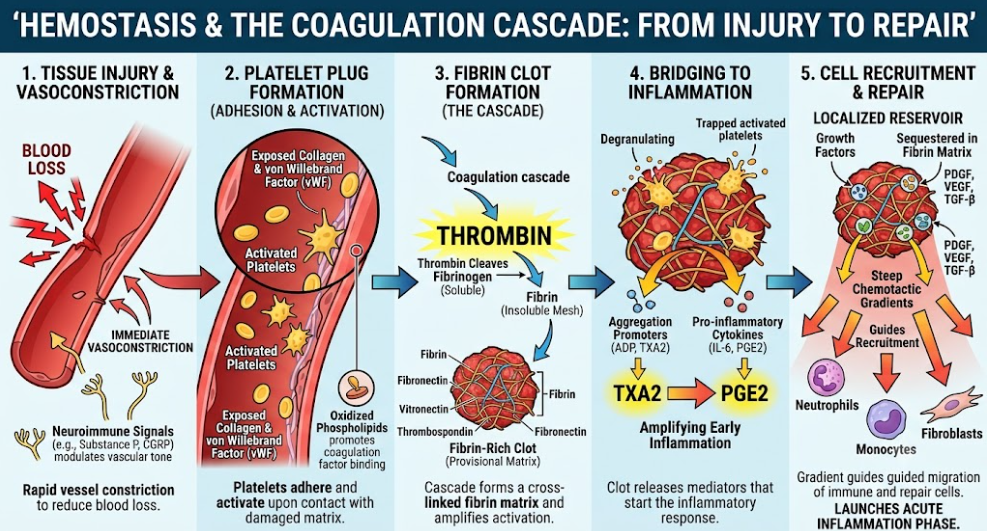
Figure 1. Temporal sequence of hemostasis and early wound repair. After tissue injury, rapid neuroimmune-mediated vasoconstriction reduces blood loss. Platelets adhere to the exposed extracellular matrix and become activated, which promotes a thrombin-mediated coagulation cascade. The resultant fibrin rich clot is a provisional matrix. In this clot, activated platelets secrete bioactive mediators (e.g., TXA2) and growth factors (e.g., PDGF, VEGF, TGF-β) thereby generating steep chemotactic gradients to direct the precise recruitment of leukocytes and fibroblasts for the initiation of the acute inflammatory phase.
Moreover, the hemostatic clot serves some secondary but biologically important functions: it physically protects the breached tissue from bacterial invasion, it activates the complement system via trapped plasma proteins, and it provides binding sites for leukocyte integrins (such as αMβ2 or Mac-1) to facilitate cell extravasation. Crucially, the Spatiotemporal dynamics of hemostasis require a stringent temporal “switch-off” mechanism.(Scully et al., 2020) Once there is sufficient and stable clot formation the whole process of coagulation has to be actively stopped to avoid over the pathological thrombosis and local tissue ischemia. There are several negative feedback mechanisms that mediate this arrest. Prostacyclin is released from the endothelium and actively inhibits further platelet aggregation.(Etulain, 2018) Antithrombin III directly inhibits the activity of thrombin. Activated protein C systemically degrades coagulation factors V and VII.(Cognasse et al., 2005) This precise temporal downregulation ensures that coagulation remains strictly localized, allowing the wound microenvironment to safely and efficiently shift its biological focus to leukocyte infiltration and innate immune defense.(Shiraki et al., 2004)
The main cellular players in the process of hemostasis are the platelets. They are the first responders at the site of tissue injury. Upon disruption of the vasculature, they immediately encounter the exposed subendothelial extracellular matrix (ECM).(Dowling et al., 2025) They are activated by binding of ECM proteins, including collagen, fibronectin and von Willebrand factor to specific platelet receptors such as glycoprotein VI.(L. Yang et al., 2025) This is followed by thrombin generation from the coagulation cascade that induces a profound conformational change leading to full platelet activation, aggregation and reinforcement of the provisional fibrin scaffold.(Etulain, 2018) But the physiological function of platelets is much more than simple coagulation and plugging the wound.(Smith et al., 2015) These are early master regulators of the repair process and set the spatiotemporal clock for all subsequent phases. The platelets that are activated are quickly degranulated once caught in the clot, releasing a large and diverse secretome from their alpha and dense granules. This payload’s composition includes potent growth factors such as Platelet-Derived Growth Factor (PDGF), Vascular Endothelial Growth Factor (VEGF), Transforming Growth Factor-β (TGF-β), Epidermal Growth Factor (EGF) and Insulin-like Growth Factor 1 (IGF-1).(Locatelli, Colciago, Castiglioni, & Maier, 2021) These secreted factors are key chemoattractants that establish spatial gradients to attract neutrophils, monocytes and fibroblasts to the wound bed.(Smith et al., 2015) Furthermore, these growth factors directly promote the proliferation and migration of resident skin cells such as keratinocytes and endothelial cells, thus preparing the microenvironment for subsequent re-epithelialization and angiogenesis.(Talbott, Mascharak, Griffin, Wan, & Longaker, 2022)
Platelets not only orchestrate the recruitment of cells but also play important immunomodulatory and direct antimicrobial roles. They express Toll-like receptors (TLRs) that allow active recognition of pathogen-associated molecular patterns (PAMPs) and control of early infection.(Cognasse et al., 2005) Platelets respond by releasing specific chemokines including CXCL4 (Platelet Factor 4) and CXCL7, as well as a variety of antimicrobial peptides such as defensins and thrombocidins. Platelets modulate the innate immune defenses, control the initial bacterial invasion and regulate vascular permeability before the massive influx of leukocytes by releasing these mediators and vasoactive amines.(Scopelliti, Cattani, Dimartino, Mirisola, & Cavani, 2022)
Very recently work has also highlighted the very sophisticated intercellular communication mechanisms employed by platelets in the early stages of tissue repair. Platelets transport specific microRNAs and cytokines to neighboring stromal and immune cells via extracellular vesicles, which is beneficial in regenerative processes and reduces the risk of excessive inflammation and hypertrophic scar formation.(Johnson et al., 2023) Interestingly, activated platelets have been shown to donate functional mitochondria to mesenchymal stem cells (MSCs) using clathrin-mediated endocytosis.(Verma, Kumar, Garg, & Verma, 2023) This intercellular mitochondrial transfer metabolically reprograms the MSCs, i.e. activates citrate metabolism and de novo lipogenesis, which ultimately enhances their secretion of critical reparative factors such as VEGF and hepatocyte growth factor (HGF) to enhance angiogenesis. These multifaceted structural, biochemical and metabolic contributions by platelets bridge the hemostatic and inflammatory phases to create a highly coordinated environment essential for successful tissue regeneration(Everts et al., 2024) Furthermore, the critical need of macrophages in this particular spatiotemporal window has been experimentally validated in murine models. For example, the role of macrophages in the transition of inflammation to tissue repair is indispensable as demonstrated by in vivo studies where the targeted depletion of macrophages in mice resulted in severely delayed wound closure and defective granulation tissue formation.(Zhu, Ding, Ma, Iwashina, & Tredget, 2016) (Figure 2.)
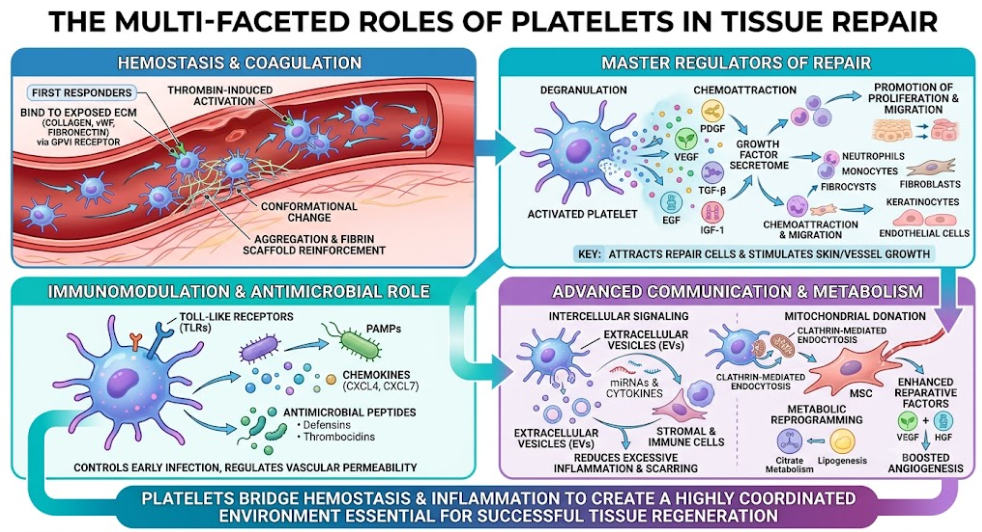
Figure 2. Key roles of platelets in tissue repair. First, platelets stop the bleeding. Then they make a fibrin matrix. The subsequent activation results in the release of growth factors that determine the spatiotemporal clock of phases of repair . Platelets also contribute to antimicrobial defence and immune regulation. Regenerative responses are metabolically activated by specialized signaling mechanisms, such as the transfer of mitochondria to mesenchymal stem cells. The interconnected panels illustrate how the biochemical and metabolic contributions by platelets establish a highly coordinated environment that is critical for recovery.
The inflammatory phase is the body’s first innate defense against pathogenic invasion and is also important for clearing cellular debris. This highly orchestrated phase is almost instantaneously initiated by injury-induced danger signals. These are damage-associated molecular patterns (DAMPs), such as intracellular DNA, ATP and hydrogen peroxide (H2O2) passively released from necrotic cells, and pathogen-associated molecular patterns (PAMPs), such as bacterial lipopolysaccharides (LPS) and peptidoglycans.(Zhu et al., 2016) At the site of injury, sensory nerve endings simultaneously rapidly sense these noxious stimuli and release neuropeptides, including substance P and calcitonin gene-related peptide (CGRP), creating a neuroimmune bidirectional loop that immediately modulates local vascular tone and primes resident immune cells.(He et al., 2025)
Resident tissue cells such as mast cells and macrophages sense these injury cues through pattern recognition receptors (PRRs), especially Toll-like receptors (TLRs).(Mogensen Trine, 2009) These ligands are specifically recognized (e.g. TLR1, TLR2 and TLR5 for bacterial components or TLR3 or TLR7 for viral RNA) and this recruits key adaptor proteins such as MyD88 and TRIF.(Kaisho & Akira, 2006; F. X. Zhang et al., 1999) This activates downstream intracellular signaling cascades, notably the nuclear factor-κB (NF-κB) and mitogen-activated protein kinase (MAPK) pathways.(Rodrigues, Kosaric, Bonham, & Gurtner, 2019) In addition, transcription-independent pathways, driven by rapid intracellular Ca2+ influx and localized generation of reactive oxygen species (ROS), are activated within minutes to compensate for any delay in transcriptional machinery.(Lansdown, 2002)
This signaling cascade rapidly upregulates the expression of pro-inflammatory cytokines and chemokines, triggering a highly coordinated leukocyte adhesion cascade. Cytokines induce local vasodilation and expression of endothelial selectins (E- and P-selectins) that allow for initial tethering and “slow rolling” of circulating leukocytes.(Zarbock, Ley, McEver, & Hidalgo, 2011) Chemokines, especially CXCL8 (IL-8) and leukotriene B4 (LTB4), then activate leukocyte integrins (e.g., Mac-1) to induce firm cellular arrest against intercellular adhesion molecules (ICAMs) on the endothelium and then diapedesis (extravasation) into the wound space.(Lämmermann et al., 2008; Mezu-Ndubuisi & Maheshwari, 2021)
Neutrophils, the “first responders”, arrive within hours and employ multiple microbicidal mechanisms to clear pathogens and necrotic tissue. These include phagocytosis, a large oxidative burst of ROS and degranulation of proteolytic enzymes such as elastase, matrix metalloproteinases (MMPs) and myeloperoxidase. In addition, neutrophils produce neutrophil extracellular traps (NETs), extruded webs of chromatin decorated with antimicrobial peptides, which are effective in trapping and killing microbes.(Boniakowski, Kimball, Jacobs, Kunkel, & Gallagher, 2017)
Circulating monocytes infiltrate the wound bed following neutrophils. The local inflammatory environment and metabolic reprogramming towards high glycolytic activity induce the differentiation of these monocytes into classically activated, pro-inflammatory M1-like macrophages.(Loi et al., 2016) Macrophages and neutrophils are the major sources of important cytokines, such as Tumor Necrosis Factor-α (TNF-α), Interleukin-1β (IL-1β), and Interleukin-6 (IL-6). Most importantly, TNF-α and IL-1β induce a strong positive feedback loop, directly re-activating NF-κB-mediated gene expression and further amplifying the acute inflammatory response.(S. A. Eming et al., 2014; He et al., 2025)
These cellular and molecular events are tightly regulated by a very specific spatiotemporal clock . Spatially, pro-inflammatory macrophages are activated early on at the wound edge, where they are instructed to release factors like CXCL1 and epiregulin (EREG) that create a gradient for migration of nearby keratinocytes.(Almet, Liu, Nie, & Plikus, 2025) Early peaks of mediators such as retinoic acid (RA) temporally drive local tissue progenitors to differentiate into pro-inflammatory fibroblasts that secrete CCL2 (MCP-1) to massively amplify monocyte recruitment during the first three days post-injury.(Rodero et al., 2014) This spatiotemporally controlled “cytokine storm” and proteolytic activity are strictly required for early microbial clearance, but need to be tightly controlled.(P. Krzyszczyk et al., 2018) Excess ROS and proteases (e.g., free elastase) can non-selectively digest the provisional extracellular matrix, newly formed blood vessels, and important growth factors.(Basu, Binder, Suto, Anderson, & Srivastava, 2000) Therefore, the timely resolution of inflammation, which is facilitated by neutrophil apoptosis, subsequent macrophage efferocytosis, and a metabolic shift to oxidative phosphorylation in M2-like macrophages, is an absolute prerequisite to prevent chronic wound formation and allow the successful transition into the proliferative phase.(Jetten et al., 2014) (Figure 3.)
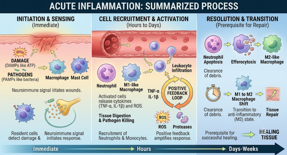
Figure 3. Three-phase chart of acute inflammation from onset to resolution. It describes how the early signals cause a cascade of recruitment of leukocytes from the blood to fight the pathogens. During the resolution phase, cellular changes are induced to clear debris and transition to M2-like macrophages, enabling tissue repair and preventing chronic inflammation over days and weeks.
In the early phases of Wound healing, a highly orchestrated spatiotemporal interplay between the coagulation cascade, platelet function, and acute inflammation is necessary for the successful restoration of tissue integrity.(Y. Zhang et al., 2026) The first coagulation cascade achieves hemostasis and quickly creates a provisional fibrin-rich scaffold that acts as a critical spatial boundary and structural matrix for the healing process. This three-dimensional matrix offers specific binding sites to guide circulating immune cells to infiltrate exactly the site of injury, preventing chaotic spread of inflammation to healthy tissues.(P. Krzyszczyk et al., 2018) Once attached to this provisional scaffold, platelets are the key temporal pacemakers of the repair. They release concentrated bursts of bioactive mediators and potent growth factors (e.g. PDGF and VEGF) through rapid degranulation to establish steep chemotactic gradients.(A. M. Soliman & Barreda, 2022) These temporal signals efficiently recruit innate immune cells at just the right time, effectively bridging the hemostatic and inflammatory phases. Neutrophils and monocytes are recruited to the site and they cause a transient but necessary “cytokine storm” mediated mainly by classically activated M1 macrophages secreting pro-inflammatory mediators such as TNF-α, IL-1β and IL-6.(Agramunt, Kang, & Rinkevich, 2026) This potent, localized inflammatory response is essential to the removal of pathogenic invasion and the clearance of necrotic cellular debris. But literature emphasizes that this acute inflammation should actively be resolved in time to safely enter the proliferative tissue building phase. This temporal resolution is very sensitive to the clearance of apoptotic neutrophils, which promotes the metabolic reprogramming of macrophages from an M1 destructive state to an M2 pro-reparative phenotype.(W. Yang et al., 2019) When these spatiotemporal clocks become desynchronized, often as a result of persistent infection or underlying metabolic disorders such as diabetes, the body produces excess destructive molecules such as reactive oxygen species (ROS) and matrix metalloproteinases (MMPs).(Werdin, Tenenhaus, & Rennekampff, 2008) These proteases then degrade the spatial fibrin scaffold indiscriminately and destroy the essential endogenous growth factors previously provided by the platelets.(A. M. Soliman & Barreda, 2022) This total uncoupling of spatial architecture and temporal signaling then locks the tissue in a never-ending, self-sustaining inflammatory loop that clinically manifests as chronic, non-healing wounds. Understanding the delicate spatiotemporal balance of this fundamental triad is therefore critical to understanding the pathogenesis of impaired healing and highlights the need for advanced therapies that target these precise microenvironmental clocks.(He et al., 2025) There is strong experimental and clinical evidence that these spatiotemporal clocks are decoupled in disease. In vivo studies in diabetic mouse models, such as ob/ob and db/db mice, suggest that macrophages are permanently trapped in the pro-inflammatory M1 state and are unable to efferocytose in a timely fashion.(Paulina Krzyszczyk, Rene Schloss, Andre Palmer, & François Berthiaume, 2018) Also, ex vivo studies on wound fluid and tissue biopsies from human patients with chronic venous and diabetic ulcers consistently reveal elevated levels of proteases, particularly MMP-2 and MMP-9, and degraded growth factors.(Wilkinson & Hardman, 2020) Complete uncoupling of spatial architecture from temporal signaling leads to a persistent inflammatory loop in the tissue that clinically manifests as chronic non-healing wounds.
The Spatiotemporal dynamics of the early Wound healing response highlight a beautifully orchestrated biological system. The coagulation cascade is immediately activated and platelet degranulation occurs to prepare the wound environment for acute inflammation. Recruited immune cells then release pro-inflammatory cytokines in a precise temporal manner to ensure a sterile wound bed ready for tissue regeneration. Insights into the interplay between these mechanisms are critical for targeted therapies for chronic, non-healing wounds in which these precise spatiotemporal clocks are disrupted.
Agramunt, J., Kang, Y., & Rinkevich, Y. (2026). Spatiotemporal dynamics of mammalian wound healing. Cell Discov, 12(1), 4. doi:10.1038/s41421-025-00865-2
Almet, A. A., Liu, Y., Nie, Q., & Plikus, M. V. (2025). Integrated Single-Cell Analysis Reveals Spatially and Temporally Dynamic Heterogeneity in Fibroblast States during Wound Healing. Journal of Investigative Dermatology, 145(3), 645-659.e625. doi:10.1016/j.jid.2024.06.1281
Basu, S., Binder, R. J., Suto, R., Anderson, K. M., & Srivastava, P. K. (2000). Necrotic but not apoptotic cell death releases heat shock proteins, which deliver a partial maturation signal to dendritic cells and activate the NF-κB pathway. International Immunology, 12(11), 1539-1546. doi:10.1093/intimm/12.11.1539
Boniakowski, A. E., Kimball, A. S., Jacobs, B. N., Kunkel, S. L., & Gallagher, K. A. (2017). Macrophage-Mediated Inflammation in Normal and Diabetic Wound Healing. The Journal of Immunology, 199(1), 17-24. doi:10.4049/jimmunol.1700223
Cognasse, F., Hamzeh, H., Chavarin, P., Acquart, S., Genin, C., & Garraud, O. (2005). Evidence of Toll-like receptor molecules on human platelets. Immunology & Cell Biology, 83(2), 196-198. doi:https://doi.org/10.1111/j.1440-1711.2005.01314.x
Dowling, C., Chu, L., Etkin, Y., & Oropallo, A. (2025). Assessment and management of chronic venous, arterial, and diabetic wounds in older adults. Semin Vasc Surg, 38(3), 281-290. doi:10.1053/j.semvascsurg.2025.06.007
Eming, S. A., Martin, P., & Tomic-Canic, M. (2014). Wound repair and regeneration: mechanisms, signaling, and translation. Sci Transl Med, 6(265), 265sr266. doi:10.1126/scitranslmed.3009337
Eming, S. A., Wynn, T. A., & Martin, P. (2017). Inflammation and metabolism in tissue repair and regeneration. Science, 356(6342), 1026-1030. doi:doi:10.1126/science.aam7928
Etulain, J. (2018). Platelets in wound healing and regenerative medicine. Platelets, 29(6), 556-568. doi:10.1080/09537104.2018.1430357
Everts, P. A., Lana, J. F., Alexander, R. W., Dallo, I., Kon, E., Ambach, M. A., . . . Podesta, L. (2024). Profound Properties of Protein-Rich, Platelet-Rich Plasma Matrices as Novel, Multi-Purpose Biological Platforms in Tissue Repair, Regeneration, and Wound Healing. Int J Mol Sci, 25(14), 7914.
Garbuzenko, E., Nagler, A., Pickholtz, D., Gillery, P., Reich, R., Maquart, F. X., & Levi-Schaffer, F. (2002). Human mast cells stimulate fibroblast proliferation, collagen synthesis and lattice contraction: a direct role for mast cells in skin fibrosis. Clinical & Experimental Allergy, 32(2), 237-246. doi:https://doi.org/10.1046/j.1365-2222.2002.01293.x
He, X., Han, Z., Ruan, Y., Wang, Z., Liao, B., Li, X., . . . Bai, D. (2025). Smart responsive biomaterials for spatiotemporal modulation of functional tissue repair. Materials Today Bio, 33, 102063. doi:https://doi.org/10.1016/j.mtbio.2025.102063
Jetten, N., Roumans, N., Gijbels, M. J., Romano, A., Post, M. J., de Winther, M. P. J., . . . Xanthoulea, S. (2014). Wound Administration of M2-Polarized Macrophages Does Not Improve Murine Cutaneous Healing Responses. PLOS ONE, 9(7), e102994. doi:10.1371/journal.pone.0102994
Jin, C., Jin, Y., Ding, Z., Nuch, K. S., Han, M., Shim, J., . . . Heo, C. Y. (2025). Cellular and Molecular Mechanisms of Wound Repair: From Biology to Therapeutic Innovation. Cells, 14(23), 1850.
Johnson, J., Law, S. Q. K., Shojaee, M., Hall, A. S., Bhuiyan, S., Lim, M. B. L., . . . Lichtfuss, G. (2023). First-in-human clinical trial of allogeneic, platelet-derived extracellular vesicles as a potential therapeutic for delayed wound healing. Journal of Extracellular Vesicles, 12(7), 12332. doi:https://doi.org/10.1002/jev2.12332
Kaisho, T., & Akira, S. (2006). Toll-like receptor function and signaling. Journal of Allergy and Clinical Immunology, 117(5), 979-987. doi:10.1016/j.jaci.2006.02.023
Krzyszczyk, P., Schloss, R., Palmer, A., & Berthiaume, F. (2018). The Role of Macrophages in Acute and Chronic Wound Healing and Interventions to Promote Pro-wound Healing Phenotypes. Front Physiol, 9, 419. doi:10.3389/fphys.2018.00419
Krzyszczyk, P., Schloss, R., Palmer, A., & Berthiaume, F. (2018). The Role of Macrophages in Acute and Chronic Wound Healing and Interventions to Promote Pro-wound Healing Phenotypes. Front Physiol, Volume 9 – 2018. doi:10.3389/fphys.2018.00419
Lämmermann, T., Bader, B. L., Monkley, S. J., Worbs, T., Wedlich-Söldner, R., Hirsch, K., . . . Sixt, M. (2008). Rapid leukocyte migration by integrin-independent flowing and squeezing. Nature, 453(7191), 51-55. doi:10.1038/nature06887
Lansdown, A. B. (2002). Calcium: a potential central regulator in wound healing in the skin. Wound Repair Regen, 10(5), 271-285. doi:10.1046/j.1524-475x.2002.10502.x
Locatelli, L., Colciago, A., Castiglioni, S., & Maier, J. A. (2021). Platelets in Wound Healing: What Happens in Space? Frontiers in Bioengineering and Biotechnology, Volume 9 – 2021. doi:10.3389/fbioe.2021.716184
Loi, F., Córdova, L. A., Pajarinen, J., Lin, T.-h., Yao, Z., & Goodman, S. B. (2016). Inflammation, fracture and bone repair. Bone, 86, 119-130. doi:https://doi.org/10.1016/j.bone.2016.02.020
Mezu-Ndubuisi, O. J., & Maheshwari, A. (2021). The role of integrins in inflammation and angiogenesis. Pediatric Research, 89(7), 1619-1626. doi:10.1038/s41390-020-01177-9
Mogensen Trine, H. (2009). Pathogen Recognition and Inflammatory Signaling in Innate Immune Defenses. Clinical Microbiology Reviews, 22(2), 240-273. doi:10.1128/cmr.00046-08
Razzell, W., Wood, W., & Martin, P. (2011). Swatting flies: modelling wound healing and inflammation in Drosophila. Dis Model Mech, 4(5), 569-574. doi:10.1242/dmm.006825
Rodero, M. P., Licata, F., Poupel, L., Hamon, P., Khosrotehrani, K., Combadiere, C., & Boissonnas, A. (2014). In Vivo Imaging Reveals a Pioneer Wave of Monocyte Recruitment into Mouse Skin Wounds. PLOS ONE, 9(10), e108212. doi:10.1371/journal.pone.0108212
Rodrigues, M., Kosaric, N., Bonham, C. A., & Gurtner, G. C. (2019). Wound Healing: A Cellular Perspective. Physiological Reviews, 99(1), 665-706. doi:10.1152/physrev.00067.2017
Scopelliti, F., Cattani, C., Dimartino, V., Mirisola, C., & Cavani, A. (2022). Platelet Derivatives and the Immunomodulation of Wound Healing. Int J Mol Sci, 23(15), 8370.
Scully, D., Sfyri, P., Wilkinson, H. N., Acebes-Huerta, A., Verpoorten, S., Muñoz-Turrillas, M. C., . . . Matsakas, A. (2020). Optimising platelet secretomes to deliver robust tissue-specific regeneration. Journal of Tissue Engineering and Regenerative Medicine, 14(1), 82-98. doi:https://doi.org/10.1002/term.2965
Shiraki, R., Inoue, N., Kawasaki, S., Takei, A., Kadotani, M., Ohnishi, Y., . . . Yokoyama, M. (2004). Expression of Toll-like receptors on human platelets. Thrombosis Research, 113(6), 379-385. doi:10.1016/j.thromres.2004.03.023
Smith, S. A., Travers, R. J., & Morrissey, J. H. (2015). How it all starts: Initiation of the clotting cascade. Critical Reviews in Biochemistry and Molecular Biology, 50(4), 326-336. doi:10.3109/10409238.2015.1050550
Soliman, A. M., & Barreda, D. R. (2022). Acute Inflammation in Tissue Healing. Int J Mol Sci, 24(1). doi:10.3390/ijms24010641
Soliman, A. M., & Barreda, D. R. (2023). Acute Inflammation in Tissue Healing. Int J Mol Sci, 24(1), 641.
Takeo, M., Lee, W., & Ito, M. (2015). Wound healing and skin regeneration. Cold Spring Harb Perspect Med, 5(1), a023267. doi:10.1101/cshperspect.a023267
Talbott, H. E., Mascharak, S., Griffin, M., Wan, D. C., & Longaker, M. T. (2022). Wound healing, fibroblast heterogeneity, and fibrosis. Cell Stem Cell, 29(8), 1161-1180. doi:10.1016/j.stem.2022.07.006
Vardakostas, D., Moustogiannis, A., Garoufalia, Z., Karatza, E., Philippou, A., Kouraklis, G., . . . Mantas, D. (2025). Expression of Tissue Remodeling- and Inflammation-Related Factors During the Wound-Healing Process in Humans. Journal of Personalized Medicine, 15(1), 14.
Verma, R., Kumar, S., Garg, P., & Verma, Y. K. (2023). Platelet-rich plasma: a comparative and economical therapy for wound healing and tissue regeneration. Cell and Tissue Banking, 24(2), 285-306. doi:10.1007/s10561-022-10039-z
Wang, H., Yang, L., Jiang, H., Li, Z., Zhou, X., Wang, B., . . . He, C. (2026). Skin-mimetic bilayer hydrogel enhances spatiotemporal coordination of neuro-immune-vascular interactions to accelerate diabetic wound healing. Bioactive Materials, 61, 758-773. doi:https://doi.org/10.1016/j.bioactmat.2026.02.054
Wang, P. H., Huang, B. S., Horng, H. C., Yeh, C. C., & Chen, Y. J. (2018). Wound healing. J Chin Med Assoc, 81(2), 94-101. doi:10.1016/j.jcma.2017.11.002
Weller, K., Foitzik, K., Paus, R., Syska, W., Maurer, M., Weller, K., . . . Maurer, M. (2006). Mast cells are required for normal healing of skin wounds in miceMast cells are required for normal healing of skin wounds in mice. The FASEB Journal, 20(13), 2366-2368. doi:https://doi.org/10.1096/fj.06-5837fje
Werdin, F., Tenenhaus, M., & Rennekampff, H.-O. (2008). Chronic wound care. The Lancet, 372(9653), 1860-1862. doi:10.1016/S0140-6736(08)61793-6
Wilkinson, H. N., & Hardman, M. J. (2020). Wound healing: cellular mechanisms and pathological outcomes. Open Biol, 10(9), 200223. doi:10.1098/rsob.200223
Wu, D., Liu, R., Cen, X., Dong, W., Chen, Q., Lin, J., . . . Qin, D. (2025). Preclinical study of engineering MSCs promoting diabetic wound healing and other inflammatory diseases through M2 polarization. Stem Cell Res Ther, 16(1), 113. doi:10.1186/s13287-025-04248-y
Yang, L., Shi, F., Cao, F., Wang, L., She, J., He, B., . . . Cai, B. (2025). Neutrophils in Tissue Injury and Repair: Molecular Mechanisms and Therapeutic Targets. MedComm, 6(5), e70184. doi:https://doi.org/10.1002/mco2.70184
Yang, W., Tao, Y., Wu, Y., Zhao, X., Ye, W., Zhao, D., . . . Tang, L. (2019). Neutrophils promote the development of reparative macrophages mediated by ROS to orchestrate liver repair. Nature Communications, 10(1), 1076. doi:10.1038/s41467-019-09046-8
Zarbock, A., Ley, K., McEver, R. P., & Hidalgo, A. (2011). Leukocyte ligands for endothelial selectins: specialized glycoconjugates that mediate rolling and signaling under flow. Blood, 118(26), 6743-6751. doi:10.1182/blood-2011-07-343566
Zhang, F. X., Kirschning, C. J., Mancinelli, R., Xu, X.-P., Jin, Y., Faure, E., . . . Arditi, M. (1999). Bacterial Lipopolysaccharide Activates Nuclear Factor-κB through Interleukin-1 Signaling Mediators in Cultured Human Dermal Endothelial Cells and Mononuclear Phagocytes *. Journal of Biological Chemistry, 274(12), 7611-7614. doi:10.1074/jbc.274.12.7611
Zhang, Y., Guo, D., Song, L., Su, G., Lin, Z., Zhang, H., . . . Zou, Z. (2026). Inflammation and wound healing: a comprehensive overview of mechanisms, therapeutic strategies, and translational perspectives. Biomarker Research, 14(1), 75. doi:10.1186/s40364-026-00935-x
Zhu, Z., Ding, J., Ma, Z., Iwashina, T., & Tredget, E. E. (2016). Systemic depletion of macrophages in the subacute phase of wound healing reduces hypertrophic scar formation. Wound Repair and Regeneration, 24(4), 644-656. doi:https://doi.org/10.1111/wrr.12442