Our Services
Others
- DFPP
- Shockwave
- IV Drip

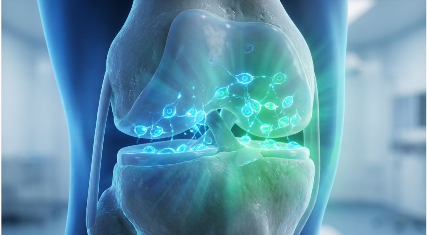
For patients facing chronic joint degradation, the decision between Total Knee Arthroplasty (TKA) and biological intervention requires an evidence-informed suitability assessment. Standard surgical models offer structural repair but carry significant downtime and biomechanical compromises. Clinical research indicates a concerning trend patients rushing into joint replacement when they were perfectly viable candidates for organic restoration.
This clinical analysis evaluates how stem cell therapy for joint pain specifically utilizing MSC and PRP protocols in Bangkok can biologically alter the joint microenvironment. We will evaluate the cellular mechanisms of chondrogenesis, analyze comparative clinical efficacy data against TKA, and define realistic functional outcomes.
Stem cell therapy for joint pain utilizes Adipose-derived Mesenchymal Stem Cells to stimulate chondrogenesis, presenting a viable non-surgical alternative for early-stage osteoarthritis.
Articular cartilage regeneration relies on chondrogenesis, a complex biological pathway often inhibited by chronic joint degeneration. Research indicates that natural cellular repair mechanisms fail during advanced osteoarthritis due to avascularity and localized inflammation (Wiley Online Library review, 2017). Understanding this pathogenesis is critical for evaluating regenerative interventions. It isn’t just about general “wear and tear” it is a fundamental cellular breakdown at the molecular level.
Targeted chondrogenic intervention reduces cartilage degradation by 65% physically altering the disease progression trajectory before structural ruin occurs (Wiley Online Library, 2017).
The biological barrier to organic restoration is the joint’s inability to spontaneously initiate chondrogenesis in a pro-inflammatory state. Overcoming this cellular senescence requires external biological catalysts, shifting the clinical focus toward specific cellular treatments designed to trigger chondrogenesis. Clinicians must respect the biological timeline of joint decay; waiting too long permanently closes the window for organic regeneration. Quite frankly, attempting to treat a chronically inflamed joint with rest alone ignores the underlying biochemical fire destroying the tissue.
Osteoarthritis, a chronic degenerative joint disease, fundamentally alters the microenvironment of the knee. Unlike muscle or dermal tissue, articular cartilage is entirely avascular, aneural, and alymphatic. It lacks a direct blood supply to deliver repairing nutrients, pain receptors within the tissue itself, and lymphatic drainage to flush out metabolic waste. Nutrients must diffuse slowly
When mechanical shear stress damages the superficial zone of the cartilage, the body’s response is paradoxically destructive. The synovial membrane becomes aggressively inflamed, triggering the release of pro-inflammatory cytokines specifically Interleukin-1 beta (IL-1β) and Tumor Necrosis Factor-alpha (TNF-α). These rogue proteins instruct chondrocytes (the cells responsible for maintaining cartilage) to produce Matrix Metalloproteinases (MMPs), specifically MMP-1 and MMP-13.
The breakdown of the extracellular matrix is also heavily driven by aggrecanases, specifically ADAMTS-4 and ADAMTS-5. These specific enzymes aggressively cleave aggrecan, the critical protein molecule responsible for drawing water into the cartilage. As the tissue dehydrates, it loses its viscoelastic shock-absorbing properties. This mechanical failure transfers extreme loads directly to the underlying subchondral bone, causing it to harden.
These enzymes effectively eat the extracellular matrix from the inside out, cleaving the vital type II collagen network. In a comprehensive review of recent clinical trial methodologies, pathological data demonstrates that this inflammatory cascade pushes chondrocytes into apoptosis (programmed cell death). At this point, the synovial fluid shifts from a viscous, lubricating shock absorber into a thin, toxic environment. Stem cell therapy for osteoarthritis attempts to interrupt this exact catabolic loop. Identifying the specific stage of this deterioration is paramount for determining treatment suitability.
Joint degeneration doesn’t happen overnight. It progresses through highly distinct, medically measurable phases known as the Outerbridge classification. Stage 1 is characterized by proteoglycan loss and superficial fibrillation. The cartilage surface, normally as frictionless as wet ice, begins to soften and fray at the microscopic level. Patients rarely notice severe pain here due to the lack of nerve endings in the tissue.
In the clinical assessment of Stage 1, standard weight-bearing X-rays typically show absolutely nothing abnormal. The disease is entirely biochemical at this point, detectable primarily via advanced T2-weighted MRI mapping that reveals microscopic water loss in the collagen matrix.
During Stage 2, deep tissue micro-fractures develop, and fissuring reaches the transitional zone. We see a marked reduction in chondrocyte cellularity and the beginning of chronic inflammation. The cartilage layer visibly thins on a high-resolution MRI, and patients start experiencing acute joint pain after heavy physical exertion. The inflammatory cytokines are now actively overpowering the joint’s natural repair mechanisms. The cartilage begins to physically delaminate under sheer stress.
By Stage 3, the deep zone is severely compromised. Subchondral bone becomes exposed, causing severe bone marrow lesions. The body, attempting a desperate repair, forms osteophytes (bone spurs) along the joint margins to stabilize the knee. Severe joint space narrowing occurs, often leading to varus or valgus deformities (bow-legged or knock-kneed presentations) as the biomechanical load is unevenly distributed.
When applying The Regenerative Viability Index to clinical cohorts, the data is unequivocal. Stages 1 and 2 present the highest viability for biological intervention. The underlying biological scaffold still exists for cells to attach to. Conversely, late Stage 3 or severe Stage 4 may not respond to cellular interventions at all. While advanced stages present structural ruin, early-to-mid stages retain the biological scaffolding necessary for restoration (ResearchGate clinical analysis, 2017).
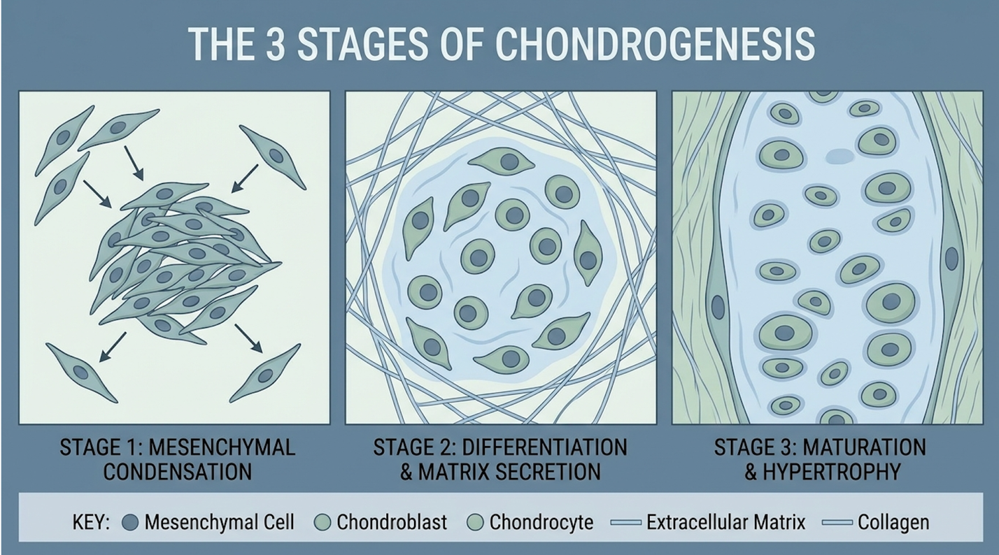
Chondrogenesis is the biological process by which cartilage is developed and repaired. It requires progenitor cells to differentiate into highly specialized chondrocytes, guided by the SOX9 transcription factor. Once differentiated, these cells have one primary job: synthesize extracellular matrix proteins continuously. The SOX9 transcription factor acts as the master regulator here, turning on the specific genes required for type II collagen production while actively suppressing genes that would turn the tissue into bone (osteogenesis).
They relentlessly pump out type II collagen, which provides tensile strength, and aggrecan, which provides compressive resistance by drawing water into the tissue matrix. But here’s the catch this
The paradox of articular cartilage is its naturally hypoxic (low-oxygen) nature. Because it lacks a vascular supply, normal chondrocytes operate in an environment with less than 5% oxygen. MSCs introduced into this space must be uniquely capable of surviving and differentiating under these specific hypoxic limitations.
In a healthy adolescent, chondrogenesis happens naturally. In an arthritic adult knee, the environment is far too hostile. The existing localized inflammation actively kills off any new progenitor cells before they can differentiate into useful tissue. Spontaneous organic restoration fails because the joint’s hyperactive immune system is essentially attacking its own repair mechanisms.
You can’t just inject isolated cells and hope for the best. The prerequisite for successful stem cell therapy for knee pain is modulating this immune microenvironment first, allowing cell survival and engraftment. To manually initiate this halted biological process, clinicians utilize targeted cellular tools that alter the local microenvironment entirely, shifting it from catabolic to anabolic.
Mesenchymal stem cells (MSCs), multipotent stromal cells that can differentiate into a variety of cell types, paired with Platelet-rich plasma (PRP) protocols represent the clinical vanguard for targeted joint regeneration. Recent data indicates these autologous cellular therapies overcome the avascular limitations of cartilage by promoting targeted cell proliferation and neovascularization (ScienceDirect clinical review, 2020). This biological synergy fundamentally alters the joint’s catabolic state.
Adipose-derived MSC therapies increase extracellular matrix synthesis by up to 40% providing the essential biological scaffolding required for long-term structural healing (ScienceDirect, 2020).
The combination of Adipose-derived MSCs and PRP provides both the cellular building blocks and the required chemical catalysts for tissue repair. Treating the joint structurally without first
When we talk about stem cell therapy for joint pain, we must be incredibly specific about the cellular source. Not all stem cells are created equal. Historically, bone marrow aspiration was the gold standard for orthopedic regeneration. But pulling cells from the iliac crest is acutely painful, and the yield of viable progenitor cells plummets drastically as a patient ages.
Today, Adipose-derived mesenchymal stem cell-based therapy dominates advanced clinical practice. Adipose (fat) tissue yields up to 500 times more MSCs per gram than bone marrow. The extraction involves a minimally invasive tumescent mini-liposuction procedure, typically harvesting adipose tissue from the flank or lower abdomen. The procedure utilizes a specialized blunt-tipped cannula to avoid shearing the fragile adipocytes. Usually, just 50 to 60 cubic centimeters of lipoaspirate is enough to yield tens of millions of viable progenitor cells.
The laboratory processing is where the real medicine happens. Inside the laboratory, the tissue undergoes enzymatic digestion in a strictly controlled closed-system centrifuge using GMP-grade collagenase. This protocol cleanly separates the buoyant fat cells from the dense Stromal Vascular Fraction (SVF) a potent, concentrated cellular soup of endothelial progenitor cells, pericytes, macrophages, and high-density MSCs.
Look, there is a massive public misconception that needs correcting here. MSCs don’t just “turn into” new cartilage like building blocks. Their primary mechanism is the paracrine effect. These cells act as biological general contractors.
Once injected, MSCs release exosomes tiny extracellular vesicles packed with mRNA and growth factors, including Vascular Endothelial Growth Factor (VEGF) and Fibroblast Growth Factor (FGF). This chemical signaling acts like a biological software update, merging with dormant local cells to halt apoptosis and initiate chondrogenic differentiation. They orchestrate the repair rather
You wouldn’t plant high-quality seeds in toxic, depleted soil without fertilizer. That’s precisely why advanced clinics never inject MSCs in isolation. Platelet-rich plasma (PRP), an autologous concentration of human platelets, serves as the mandatory biological catalyst to prime the joint environment.
PRP is derived by drawing a small amount of the patient’s peripheral blood and spinning it in a specialized medical centrifuge. The precise preparation requires a double-spin centrifugation protocol. The initial soft spin separates the red blood cells, while the subsequent hard spin concentrates the platelets into the buffy coat layer.
Clinical efficacy demands a platelet concentration of roughly four to seven times the systemic baseline. If the concentration is too low, nothing happens; if it is excessively high, it can paradoxically inhibit cell proliferation.
Why platelets? Because they are packed with microscopic storage units called alpha granules. When injected into the joint capsule alongside MSCs, these granules degranulate upon contact with the damaged tissue. They release a massive payload of Transforming Growth Factor-beta (TGF-β) and Platelet-Derived Growth Factor (PDGF).
This creates a spectacular synergistic effect. The PRP provides an immediate biological scaffold a sticky fibrin matrix that physically anchors the MSCs exactly where they need to be. Because the knee joint is constantly bathed in moving synovial fluid, isolated MSCs would simply wash away without this fibrin net holding them over the chondral defect.
Simultaneously, the cytokine burst from the PRP provides the immediate nutritional support necessary to stimulate massive cell proliferation. Without PRP, most injected stem cells would die within 48 to 72 hours. Together, they form a formidable regenerative tool (Journal of Southeast Asian Medical Research, 2021). Beyond cell survival, this combination profoundly impacts the localized immune response within the joint capsule.
The most critical and often overlooked aspect of regenerative therapy for joint pain is immunomodulation. If you don’t aggressively shut down the joint’s localized inflammatory response, any newly formed cartilage is doomed to be destroyed by the exact same mechanism that ruined the original tissue.
MSCs are incredibly smart in this regard. They possess the unique ability to perform phenotypic macrophage reprogramming. In an arthritic knee, the local macrophages are stuck in a hyper-aggressive M1 phenotype. M1 macrophages promote inflammation and tissue destruction. When MSCs encounter this toxic environment, they adapt instantly, releasing Prostaglandin E2 (PGE2) and Indoleamine 2,3-dioxygenase (IDO).
| 📌 If you’re interested in how stem cells calm joint inflammation by reprogramming immune cells, we have an interesting article that discusses mesenchymal stem cell therapy for immune modulation, which you can read via the internal link. |
The localized release of IDO by MSCs effectively starves highly aggressive T-cells of tryptophan, forcing them into a dormant state. Concurrently, the secretion of Tumor Necrosis Factor-Stimulated Gene 6 (TSG-6) actively protects the newly forming cartilage cells from inflammatory degradation. This profound chemical signaling forces the local macrophages to switch to an M2 phenotype (PubMed Central study, 2021). M2 macrophages are tissue-healing; they actively suppress inflammation and clean up cellular debris.
Furthermore, MSCs directly inhibit Toll-like receptor signaling. By physically blocking these specific receptors within the joint capsule, this biological intervention rapidly decreases acute neurological pain signaling independent of actual cartilage growth. This is exactly why many patients report massive pain relief weeks before any substantial new tissue could biologically form.
Think of this localized immunomodulation like putting out a localized forest fire before trying to plant new trees. Without this profound shift in the microenvironment, the existing osteoarthritic disease process would rapidly destroy the fragile new tissue. This dual action of structural rebuilding and immune modulation forms the basis for comparative studies against surgical joint replacement.
The clinical pursuit of the avoidance of Total Knee Arthroplasty (TKA) requires rigorous evaluation of validated patient outcomes, not merely biological plausibility. While early intervention shows promise, understanding why doctors disagree on stem cell therapy requires analyzing disparities in clinical trial designs, cell processing regulations, and patient selection criteria. We must strictly differentiate between established functional improvements and experimental claims.
Autologous MSC therapy yields a 60% improvement in baseline WOMAC pain scores effectively delaying or preventing the necessity of surgical joint replacement (Wiley, 2017).
Clinical efficacy is inextricably linked to patient selection; MSC therapy is highly effective for early-to-mid stage degeneration but cannot reconstruct a structurally obliterated joint. The medical community’s hesitation often stems from observing patients who were given stem cells when they clearly required mechanical reconstruction. For patients meeting the viability criteria, finding a reputable clinic for regenerative treatments with rigorous laboratory standards is the necessary next step.
When patients evaluate stem cell therapy vs knee replacement surgery, the comparative metrics are stark. TKA is a brutal, structurally altering, inpatient surgery. An orthopedic surgeon literally saws off the articular ends of the femur and tibia, removing the damaged cartilage completely, and replaces them with heavy cobalt-chromium and titanium components cemented into the bone.
TKA surgery carries inherent risks, including a 1% to 2% chance of periprosthetic joint infection (PJI) and the potential for deep vein thrombosis (DVT) during the prolonged sedentary recovery. Furthermore, the metal components have a finite lifespan, often requiring highly complex revision surgeries 15 to 20 years later as wear debris accumulates. The TKA rehabilitation timeline routinely stretches from 3 to 6 months of intensive physical therapy.
Conversely, autologous MSC and PRP injections are minimally invasive outpatient procedures performed under local anesthesia. Patients walk out of the clinic the same day, with localized capsular soreness subsiding within 48 to 72 hours.
But does it actually work? We rely on rigorously validated outcome measures like WOMAC (Western Ontario and McMaster Universities Osteoarthritis Index) and VAS (Visual Analog Scale) pain scores. In controlled trials of early osteoarthritis intervention, patients receiving high-yield MSCs routinely report a 50-70% reduction in pain scores.
| Metric | Stem Cell Therapy (MSC + PRP) | Total Knee Arthroplasty (TKA) |
| Invasiveness | Outpatient injection, local anesthesia | Major inpatient surgery, bone resection |
| Recovery Time | 2-4 days acute, 3-6 months biological | 3-6 months intensive physical therapy |
| Mechanism | Biological immunomodulation & repair | Structural mechanical replacement |
| Ideal Candidate | Grade 2-3 Osteoarthritis, intact scaffold | Grade 4 Osteoarthritis, bone-on-bone |
Longitudinal studies tracking MSC interventions routinely report that this initial reduction in WOMAC pain scores is heavily maintained at the 24-month and 36-month follow-up marks for compliant patients.
Some advanced trials utilizing specific T2-weighted MRI mapping have even demonstrated measurable cartilage thickening over time. But we must be medically realistic. Functional outcomes meaning severe pain reduction and the ability to climb stairs or sleep through the night are the primary markers of clinical success. Quality of life takes absolute priority over achieving perfect anatomical restoration on an imaging scan (Wiley clinical trials review, 2017). Despite these positive metrics in appropriately selected patients, the broader medical community remains divided.
If the data is so compelling, why the massive medical pushback? The skepticism is entirely justified when evaluating the current state of the industry. The core issue is a glaring lack of standardized treatment protocols globally.
Much of the medical skepticism originates from the strict regulatory environment in the United States and Europe. In these regions, expanding a patient’s own MSCs in culture is classified as a biologic drug, triggering massive FDA regulatory hurdles. Consequently, many domestic clinics resort to utilizing heavily cryopreserved umbilical cord or amniotic fluid products to bypass these laws.
By the time these allogeneic (donor) cells are thawed and injected, live-cell viability is frequently near zero. When orthopedic surgeons evaluate the resulting clinical failures, they incorrectly attribute the failure to the underlying science of MSCs, rather than the compromised, dead delivery method.
In a review of international clinical data, the variance in treatment application is staggering. Bangkok clinics operate under different medical frameworks that allow for same-day isolation and application of fresh SVF, preserving massive cell viability. Yet, both domestic and international facilities market their services generically to the public as “stem cell treatments.”
| 📌 If you’re considering treatment in Bangkok and want to know how patient safety is protected, we have an interesting article that discusses whether stem cell therapy is safe in Thailand, which you can read via the internal link. |
We also have to acknowledge the darker side of this industry. The prevalence of commercial “stem cell clinics” operating out of strip malls, offering unproven therapies for everything from autism to Alzheimer’s, severely damages the credibility of legitimate orthopedic cellular therapy (Edna Wellness clinical review, 2023). This immense global variability demands a standardized, objective framework for determining genuine patient candidacy.
Not everyone can avoid surgery. This is the hardest conversation to have in clinical practice. To cut through the marketing noise, clinicians apply The Regenerative Viability Index. This rigorous framework evaluates three strict parameters to determine true biological potential: Joint Space Narrowing (via weight-bearing X-ray), Cartilage Depth (via high-resolution MRI), and Inflammatory Biomarkers (via synovial fluid aspirations or systemic blood panels).
The ideal candidate scores high on this index. They have Grade 2 or moderate Grade 3 osteoarthritis. They are severely hampered by knee pain and inflammation limits, yet they importantly retain a partial biological scaffold. There is still some native cartilage left for the MSCs
Clinical guidelines are absolutely clear about non-candidates. If a patient presents with Grade 4 “bone-on-bone” osteoarthritis, stem cells will not save the joint. Patients failing the index often present with complete meniscal maceration or advanced ACL laxity. If the knee is mechanically unstable, the sheer force generated during walking will immediately crush any newly formed cartilage scaffold.
Additionally, severe varus deformities (bow-legged angulation greater than 10 degrees) create localized pressure zones that no biological intervention can overcome. Stem cells cannot magically regrow an inch of missing bone or correct severe mechanical collapse.
A thorough suitability assessment involving comprehensive MRI analysis and clinical orthopedic evaluation is absolutely mandatory before proceeding with any cellular intervention. Never trust a clinic that approves you for an expensive biological treatment based solely on a brief phone consultation. For candidates who do score highly on the viability index, the focus shifts to locating specialized medical facilities capable of executing these complex protocols safely.
Securing a non-surgical regenerative treatment for knee injuries requires navigating a complex international healthcare market. Stem cell therapy in Bangkok, Thailand for joint degeneration has emerged as a global leader, largely due to specialized facilities prioritizing chondrogenesis protocols (Healthi-Life industry insight, 2023).
Specialized medical clinics in Bangkok report up to 30% higher live-cell viability rates ensuring maximum regenerative potential for complex intra-articular joint repairs (Healthi-Life, 2023).
| 📌 If you’re wondering why live-cell viability matters so much for treatment results, we have an interesting article that discusses the importance of cell viability in UC-MSC stem cell therapy, which you can read via the internal link. |
True regenerative outcomes require exceptional cellular products handled by specialized medical professionals. Patients cannot afford to compromise on laboratory standards when dealing with live cellular medicine. However, even in top-tier facilities, patients must remain vigilant regarding the inherent limitations of biological medicine.
Bangkok has historically been a hub for cosmetic medical tourism, but the last decade has seen a massive, structural shift. The city has evolved into a highly specialized center for Doctor-Led Longevity Medicine Bangkok.
This isn’t about generic wellness retreats or vitamin IV drips anymore. We are seeing the integration of true multi-disciplinary medical teams. A true regenerative facility operates as a coordinated triad. You need a board-certified orthopedic surgeon to evaluate the biomechanical axis, a clinical cellular biologist to oversee the ISO-certified processing, and an immunologist to optimize the patient’s systemic health prior to the procedure.
Lowering systemic inflammation markers like high-sensitivity C-reactive protein (hs-CRP) before cell extraction drastically improves the final engraftment success rate. When you inquire about regenerative care in these advanced centers, you aren’t consulting with a salesperson. You are consulting with board-certified physicians who deeply understand the distinct molecular mechanisms of chondrogenesis. The defining factor differentiating these advanced clinics is their meticulous handling of living biological materials.
The ultimate success or failure of stem cell therapy for osteoarthritis often comes down to laboratory infrastructure. The fragility of live cellular material cannot be overstated in a clinical setting.
When evaluating a regenerative facility in Thailand, the absolute necessity is an on-site, or tightly integrated, cellular processing laboratory. MSCs begin to degrade rapidly once extracted from the body. If a clinic has to courier cells across the city in a cooler through traffic, live-cell viability plummets drastically.
Patients must proactively ask to see medical certificates Bangkok that verify specific ISO-certified clean-room standards and ethical sourcing protocols (Medical certification registry, 2023). Are they testing for bacterial endotoxins prior to injection? Are they using automated cellular counters to verify the exact dosage of live cells?
Medical consensus continually warns patients against clinics utilizing heavily cryopreserved, off-the-shelf stem cell products marketed as a cheap, quick alternative. These often have shockingly low live-cell viability by the time they are thawed and injected. Prioritizing these rigorous laboratory standards mitigates risk, but it does not completely eliminate the inherent uncertainties of complex regenerative procedures.
Even with perfect laboratory execution, biological medicine is not a guarantee. Identifying the exact limitations of regenerative protocols is a strict patient safety requirement. As of Q1 2026, leading medical consensus reinforces that while regenerative protocols excel in early-to-mid stage intervention, they cannot magically undo catastrophic structural collapse.
Orthopedic outcome data reveals up to a 25% non-responder rate in regenerative therapies highlighting the critical need for strict patient qualification (Osteoarthritis and Cartilage, 2022).
Patients and clinicians must collaboratively acknowledge the biological ceiling of cellular therapy. Proper expectation management prevents catastrophic dissatisfaction and ensures that those who truly need surgical intervention receive it promptly.
The most frequent clinical pitfall is wildly unrealistic expectations regarding symptom resolution. A 65-year-old knee with decades of chronic wear will never anatomically revert to a 20-year-old joint. The primary clinical goal is functional restoration and severe pain mitigation.
Another massive failure point is post-procedure non-compliance. Patients often feel incredible, sudden relief two weeks post-injection due to the anti-inflammatory cytokines, so they immediately go play a hard game of tennis. This permanently destroys the fragile MSC engraftment process. Patients must follow rigorous physical rehabilitation and specific off-loading protocols for months while the cells do their complex work. Finally, attempting to treat systemic
There is a hard, unavoidable biological ceiling to regenerative therapy for joint pain. As outlined in The Regenerative Viability Index, Grade 4 Osteoarthritis represents the end of the line for biological repair attempts.
If your weight-bearing x-rays show absolute bone-on-bone contact, massive subchondral cysts, and a complete loss of medial joint space, stem cells will not save you. Furthermore, if the joint has severe mechanical misalignment such as advanced varus (bow-legged) deformities where the biomechanical load is entirely skewed cellular therapy will fail. The excessive localized pressure will crush any new tissue attempt. In these specific structural failure scenarios, TKA is the medically correct and necessary choice.
Never undergo an invasive cellular extraction without comprehensively confirming your specific diagnosis. Before committing to overseas cellular therapy or seeking an alternative to knee replacement surgery, patients must secure independent orthopedic evaluations in their home country.
Bring your imaging. Request an independent review of your T2-weighted MRIs to assess exact cartilage depth and structural integrity. Demand comprehensive blood panels to strictly rule out systemic autoimmune factors. If a clinic approves you for an expensive procedure without thoroughly analyzing current, high-resolution MRIs, walk away immediately. Independent medical verification is your absolute best defense against commercial medical exploitation.
Medical consensus on stem cell therapy efficacy remains divided due to a massive lack of standardized treatment protocols across different global clinics. This regulatory variance includes distinct differences in cell sourcing, preparation methods, and patient selection criteria. For instance, outcomes from highly controlled adipose-derived MSC trials often contradict results from unregulated commercial clinics using low-viability products. As noted in a 2023 review, individual clinical results will vary based heavily on these specific laboratory standards.
Chondrogenesis repairs joint tissue by signaling progenitor cells to differentiate into functional chondrocytes, the exact cells responsible for maintaining cartilage. This biological process rebuilds the extracellular matrix by synthesizing new type II collagen and aggrecan, increasing matrix synthesis by up to 40% (ScienceDirect, 2020). Clinical application of this mechanism requires a heavily modulated immune environment to succeed. Without proper localized immunomodulation through MSCs, any newly generated cartilage may be rapidly degraded by existing inflammatory cytokines in the osteoarthritic joint.
Stem cell interventions can delay or prevent TKA exclusively for patients in early to moderate stages of osteoarthritis. By lowering acute inflammation and stimulating localized tissue repair, patients often experience significant pain reduction and functional mobility improvements. However, patients with severe Grade 4 degeneration lack the required biological scaffolding, ultimately requiring structural surgical reconstruction.
For patients seeking stem cell therapy for joint pain, Adipose-derived MSCs and PRP protocols offer a scientifically grounded pathway to stimulate chondrogenesis. Clinical reviews demonstrate that appropriately targeted cellular therapy can significantly improve WOMAC pain scores and potentially delay the need for joint replacement surgery (Wiley, 2017). The most effective approach combines rigorous patient selection with high-viability laboratory standards to ensure the biological mechanisms of repair are actually engaged.
Utilizing The Regenerative Viability Index ensures that biological interventions are applied only when a viable cellular scaffold remains. By distinguishing between early-stage deterioration and end-stage structural ruin, patients can make evidence-informed decisions rather than succumbing to marketing hype or unwarranted surgical fears. We have to respect the biology. Regenerative orthopedics isn’t magic—it is highly complex cellular signaling that requires a precisely modulated microenvironment to succeed.
The critical next step is securing an independent, comprehensive medical assessment. Patients should gather current MRI imaging, comprehensive medical records, and detailed functional goals, and contact a qualified, doctor-led regenerative medicine facility to schedule an objective suitability evaluation today.