Our Services
Others
- DFPP
- Shockwave
- IV Drip


“A detailed analysis of how smooth muscle apoptosis, ECM fibrosis, and tunical degeneration drive Corporal Veno-Occlusive Dysfunction (CVOD) in erectile dysfunction.”
Corporal Veno-Occlusive Dysfunction (CVOD), formerly called “cavernosal venous leakage”, is the leading vasculogenic etiology of erectile dysfunction (ED) in young and aging males. Penile tumescence initiation is dependent on neural-mediated arterial inflow and sinusoidal smooth muscle relaxation, and stabilization and maintenance of rigidity requires an intact mechanical blood-containment mechanism. In normal physiological circumstances, the expansion of the cavernosal smooth muscle trabecular network against the inelastic outer fibrous envelope (tunica albuginea) passively compresses the exit venules (subtunical venular plexus) to near zero venous outflow while elevating intracorporal pressure to almost mean arterial pressure. CVOD occurs when this passive occlusion fails. This review presents a detailed biomechanical, cellular and molecular account of the mechanics of blood leakage. In particular, it details the mechanisms by which progressive loss and apoptosis of cavernosal smooth muscle cells exceeding the critical ~15% threshold, intracellular oxidative stress, cellular protection via the inducible nitric oxide synthase (iNOS) pathway, phenotypic switch of cavernosal cells from a contractile to a synthetic phenotype, fibrosis of the extracellular matrix, microvascular ischemia, and degeneration of the tunica albuginea contribute to the impairment of compression of the subtunical venules. Electromyographic data of sympathetic overactivity are also integrated with biomechanical considerations of vessel lengthening, luminal constriction, and cavernosal distensibility. Knowledge of these multidimensional structural and molecular failures clarifies the pathophysiology of venous leakage and informs current diagnostic and therapeutic paradigms.
Penile erection is fundamentally a dynamic neuro-vasculogenic mechanical event governed by a precise equilibrium between fluid inflow and outflow.(Hehemann & Kashanian, 2016) The physiological sequence starts with sexual stimuli which activate the central and parasympathetic nervous systems to release nitric oxide (NO) from non-adrenergic, non-cholinergic (NANC) cavernous nerve terminals by neuronal nitric oxide synthase (nNOS).(Lue, Mueller, Jow, & Hwang, 1989) Neuronal NO rapidly diffuses into adjacent vascular and cavernosal smooth muscle cells (CSMC) where it activates soluble guanylyl cyclase (GC) to convert guanosine triphosphate (GTP) to cyclic guanosine monophosphate (cGMP).(Sârbu et al., 2016) This intracellular cascade reduces the amount of free calcium ions in the cytosol, leading to profound relaxation of both the arterial tree of the cavernosa and the trabecular smooth muscle framework of the twin expansion chambers, the corpora cavernosa.(“NIH Consensus Conference. Impotence. NIH Consensus Development Panel on Impotence,” 1993)
Getting and keeping an erection is a sequential, two-step, mechanical event:
1. Tumescence (Fluid Inflow and Chamber Expansion): The corpora cavernosa become engorged with rapid arterial blood influx into the sinusoidal lacunar spaces.(M. G. Ferrini, Gonzalez-Cadavid, & Rajfer, 2017)
2. Rigidity and Maintenance (Blood Containment): As the lacunar spaces fill with blood, intracorporal pressure (ICP) rises precipitously. This increase in pressure pushes the expanding spongy trabecular tissue against the fibrous outer envelope, the tunica albuginea, which is relatively inextensible.(Udelson, 2007)
This mechanical expansion passively compresses the delicate venous drainage network just below the tunica albuginea, the subtunical venular plexus. By squashing these subtunical venules and the perforating emissary veins passing through the tunica, blood egress is reduced to near zero, trapping blood inside the cavernous chambers and elevating the ICP to baseline mean arterial pressure levels (~60–100 mmHg above flaccid pressure).(Lue, Takamura, Schmidt, Palubinskas, & Tanagho, 1983; Lue & Tanagho, 1987)
In the event of impairment of this mechanism, the condition is called arterial insufficiency. Conversely, when the storage mechanism fails due to an inability to restrict venous outflow, the condition is defined as Cavernosal Veno-Occlusive Dysfunction (CVOD), or “venous leakage”.(Kim, Owen, White, Elkelany, & Rahnema, 2015; Ludwig & Phillips, 2014; Waldinger & Olivier, 1998) Diagnostic assessments using dynamic infusion cavernosometry and cavernosography (DICC) show that CVOD is the most prevalent organic cause of ED in all age groups, detected in 67% to 89% of patients presenting with organic ED, even in young men under 40 years of age.(Donatucci & Lue, 1993; Rajfer, Valeriano, & Sinow, 2013) Clinically, men with early CVOD often present with the hallmark symptom of achieving an initial erection but rapidly losing rigidity before or during intercourse, a direct consequence of blood leaking out of the unsealed cavernosal receptacle.(Clavijo, Miner, & Rajfer, 2014)
To understand the mechanism of blood leakage in CVOD, one must first consider the functional anatomy and the physical forces involved in normal veno-occlusion.(Shafik, Shafik, El Sibai, & Shafik, 2007) The venous drainage of the corpuscle is from small venules draining the sinusoidal spaces. These venules join at the periphery of the corpus cavernosum to form a dense, complex network called the subtunical venular plexus that lies immediately between the outermost lacunar spaces and the inner surface of the tunica albuginea.(Fournier, Juenemann, Lue, & Tanagho, 1987) Small emissary veins perforate the collagenous layers of the tunica albuginea from this plexus to drain into the circumflex veins, the deep dorsal vein or the crural veins.(Lunglmayr, Nachtigall, & Gindl, 1988)
As in Figure 1. In a healthy erect state (Panel A), relaxation of the corporal smooth muscle is complete and sinusoidal distension is maximal, resulting in tight apposition of the outer sinusoidal walls to the inner surface of the tunica albuginea and complete flattening of the subtunical venules.(Fournier et al., 1987) In contrast, as shown in part B of the Figure 1, impaired smooth muscle relaxation or structural loss of muscle tissue prevents full expansion of the corporal body, leaving the subtunical venules uncompressed and allowing continuous venous leakage.(M. G. Ferrini et al., 2017)
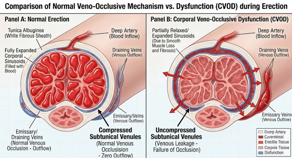
Figure 1. Cross-Sectional Hemodynamics of Normal Veno-Occlusion vs. CVOD.
Panel A shows healthy sinusoidal expansion compressing the subtunical venular plexus against the rigid tunica albuginea. Panel B shows incomplete sinusoidal expansion with uncompressed venules and persistent venous leak.
The resistance to blood outflow through a subtunical venule is exquisitely sensitive to changes in the vessel internal diameter from a biomechanical perspective. Because resistance to fluid flow in a small tube is inversely proportional to the fourth power of its luminal diameter, a small mechanical compression of the inner lumen produces an exponential increase in resistance to flow.(Udelson, L’Esperance, Morales, Patel, & Goldstein, 2000)
Also, veno-occlusion works by a dual mechanism of action where direct compression and stretching of tissues work together. As the corpora cavernosa expand during tumescence, the subtunical venules are greatly lengthened in the longitudinal direction, from 1.2 to 1.7 times their flaccid resting length. (Udelson, 2007) The physical principle of luminal constructability states that when a biological vessel is stretched longitudinally its transverse diameter decreases. In biological veins this longitudinal stretching causes the internal lumen to constrict in the transverse direction to the stretch, which increases the flow resistance drastically, even before the vein is completely mechanically squashed against the tunica albuginea. In the case of sub-optimal trabecular expansion, the outflow resistance is completely lost and neither effective longitudinal stretch nor direct mechanical compression can occur.(Udelson, 2007)
The principal abnormality in organic CVOD is not a primary anatomic abnormality of the draining veins themselves but rather a structural and cellular failure of the corporal spongy tissue (trabecular meshwork and cavernosal smooth muscle) to generate adequate expansibility and intracavernous pressure to collapse the subtunical venules.(M. G. Ferrini et al., 2017)
The functional bulk of the corpora cavernosa is composed of cavernosal smooth muscle cells (CSMC) in a collagenous and elastic extracellular matrix. Relaxation of CSMC increases tissue compliance and provides the driving force for lacunar enlargement.(Lue & Tanagho, 1987) Histomorphometry and clinical correlation studies have shown that aging, vascular risk factors, and neural injuries induce programmed cell death (apoptosis) of CSMCs mainly through intracellular reactive oxygen species (ROS) and oxidative stress.(Grünewald & Beal, 1999) Quantitative tissue analyses demonstrate that a man can tolerate a small decrease in CSMC mass, but when about 15% or more of the functional corporal smooth muscle mass is lost, symptomatic venous leakage is an inevitable consequence.(Clavijo et al., 2014)
As SMCs undergo apoptosis, they are replaced by non-yielding collagenous connective tissue (fibrosis). This loss of smooth muscle content directly decreases corporal expandability, the ability of the spongy tissue to stretch and receive blood at low pressures. When trabecular fibrosis reduces the expandability, the corporal tissue becomes rigid and non-compliant.(M. G. Ferrini et al., 2017; Udelson, 2007) So, normal arterial inflow cannot produce the volume expansion needed to wedge the outer trabeculae against the tunica albuginea. The venules of the subtunica are widely patent and this results in severe leakage of blood.(“NIH Consensus Conference. Impotence. NIH Consensus Development Panel on Impotence,” 1993)
As in Figure 2. Aging and vascular risk factors induce intracellular oxidative stress in the mitochondria of cavernosal smooth muscle cells. This oxidative stress as shown causes cell apoptosis and loss of smooth muscle leading to incomplete expansion of the corporal bodies and then CVOD. In turn, the smooth muscle cell endogenously upregulates expression of inducible nitric oxide synthase (iNOS) to generate intracellular NO, a protective, antiapoptotic mechanism to combat oxidative damage and retard progressive fibrosis.(M. Ferrini, Magee, Vernet, Rajfer, & González-Cadavid, 2001; Pannu & Singh, 2006)
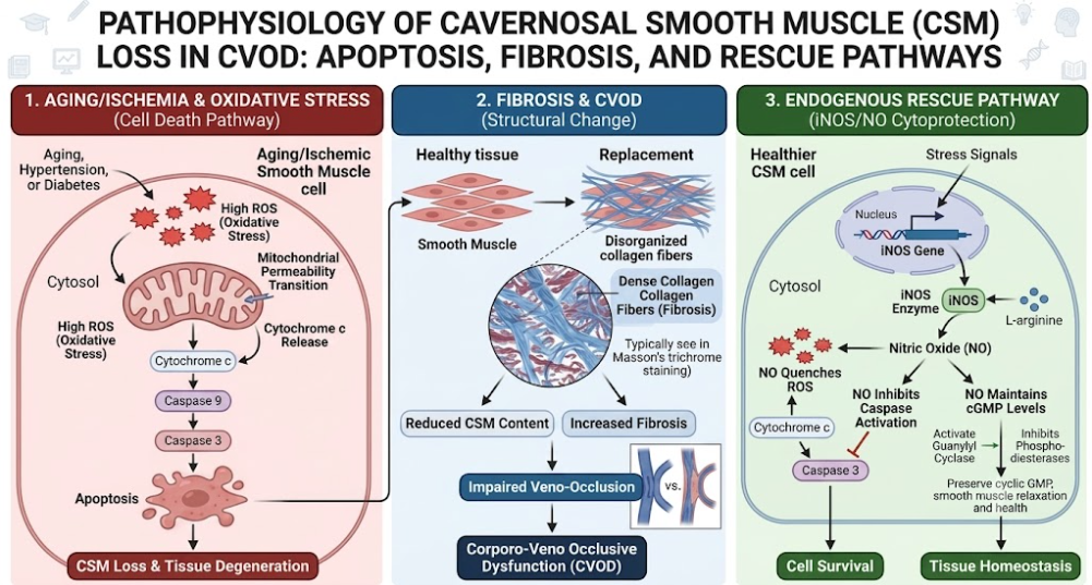
Figure 2. CSMC Apoptosis and the Endogenous iNOS Rescue Pathway.
Mitochondrial oxidative stress induces smooth muscle apoptosis and fibrosis, whereas cytosolic iNOS expression produces intracellular NO to scavenge ROS and maintain smooth muscle integrity.
Unlike neuronal nitric oxide synthase (nNOS) found in the nerve terminals outside of the smooth muscle cell, which produces NO to initiate erection, the inducible nitric oxide synthase (iNOS) comes from the cytosol of the CSMC itself. In response to stress adaptation to chronic oxidative stress and microvascular ischemia, CSMCs upregulate iNOS gene expression. (Gonzalez-Cadavid & Rajfer, 2004) iNOS-derived intracellular NO acts directly in the cytoplasm of CSMC to quench mitochondrial ROS and inhibit proapoptotic signaling cascades.(M. Ferrini et al., 2001) Long-term upregulation of this intracellular NO-cGMP pathway in experimental models slows down smooth muscle degradation, promotes smooth muscle preservation and can partially reverse corporal fibrosis and veno-occlusive failure.(Pannu & Singh, 2006)
The smooth muscle cells of the adult corpora cavernosa are not terminally differentiated, but rather show phenotypic plasticity. Under physiological conditions, CSMCs maintain a contractile phenotype with high expression levels of contractile markers, including Calponin-1 and alpha-smooth muscle actin, that enable normal tone regulation and relaxation compliance.(M. G. Ferrini et al., 2009; Wei et al., 2012)
Phenotypic modulation of CSMCs to a proliferative/synthetic phenotype is induced by cavernous nerve injury, chronic hypoxia or severe microvascular ischemia. This phenotypic switch is characterized by a significant downregulation of Calponin-1 protein and mRNA, and an upregulation of Osteopontin, a marker of proliferative, fibrotic smooth muscle cells.(Qian et al., 2020) CSMCs with synthetic-phenotype loss contractility and compliance of relaxation with excessive secretion of abnormal extracellular matrix components. This results in a dramatic reduction in the smooth muscle-collagen ratio (from ~6.28% down to ~2.12%) causing severe trabecular fibrosis and irreversible CVOD.(M. G. Ferrini et al., 2009; Wei et al., 2012)
The extracellular matrix of healthy corporal tissue is a balanced network of Type I, Type III, and Type IV collagen fibers interspersed with elastic fibers. Pathological ECM remodeling in CVOD is driven by persistent smooth muscle apoptosis and synthetic activity of CSMCs.(Azadzoi, Park, Andry, Goldstein, & Siroky, 1997) Collagen deposition increases substantially and the proportion of functional elastic fibers decreases. The excess connective tissue stiffens the trabecular walls and forms a physical barrier preventing the sinusoids from expanding even though there is blood flow within the lacunae.(Azadzoi et al., 1997; Qian et al., 2020)
Adequate perfusion of tissue is deeply linked to the health of corporal tissue. Arteriogenic occlusive disease (e.g. iliac or pudendal atherosclerosis) reduces the inflow of arterial blood and causes chronic ischemia of the cavernosal tissue. Laser Doppler flowmetry studies in ischemic models demonstrate that the maximal intracavernosal blood flow during erection is directly responsible for the equilibrium intracorporal pressure and inverse pressure decay/leakage rate. Chronic ischemia deprives CSMCs of oxygen resulting in pro-fibrotic signaling, collagen deposition and smooth muscle degeneration, and secondarily induces CVOD.(Azadzoi et al., 1997)
Systemic viral infections such as SARS-CoV-2 (COVID-19) have also been implicated in CSMC damage due to endothelial injury. SARS-CoV-2 infects endothelial cells through membrane bound ACE- 2 receptors resulting in endotheliitis and microvascular thrombosis.(Hosseinzadeh Zoroufchi, Doustmohammadi, Mokhtari, & Abdollahpour, 2021) Corpus cavernosum electromyography (cc-EMG) studies show that patients with a history of symptomatic COVID-19 have significantly lower pre-injection electrical amplitudes (166.0 uV versus 255.0 uV in healthy controls) and severely impaired smooth muscle relaxation capacity (Relaxation Degree 0.32 versus 0.58). This microvascular endothelial injury results in downstream cavernosal smooth muscle injury, impairing the tissue’s ability to relax and compress the subtunical venules.(Hosseinzadeh Zoroufchi et al., 2021)
The cavernosal smooth muscle is the active force for expansion, while the tunica albuginea is the passive, non-compliant outer wall that is required to compress the subtunical venules. An underappreciated primary cause of CVOD are pathological changes in the tunica albuginea itself.(Fitzpatrick, 1982)
Histological study of the tunica albuginea in control subjects with normal potency shows thick densely packed circularly and longitudinally disposed bundles of collagen impregnated by abundant elastic fibers. In contrast, biopsies taken from patients with venogenic erectile dysfunction show severe degeneration, atrophy and fragmentation of collagen fibres, as well as a complete lack or absence of elastic fibres.(Shafik et al., 2007)
At microscopic level, collagen bundles of healthy tunica albuginea show a particular bi-layered textile structure (inner circular, outer longitudinal) interspersed with elastic fibers. This structural configuration allows the tunica to change its dimensions during tumescence while providing a rigid unyielding surface to clamp passing emissary veins. In CVOD patients, collagen fibers are severely degenerated, atrophied and fragmented and lose their circular alignment.
In papaverine-induced erection, control subjects achieve a normal mean ICP of approximately 98.4 cmH2O, whereas patients with VED reach a transient peak pressure of approximately 65.2 cmH2O before rapidly declining to flaccid baseline levels (approximately 5.9 cmH2O). (Shafik et al., 2007)
Elastic fibers are normally present within the tunica albuginea, which allow the sheath to stretch by up to 150% of its length when flaccid, during tumescence, before becoming rigid.(Qian et al., 2020) In CVOD patients, elastic fibers are very sparse or completely absent. The loss of integrity of the elastic fibers results in structural subluxation and floppiness of the tunica albuginea sheath. When the internal lacunar space is enlarged, the subluxated and floppy tunica is stretched out of shape rather than providing a firm, unyielding surface. Thus, the subtunical venular plexus and perforating emissary veins are not compressed and there is continuous and uncontrolled venous leakage.(Ebbehøj & Wagner, 1979; Fournier et al., 1987; Rajfer, Rosciszewski, & Mehringer, 1988)
Pathological remodeling of the tunica albuginea involves structural disorganization as well as changes in the relative ratios of collagen subtypes. Type I collagen is made of thick rigid fibers giving tensile strength and Type III collagen confers elastic compliance. Cavernous nerve injury and chronic hypoxia significantly increase the Collagen I/III ratio (by >43%) and decrease Type IV basement membrane collagen. This shift to rigid type I collagen reduces the elasticity and compliance of the tunica albuginea and compromises its ability to dynamically adapt during erection, thus further compounding veno-occlusive failure.(Qian et al., 2020)
The essential diagnostic problem in sexual medicine is to differentiate true organic CVOD (due to irreversible structural damage to the smooth muscle or tunica) from functional or “misdiagnosed” CVOD.(Glina & Ghanem, 2013)
The CSMC are kept contracted by an active sympathetic adrenergic tone to maintain penile flaccidity. During sexual stimulation sympathetic activity must be inhibited to allow NANC mediated NO relaxation.(Sârbu et al., 2016) In young ED patients with no organic causes, psychological stress, performance anxiety or autonomic nervous system dysfunction can lead to severe sympathetic overactivity.(Meng et al., 2025)
In the setting of adequate arterial inflow, excessive adrenergic stimulation inhibits cavernosal smooth muscle relaxation. The smooth muscle is semi-contracted so that the sinusoids cannot expand sufficiently to compress the subtunical venules. These patients, who undergo a single standard stress CDDU examination, exhibit high end-diastolic velocity (EDV ≥ 6 cm/s) and are misdiagnosed as having CVOD (“misdiagnosed CVOD”).(Glina & Ghanem, 2013; Meng et al., 2025)
As in the Figure 3., Spontaneous Cavernous Activity (SCA) is recorded with Corpus Cavernosum Electromyography (CC-EMG) and reflects the intrinsic autonomic and myogenic status of the smooth muscle. In previous study shows the patients with psychogenic ED (misdiagnosed CVOD caused by sympathetic overactivity) have shown significantly elevated SCA amplitudes (305.65 uV) compared to patients with true organic CVOD (172.07 uV) who have suppressed SCA due to structural muscle degeneration.(Meng et al., 2025; Truss et al., 1993)
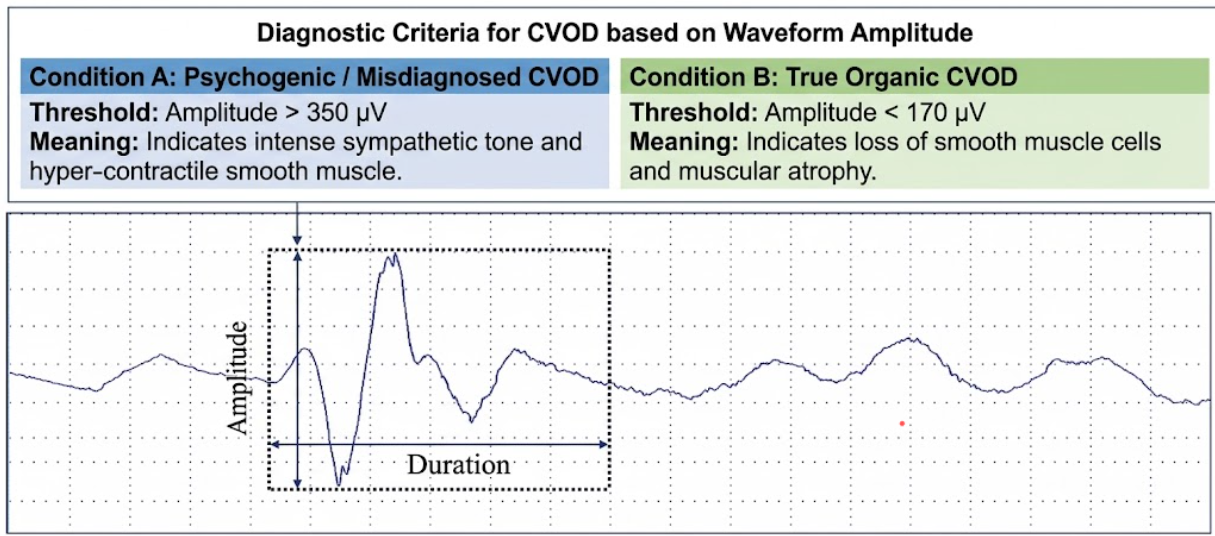
Figure 3. Spontaneous Cavernous Activity (SCA) is recorded with Corpus Cavernosum Electromyography (CC-EMG)
Electromyographic waveform analysis for differentiating psychogenic (or misdiagnosed) corporal veno-occlusive dysfunction (CVOD) from true organic CVOD. Condition A (amplitude > 350 uV) indicates intense sympathetic tone and hyper-contractile smooth muscle, whereas Condition B (amplitude < 170 uV) reflects loss of smooth muscle cells and muscular atrophy.
The pathophysiological paradigm of Corporal Veno-Occlusive Dysfunction (CVOD) has evolved considerably from a simplistic anatomical notion of “abnormal venous channels” to a complex biomechanical, structural and molecular failure of the penile blood containment system.(M. G. Ferrini et al., 2017) Veno-occlusive competence depends more on the functional compliance and structural integrity of the cavernosal spongy tissue and of its surrounding fibrous sheath, the tunica albuginea than on the native architecture of the exit veins.(Shafik et al., 2007; Udelson, 2007)
A critical threshold of loss of cavernosal smooth muscle cells (CSMCs) is a central concept in the cellular pathogenesis of CVOD. Quantitative histomorphometry studies have shown that small decreases in smooth muscle mass can be tolerated by the human corpus cavernosum, but that when smooth muscle loss exceeds approximately 15%, the remaining muscle tissue cannot generate the expansion needed to compress the subtunical venular plexus against the tunica albuginea.(Clavijo et al., 2014; M. G. Ferrini et al., 2017) The loss of cells is mainly due to intracellular reactive oxygen species (ROS) and mitochondrial oxidative stress due to aging, chronic microvascular ischemia and metabolic risk factors. Interestingly, CSMCs have an endogenous molecular defense against this apoptotic cascade by upregulating inducible nitric oxide synthase (iNOS) in the cytosol. iNOS-derived cytosolic NO provides an anti-apoptotic counter-measure that quenches ROS and retards progressive fibrosis, representing a potential therapeutic target for halting structural degradation.(M. Ferrini et al., 2001)
CSMC phenotypic switching is the phenomenon that completes the picture of cell loss. CSMCs modulate from functional contractile phenotype (characterized by Calponin-1 and alpha-smooth muscle actin) to proliferative, fibrotic synthetic phenotype (characterized by Osteopontin expression) under chronic hypoxia or nerve injury conditions.(Qian et al., 2020) Synthetic CSMCs actively secrete an excess of extracellular matrix, altering the structural composition of the trabeculae. Such ECM remodeling is associated with a dramatic reduction in the smooth muscle: collagen ratio (from ~6.28% to ~2.12%) and a replacement of elastic Type III collagen with rigid Type I collagen. Severe trabecular fibrosis limits cavernosal expandability and prevents blood pooling and sinusoidal expansion.(M. G. Ferrini et al., 2009; M. G. Ferrini et al., 2007)
At the tissue level, the tunica albuginea is crucial in providing the rigid, non-compliant backdrop required for veno-occlusion. In CVOD patients, microscopic evaluation shows severe degeneration, atrophy and fragmentation of collagen bundles and total absence or lack of elastic fibers. This structural disorganization results in subluxation and floppiness of tunica albuginea. When intra-lacunar pressure rises, a floppy tunica is abnormally distended instead of a firm surface, and the subtunical venules and the perforating emissary veins are kept patent and liable to continuous leakage of blood.(Shafik et al., 2007) Moreover, biomechanical mechanics demonstrate that normal veno-occlusion depends not only on direct compression but also on longitudinal vessel stretching. As corpora increase, subtunical venules are stretched longitudinally, resulting in orthogonal luminal narrowing due to luminal constructability. Loss of tunical floppiness or muscle loss precludes trabecular expansion, leading to simultaneous loss of direct mechanical compression and stretch-induced luminal narrowing.(Udelson, 2007)
From a clinical point of view, the difference between true organic CVOD and functional, psychogenic CVOD remains an important challenge. Performance anxiety or stress causes sympathetic overactivity that results in tonic contraction of CSMCs, which inhibits the relaxation required for sinusoidal expansion. This relaxation failure results in a high end-diastolic velocity (EDV ≥ 6 cm/s) on a single Color Duplex Doppler Ultrasound (CDDU) examination and a false positive diagnosis of CVOD (“misdiagnosed CVOD”). Corpus cavernosum electromyography (CC-EMG) evaluating spontaneous cavernous activity (SCA) provides a non-invasive tool to address this diagnostic dilemma. SCA amplitudes are significantly higher in patients with misdiagnosed CVOD driven by sympathetic overactivity (~305.65 uV) than in patients with true organic CVOD (~172.07 uV) where the electrical potentials are suppressed due to smooth muscle cell loss and muscular atrophy. Such functional differences help to avoid unnecessary venous surgery and to initiate proper psychological and pharmacological interventions.(Meng et al., 2025)
Corporal Veno-Occlusive Dysfunction (CVOD) is a biomechanical and structural failure of the penile blood-containment mechanism, and not a primary disease of the exit veins. CVOD results from the synergistic breakdown of cavernosal smooth muscle cells and the tunica albuginea. The primary pathophysiology involves apoptosis of cavernosal smooth muscle beyond the critical threshold of ~15% loss, cellular phenotypic switching from the contractile to the synthetic fibrotic state, extracellular matrix remodeling featuring an increased Collagen I/III ratio, and degenerative atrophy of the tunica albuginea resulting in structural subluxation and floppiness which precludes passive compression of the subtunical venular plexus. Furthermore, intracellular oxidative stress causes an endogenous up-regulation of iNOS as a protective anti-apoptotic counter-pathway, while microvascular ischemia and viral endotheliitis further aggravate smooth muscle injury. Diagnostic differentiation with Corpus Cavernosum Electromyography (CC-EMG) and Spontaneous Cavernous Activity (SCA) is needed to differentiate true myogenic failure from false-positive CVOD due to sympathetic overactivity from anxiety. In summary, a profound knowledge of these cellular, molecular, and biomechanical mechanisms serves as a solid basis for enhancing the precision of diagnosis, avoiding unnecessary surgical interventions, and advancing targeted regenerative therapies for the maintenance of smooth muscle mass and veno-occlusive functionality in men with vasculogenic erectile dysfunction.
Azadzoi, K. M., Park, K., Andry, C., Goldstein, I., & Siroky, M. B. (1997). Relationship between cavernosal ischemia and corporal veno-occlusive dysfunction in an animal model. J Urol, 157(3), 1011-1017.
Clavijo, R. I., Miner, M. M., & Rajfer, J. (2014). Erectile Dysfunction and Essential Hypertension: The Same Aging-related Disorder? Rev Urol, 16(4), 167-171.
Donatucci, C. F., & Lue, T. F. (1993). Erectile dysfunction in men under 40: etiology and treatment choice. International journal of impotence research, 5(2), 97-103.
Ebbehøj, J., & Wagner, G. (1979). Insufficient penile erection due to abnormal drainage of cavernous bodies. Urology, 13(5), 507-510. doi:10.1016/0090-4295(79)90459-x
Ferrini, M., Magee, T. R., Vernet, D., Rajfer, J., & González-Cadavid, N. F. (2001). Aging-related expression of inducible nitric oxide synthase and markers of tissue damage in the rat penis. Biol Reprod, 64(3), 974-982. doi:10.1095/biolreprod64.3.974
Ferrini, M. G., Gonzalez-Cadavid, N. F., & Rajfer, J. (2017). Aging related erectile dysfunction-potential mechanism to halt or delay its onset. Transl Androl Urol, 6(1), 20-27. doi:10.21037/tau.2016.11.18
Ferrini, M. G., Kovanecz, I., Sanchez, S., Umeh, C., Rajfer, J., & Gonzalez-Cadavid, N. F. (2009). Fibrosis and loss of smooth muscle in the corpora cavernosa precede corporal veno-occlusive dysfunction (CVOD) induced by experimental cavernosal nerve damage in the rat. J Sex Med, 6(2), 415-428. doi:10.1111/j.1743-6109.2008.01105.x
Ferrini, M. G., Kovanecz, I., Sanchez, S., Vernet, D., Davila, H. H., Rajfer, J., & Gonzalez-Cadavid, N. F. (2007). Long-term continuous treatment with sildenafil ameliorates aging-related erectile dysfunction and the underlying corporal fibrosis in the rat. Biol Reprod, 76(5), 915-923. doi:10.1095/biolreprod.106.059642
Fitzpatrick, T. J. (1982). The penile intercommunicating venous valvular system. J Urol, 127(6), 1099-1100. doi:10.1016/s0022-5347(17)54249-4
Fournier, G. R., Jr., Juenemann, K. P., Lue, T. F., & Tanagho, E. A. (1987). Mechanisms of venous occlusion during canine penile erection: an anatomic demonstration. J Urol, 137(1), 163-167. doi:10.1016/s0022-5347(17)43911-5
Glina, S., & Ghanem, H. (2013). SOP: corpus cavernosum assessment (cavernosography/cavernosometry). J Sex Med, 10(1), 111-114. doi:10.1111/j.1743-6109.2012.02795.x
Gonzalez-Cadavid, N. F., & Rajfer, J. (2004). Molecular pathophysiology and gene therapy of aging-related erectile dysfunction. Experimental Gerontology, 39(11), 1705-1712. doi:https://doi.org/10.1016/j.exger.2004.06.022
Grünewald, T., & Beal, M. F. (1999). NOS knockouts and neuroprotection. Nat Med, 5(12), 1354-1355. doi:10.1038/70918
Hehemann, M. C., & Kashanian, J. A. (2016). Can lifestyle modification affect men’s erectile function? Transl Androl Urol, 5(2), 187-194. doi:10.21037/tau.2016.02.05
Hosseinzadeh Zoroufchi, B., Doustmohammadi, H., Mokhtari, T., & Abdollahpour, A. (2021). Benzodiazepines related sexual dysfunctions: A critical review on pharmacology and mechanism of action. Revista Internacional de Andrología, 19(1), 62-68. doi:https://doi.org/10.1016/j.androl.2019.08.003
Kim, E. D., Owen, R. C., White, G. S., Elkelany, O. O., & Rahnema, C. D. (2015). Endovascular treatment of vasculogenic erectile dysfunction. Asian J Androl, 17(1), 40-43. doi:10.4103/1008-682x.143752
Ludwig, W., & Phillips, M. (2014). Organic causes of erectile dysfunction in men under 40. Urol Int, 92(1), 1-6. doi:10.1159/000354931
Lue, T. F., Mueller, S. C., Jow, Y. R., & Hwang, T. I. (1989). Functional evaluation of penile arteries with duplex ultrasound in vasodilator-induced erection. Urol Clin North Am, 16(4), 799-807.
Lue, T. F., Takamura, T., Schmidt, R. A., Palubinskas, A. J., & Tanagho, E. A. (1983). Hemodynamics of erection in the monkey. J Urol, 130(6), 1237-1241. doi:10.1016/s0022-5347(17)51768-1
Lue, T. F., & Tanagho, E. A. (1987). Physiology of erection and pharmacological management of impotence. J Urol, 137(5), 829-836. doi:10.1016/s0022-5347(17)44267-4
Lunglmayr, G., Nachtigall, M., & Gindl, K. (1988). Long-term results of deep dorsal penile vein transsection in venous impotence. Eur Urol, 15(3-4), 209-212. doi:10.1159/000473435
Meng, Z., Bai, H., Liu, S., Zhi, E., Tian, R., Yao, C., . . . Chen, H. (2025). Clinical value of spontaneous cavernous activity evaluation in identifying misdiagnosed corporal venous occlusive dysfunction in psychogenic erectile dysfunction. Transl Androl Urol, 14(6), 1691-1700. doi:10.21037/tau-2025-174
NIH Consensus Conference. Impotence. NIH Consensus Development Panel on Impotence. (1993). Jama, 270(1), 83-90.
Pannu, R., & Singh, I. (2006). Pharmacological strategies for the regulation of inducible nitric oxide synthase: neurodegenerative versus neuroprotective mechanisms. Neurochem Int, 49(2), 170-182. doi:10.1016/j.neuint.2006.04.010
Qian, S. Q., Qin, F., Zhang, S., Yang, Y., Wei, Q., Wang, R., & Yuan, J. H. (2020). Vacuum therapy prevents corporeal veno-occlusive dysfunction and penile shrinkage in a cavernosal nerve injured rat model. Asian J Androl, 22(3), 274-279. doi:10.4103/aja.aja_57_19
Rajfer, J., Rosciszewski, A., & Mehringer, M. (1988). Prevalence of corporeal venous leakage in impotent men. J Urol, 140(1), 69-71. doi:10.1016/s0022-5347(17)41489-3
Rajfer, J., Valeriano, J., & Sinow, R. (2013). Early onset erectile dysfunction is usually not associated with abnormal cavernosal arterial Inflow. International journal of impotence research, 25(6), 217-220. doi:10.1038/ijir.2013.17
Sârbu, M. I., Tampa, M., Mitran, M. I., Mitran, C. I., Benea, V., & Georgescu, S. R. (2016). The Current Treatment of Erectile Dysfunction. Journal of Mind and Medical Sciences, 3(2), 118-130.
Shafik, A., Shafik, I., El Sibai, O., & Shafik, A. A. (2007). On the pathogenesis of penile venous leakage: role of the tunica albuginea. BMC Urol, 7, 14. doi:10.1186/1471-2490-7-14
Truss, M. C., Djamilian, M. H., Tan, H. K., Hinrichs, H., Feistner, H., Stief, C. G., & Jonas, U. (1993). Single potential analysis of cavernous electrical activity. Four years’ experience in more than 500 patients with erectile dysfunction. Eur Urol, 24(3), 358-365.
Udelson, D. (2007). Biomechanics of male erectile function. J R Soc Interface, 4(17), 1031-1047. doi:10.1098/rsif.2007.0221
Udelson, D., L’Esperance, J., Morales, A. M., Patel, R., & Goldstein, I. (2000). The mechanics of corporal veno-occlusion in penile erection: a theory on the effect of stretch-associated luminal constrictability on outflow resistance. International journal of impotence research, 12(6), 315-327. doi:10.1038/sj.ijir.3900628
Waldinger, M. D., & Olivier, B. (1998). Selective serotonin reuptake inhibitor-induced sexual dysfunction: clinical and research considerations. Int Clin Psychopharmacol, 13 Suppl 6, S27-33. doi:10.1097/00004850-199807006-00006
Wei, A. Y., He, S. H., Zhao, J. F., liu, Y., Liu, Y., Hu, Y. W., . . . Wu, Z. Y. (2012). Characterization of corpus cavernosum smooth muscle cell phenotype in diabetic rats with erectile dysfunction. International journal of impotence research, 24(5), 196-201. doi:10.1038/ijir.2012.16