Our Services
Others
- DFPP
- Shockwave
- IV Drip


“The literature review discusses the physiological processes of plaque formation and the biomechanical forces that can lead to plaque rupture and subsequent arterial thrombosis in cardiovascular disease.”
Coronary artery disease, however, is still the leading cause of cardiovascular morbidity and mortality worldwide, with millions of sudden cardiac events each year. Over the last few decades, the clinical and scientific paradigm of coronary events has shifted from a stenosis-centric view to a biomechanical and biological understanding of plaque vulnerability and rupture. This review assembles the current knowledge of the cellular, molecular and mechanical processes that drive the life cycle of atherosclerotic plaques from their initial lipid accumulation to an abrupt and catastrophic structural failure. The atherosclerosis is initiated by endothelial dysfunction caused by low or oscillatory wall shear stress at arterial curves and bifurcations, which leads to subendothelial retention and oxidation of low-density lipoproteins. Subsequently, macrophages are recruited and subsequently transformed into foam cells, leading to a chronic inflammatory cascade that causes a necrotic core covered by a fibrous cap derived from vascular smooth muscle cells. Plaque rupture is an event in which the local maximum circumferential stress, primarily caused by the physiological or increased blood pressure, exceeds the ultimate tensile strength of the degenerated fibrous cap. Advanced imaging and computational fluid dynamics show that plaque structural stress depends on complex spatial relationships between the necrotic core, luminal geometry and presence of cellular microcalcifications that serve as high-strain stress concentrators. When it ruptures, highly thrombogenic core components are exposed to the circulating blood, triggering a rapid and self-amplifying cascade of platelet activation and coagulation, leading to luminal thrombosis and acute myocardial infarction. The intricate relationship between structural biomechanics and molecular biology needs to be understood to develop novel plaque-stabilizing therapeutics and to improve patient risk stratification.
Cardiovascular diseases, particularly coronary artery disease, continue to stand as the leading cause of global mortality, placing an immense burden on healthcare systems and taking millions of lives each year. The clinical manifestations of coronary artery disease, which range from stable angina to acute coronary syndromes and sudden cardiac death, are fundamentally rooted in the progression of atherosclerosis within the coronary arteries.(Otsuka, Yasuda, Noguchi, & Ishibashi-Ueda, 2016) Historically, clinical interventions focused on the “stenosis hypothesis,” which assumed that the severity of luminal narrowing was the primary predictor of acute cardiac events. However, extensive post-mortem histopathological studies and in vivo intravascular imaging have challenged this concept, giving rise to the “plaque hypothesis.” This modern framework establishes that the biological vulnerability and physical stability of the atheromatous wall, rather than the degree of stenosis, determine whether a lesion remains quiescent or precipitates acute coronary occlusion. (Holmstedt, Turan, & Chimowitz, 2013)
Indeed, a significant majority of acute myocardial infarctions are triggered not by highly stenotic, flow-limiting plaques, but rather by moderately stenotic lesions that possess features of extreme physical instability. These high-risk or “vulnerable” plaques undergo structural failure, allowing their inner thrombogenic contents to interact with blood, causing rapid clot formation. Hence, it is important to study the molecular and mechanical dynamics of atherosclerosis to identify at-risk plaques before they become clinical. The present literature review offers a complete discussion of the evolution of coronary plaques, detailing the pathological continuum from subendothelial lipid deposition to catastrophic arterial thrombosis.(Noonan, Cardoso, Bobik, & Peter, 2025; Veneziano, Cocco, Gentile, Chietera, & De Luca, 2026)
The development of coronary plaques is a highly organized biological process which takes many decades and which transforms a healthy elastic muscular artery into a rigid, compromised vessel. Normal coronary arteries have three distinct layers. The innermost layer is the tunica intima, which consists of a thin subendothelial space with a monolayer of endothelial cells. The tunica media is a highly organized layer of contractile vascular smooth muscle cells and elastin-rich extracellular matrix. The outer protective fibrous sheath is the tunica adventitia. These layers work together to preserve vascular homeostasis, modulate blood flow and prevent mechanical deformation under physiological conditions.(de Villiers & Smart, 1999)
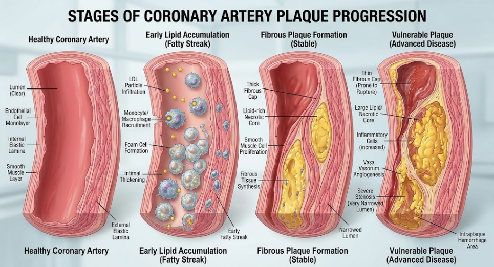
Figure 1. Evolution of plaque from normal to vulnerable coronary artery. This detailed medical illustration shows the course of coronary artery disease. It shows the progression of a healthy artery to an advanced, vulnerable thin-cap fibroatheroma (TCFA). It shows the essential steps including the initial accumulation of lipids (yellow LDL), activation of macrophages to foam cells and formation of a thin fibrous cap which can rupture and cause a blood clot. This visual guide shows the major anatomical and cellular changes that are involved in plaque instability and the risk of heart attack.
The pathological continuum of atherosclerosis is shown in Figure 1, which develops in biological stages beginning with local endothelial dysfunction that is highly dependent on fluid dynamics. Low density lipoproteins (LDLs) cross the damaged endothelial barrier and are deposited in the subendothelial intima where they are chemically modified by reactive oxygen species, such as oxidation. Oxidized LDL (oxLDL) is a powerful stimulus of inflammation. This leads to the expression of adhesion molecules on endothelial cells, which recruit circulating monocytes into the vascular wall. Monocytes are differentiated within the intima into active macrophages. These macrophages are identified by the expression of scavenger receptors that bind and ingest modified lipoproteins, resulting in the accumulation of intracellular lipid droplets and the formation of cholesterol-laden “foam cells.”(Weber & Noels, 2011; Zhang, Reddick, Piedrahita, & Maeda, 1992)
These lipid-laden foam cells accumulate and coalesce into clinically visible “fatty streaks” in the intima. This growing lipid deposit is stabilized by a dramatic phenotypic switch of vascular smooth muscle cells in the tunica media. Inflammatory cytokines promote the transition of smooth muscle cells from a quiescent, contractile phenotype to an active, synthetic phenotype with migration into the intima.(Jebari-Benslaiman et al., 2022; Staroselsky et al., 2025) Once these smooth muscle cells have migrated into the intima, they proliferate and produce structural extracellular matrix proteins, mainly interstitial collagens and elastin, which form a protective “fibrous cap” immediately below the endothelium.(Jebari-Benslaiman et al., 2022)
However, if the hypercholesterolemic and pro-inflammatory milieu persists, the plaque evolves into a more complex phase. Foam cells and smooth muscle cells of the deep layers of the intima die by programmed cell death (apoptosis) and secondary necrosis. In healthy condition, neighboring phagocytes phagocytize dying cells by the process of efferocytosis. Defective efferocytosis in advanced atherosclerosis. The structural cellular debris, free lipids, and cholesterol crystals from uncleared dead cells pool together, forming an acellular, highly thrombogenic space known as the “necrotic core.” As the necrotic core grows, it erodes into the surrounding extracellular matrix, with the only barrier between this highly reactive necrotic space and blood in the lumen being the fibrous cap that covers it.(Weber & Noels, 2011)
The critical precursor to plaque rupture is the thin-cap fibroatheroma (TCFA). Pathologically, TCFA is characterized by an advanced plaque with a large necrotic core covered by a very thin fibrous cap (<65 micrometers). The 65-micrometer threshold is an important quantitative reference point in cardiovascular pathology, based on human autopsy studies that showed that in more than 95% of ruptured coronary plaques, the cap thickness was well below this limit. A TCFA is profoundly depleted of smooth muscle cells as a consequence of chronic exposure to apoptotic triggers and inflammatory signals. Thus, the cap lacks the cellular machinery to repair and maintain the structural integrity. That delicate biological state is the precursor to mechanical failure.(Otsuka et al., 2016; Stone, Libby, & Boden, 2023)
Two major mechanical forces generated by the cardiovascular system are constantly challenging the structural integrity of an advanced coronary plaque: wall shear stress and plaque structural stress. These terms are often used interchangeably in the general literature, but they are different physical forces acting in different directions and scales in the vessel.(Noonan et al., 2025)
Wall shear stress (WSS) is a tangential frictional force applied on the vascular wall. It is generated by the viscous drag of blood flowing over the endothelial surface. In straight tubular segments of the coronary tree blood flow is laminar and results in physiological wall shear stress ranging from 1.0 to 2.5 Pascal. This normal mechanical stimulus stimulates the endothelial release of nitric oxide that acts in a protective, anti-inflammatory and vasodilator manner on the arterial wall. However, at branches, bifurcations and areas of high curvature, flow disturbance occurs resulting in low wall shear stress (usually < 1.0 Pascal). Low wall shear stress does not activate endothelial cells in a protective manner, but instead promotes leukocyte adhesion, lipid retention and infiltration of inflammatory cells.(Noonan et al., 2025)
Low wall shear stress contributes significantly to plaque initiation and to amplification of regional vulnerability, but its magnitude is orders of magnitude lower than the mechanical strength of arterial tissue. The ultimate tensile strength of a human fibrous cap is about 545 kilopascals, five orders of magnitude higher than the shear forces exerted by flowing blood. Wall shear stress cannot directly rupture or tear a plaque.(Libby, 2021)
The plaque structural stress acting in the circumferential direction within the vessel wall is the actual physical cause of plaque tearing instead. Plaque structural stress is the tensile force induced by the intra-arterial blood pressure that stretches the vessel wall during the cardiac cycle.(Libby, 2021; Staroselsky et al., 2025) Any increase in blood pressure or luminal radius will greatly increase the stress experienced by the vessel wall, as predicted by classical solid mechanics and Laplace’s law (circumferential stress is proportional to intra-arterial pressure multiplied by vessel radius and divided by wall thickness). In a healthy and uniform artery the circumferential stress is equally distributed over the elastic wall. But the formation of a complex eccentric coronary plaque changes this stress distribution.(Costopoulos et al., 2017)
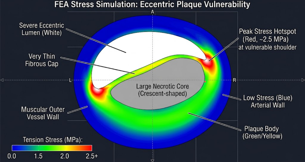
Figure 2. Finite Element Analysis of a Vulnerable Eccentric Coronary Plaque. Computational physics-style simulation displaying a continuous stress heatmap within a highly eccentric plaque. The concentration of the peak mechanical tension (brilliant red-hot spots) at the vulnerable shoulders suggests a high risk of rupture of the thin fibrous cap.
More advanced finite element analysis simulations (see Figure 2) suggest that stress is highly localized at specific sites of structural discontinuity, particularly in the “shoulder” zones where the thinner, more flexible fibrous cap meets the stiffer, normal arterial wall.(Stone et al., 2023) In vivo studies have demonstrated that plaque composition, plaque architecture and lumen geometry interact in a complex manner to determine plaque structural stress. Specifically, plaque structural stress increased with increasing luminal area (r = 0.46; p = 0.001), lumen eccentricity (r = 0.32; p = 0.001) and necrotic core volume >10% (r = 0.12; p = 0.001).(Costopoulos et al., 2017)
Moreover, investigators studying human coronary arteries for plaque mechanics have shown that the maximum plaque structural stress and the difference in plaque structural stress between systole and diastole are significantly higher in plaques that have ruptured in vivo compared to non-ruptured plaques of similar classification. The median peak structural stress is 133 kilopascals for ruptured plaques and 104 kilopascals for the stable plaques (p = 0.002).(Costopoulos et al., 2017) In addition, the cyclic mechanical loading and unloading of the plaque during the cardiac cycle generates significant differences in plaque structural stress (55 kilopascals vs. 43 kilopascals; p 0.002) in ruptured plaques compared with stable plaques. This repetitive dynamic stretching leads to mechanical fatigue in the fibrous cap, resulting in a gradual degradation of the extracellular matrix until the material is no longer able to support the mechanical load. Multivariable regression models have identified a plaque structural stress threshold of 135 kilopascals as a powerful independent predictor of plaque rupture in high-risk coronary segments.(Costopoulos et al., 2017)
Rupture of the fibrous cap is not a mechanical event but reflects ongoing inflammatory and enzymatic activity within the plaque. The thickness of the fibrous cap and its mechanical strength depend on the balance between extracellular matrix synthesis by smooth muscle cells and extracellular matrix degradation by proteases secreted by inflammatory cells.(Badimon, Padró, & Vilahur, 2012; Stone et al., 2023)
In a vulnerable plaque the immune system shifts the balance toward matrix destruction. In the plaque, T-helper 1 (Th1) lymphocytes produce interferon-γ, a potent cytokine that acts directly on vascular smooth muscle cells to inhibit their proliferation and to reduce their synthesis of new interstitial collagens. Since smooth muscle cells are the only source of collagen in the fibrous cap, this cytokine mediated inhibition eliminates the major structural repair mechanism of the cap.(Jebari-Benslaiman et al., 2022; Libby, 2002)
Simultaneously, activated macrophages and smooth muscle cells in the plaque secrete matrix metalloproteinases (MMPs), a family of zinc-dependent endopeptidases capable of degrading all major components of the vascular extracellular matrix. Macrophages secrete interstitial collagenases and gelatinases (MMP-1, MMP-9 and MMP-14) in response to pro-inflammatory cytokines such as tumor necrosis factor-α and interleukin-1β. These enzymes cleave the triple-helix structure of collagen, reducing it to fragments that are highly susceptible to further non-specific proteolytic degradation. This active enzymatic digestion thins the fibrous cap, reducing its load-bearing capacity and causing local concentrations of plaque structural stress to rise rapidly.(Badimon et al., 2012; Stone et al., 2023)
Furthermore, hypoxia in the developing necrotic core induces expression of hypoxia-inducible factors that drive neovascularization from the adventitial vasa vasorum. These newly formed microvessels are immature and fragile and do not have tight junctions. They are very leaky and rupture easily (micro-rupture) causing intraplaque hemorrhage. Intraplaque hemorrhage supplies free cholesterol from erythrocyte membranes to the core, hastening necrotic core growth. It also liberates free hemoglobin and catalytic iron, which facilitate lipid peroxidation, cellular injury and macrophage activation, further destabilizing the plaque.(Engelen, Robinson, Zurke, & Monaco, 2022; Kolodgie et al., 2003)
An interesting and complex aspect of the dynamics of the plaque rupture is the dual role of the vascular calcification. Large, confluent and sheet-like calcifications (so-called stable and fibrocalcific plaques) are mechanical stabilizers by reducing the overall wall strain. Small, spotty or cellular level microcalcifications are highly dangerous stress concentrators within the soft tissues of the cap.(Costopoulos et al., 2017; Gijsen et al., 2008)
Coronary microcalcifications originate from extracellular matrix vesicles released by smooth muscle cells and macrophages, which coalesce and crystallize to form hydroxyapatite in the subendothelial matrix. These microcalcifications (on the order of 5 to 15 micrometers in diameter) are embedded in the soft, fibrous tissue of a thin cap, creating a severe mismatch in material stiffness. Calcified hydroxyapatite has a Young’s modulus which is several orders of magnitude larger than that of the surrounding collagenous matrix.(Noonan et al., 2025; Otsuka et al., 2016)
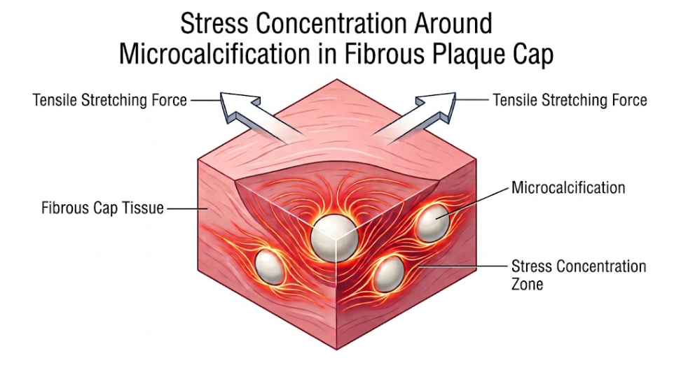
Figure 3. Stress Concentration at Microcalcification in Fibrous Plaque Cap. This biomechanical diagram shows a model of fibrous cap tissue in a coronary arterial plaque under tensile stretching force (white arrows). Embedded microcalcifications (white spheres) in this setting serve as loci of mechanical stress concentration. The lines in red and gold radiating outwards represent the areas of stress concentration, that is, the mechanical stress is highly increased and localized in these rigid inclusions. Such focal stress may cause a fibrous cap to rupture, plaque to break apart, and a thrombus to form.
During the cardiac cycle, the plaque experiences tensile circumferential stress, which causes the mechanical interface (shown in Figure 3) to have a severe stiffness mismatch that distorts the local mechanical stress field. The microcalcifications are local ‘stress risers’ that concentrate stress at the interface. Biomechanical models suggest that a single microcalcification can increase local cap stress by a factor of 2 to 7 depending on its size, geometry, and proximity to other calcified deposits.(Engelen et al., 2022)
If several microcalcifications are found clustered together in a thin fibrous cap, the overlapping stress fields between them may induce very high stress concentrations. Under cyclic pressure, the localized stress concentration is greater than the adhesive bond between the calcified inclusion and the surrounding collagenous matrix. This results in interfacial debonding or debonding at the interface between inclusion and matrix leading to microscopic voids. Under the driving force of the blood pressure, these micro-voids grow rapidly and coalesce into microfissures that propagate through the fibrous cap. This process ends with the fibrous cap tearing, ripping or fissuring, resulting in catastrophic plaque rupture. (Libby, 2021; Staroselsky et al., 2025)
The immediate onset of the thrombotic cascade is the last life-threatening sequel of coronary plaque rupture. In the intact state the vascular endothelium forms a protective barrier preventing the blood from coming into contact with the highly reactive proteins and lipids of the arterial wall. However, when the fibrous cap ruptures, the barrier is disrupted and the highly thrombogenic necrotic core is exposed directly to the circulating blood.(Falk, 2006)
This exposure causes a rapid, biphasic thrombotic response including primary hemostasis (platelet activation and aggregation) and secondary hemostasis (coagulation cascade).(Staroselsky et al., 2025)
Platelets contact subendothelial collagen and von Willebrand factor exposed at the site of rupture, initiating primary hemostasis immediately. These matrix proteins bind platelets through specific glycoprotein receptors. This adhesion leads to a dramatic conformational change of the platelets which triggers an intracellular signaling cascade leading to platelet activation. Activation leads to the release of the contents of storage granules that are rich in soluble agonists such as thromboxane A2 and adenosine diphosphate (ADP).(Młynarska et al., 2024; Staroselsky et al., 2025)
Release of mediators to recruit and activate other platelets in a paracrine and autocrine manner Thromboxane A2 is also a potent local vasoconstrictor that narrows the coronary lumen, further decreasing blood flow. ADP binds to G-protein coupled receptors on the platelet surface, the P2Y1 and P2Y12 receptors. These agonists induce a conformational change in the platelet-surface integrin glycoprotein IIb/IIIa from a low-affinity state to a high-affinity state. Activated glycoprotein IIb/IIIa binds circulating fibrinogen and von Willebrand factor, allowing cross-linking of adjacent platelets and formation of a rapidly expanding platelet plug at the rupture site.(Młynarska et al., 2024; Staroselsky et al., 2025)
Secondary hemostasis is initiated by the exposure of tissue factor, a transmembrane glycoprotein, which is abundantly expressed on macrophages, foam cells and cellular debris in the necrotic core. The circulating coagulation factor VII/VIIa binds to the exposed tissue factor and forms an active enzymatic complex and initiates the extrinsic coagulation pathway. This complex activates factor X and then creates the prothrombinase complex that rapidly converts prothrombin to active thrombin.(Amir Ajoolabady et al., 2024; Staroselsky et al., 2025)
Thrombin is a key, multidimensional player in thrombus propagation. It is a very strong activator of platelets via protease activated receptors (PARs) on the surface of platelets. Most importantly, thrombin cleaves circulating soluble fibrinogen into insoluble fibrin monomers that polymerize into long, stable fibrin strands. These strands of fibrin interdigitate with the developing platelet plug, and trap circulating red and white blood cells, thereby forming a stable, solid thrombus.(Jebari-Benslaiman et al., 2022; Młynarska et al., 2024; Staroselsky et al., 2025)
Depending on the local configuration of the stenosis, the velocity of blood flow, and the balance between systemic procoagulant and anticoagulant factors, the thrombus can expand rapidly to produce complete or near-complete occlusion of the coronary lumen. The acute occlusion interrupts the supply of oxygenated blood to the distal myocardium leading to severe myocardial ischemia, cardiomyocyte necrosis and clinical manifestations of acute myocardial infarction or sudden cardiac death.(Libby, Tabas, Fredman, & Fisher, 2014)
The transition from quantification of stenosis severity to assessment of plaque vulnerability in clinical cardiology has revolutionized the diagnosis, monitoring and treatment of coronary artery disease. Previously, angiography was used to identify highly stenotic lesions for percutaneous intervention. Clinical trials and pathological observations, however, have shown that treatment of stable high-grade stenotic lesions does not prevent the majority of myocardial infarctions. This is because myocardial infarctions often result from the abrupt rupture of moderately stenotic, nonobstructive plaques with large necrotic cores and thin fibrous caps.(Amir Ajoolabady et al., 2024; Veneziano et al., 2026)
High-risk lesions have been identified with the help of advanced clinical imaging modalities. High-resolution optical coherence tomography (OCT) can be used by the clinician to measure the thickness of the fibrous cap in vivo to identify thin-cap fibroatheromas with a resolution of 10 to 15 micrometers. Near-infrared spectroscopy (NIRS) can quantify the lipid content of the coronary wall, calculating a lipid core burden index to detect large, vulnerable necrotic pools.(A. Ajoolabady et al., 2024)
The combination of these high-resolution imaging methods with computational fluid dynamics enables clinicians to do “digital pathology” of the coronary tree. Reconstruction of patient-specific coronary artery geometries enables the calculation of local wall shear stress and plaque structural stress in vivo, and identification of sites where mechanical fatigue and high stress co-localize with biological vulnerability. The clinical utility of these biomechanical calculations is demonstrated by the results of Costopoulos et al., who show that the addition of plaque structural stress to plaque assessments significantly improves our ability to predict which plaques will rupture and cause clinical events with an optimal stress cutoff of 135 kilopascals.(Aguirre, Arbab-Zadeh, Soeda, Fuster, & Jang, 2021; Costopoulos et al., 2017)
The main cause of coronary thrombosis is plaque rupture, but it is necessary to differentiate it from plaque erosion, which is another important clinical phenotype of acute coronary syndromes. The erosion of plaque is responsible for 30% to 35% of sudden coronary death and is more common in younger patients and premenopausal women.(Ramoni et al., 2025) Eroded plaques are characterized by endothelial denudation without cap rupture (unlike rupture).(Russo et al., 2025) These lesions are typically devoid of large lipid pools and necrotic cores, and comprise a matrix rich in proteoglycan and glycosaminoglycan, with relative preservation of smooth muscle cells and minimal infiltration of inflammatory cells.(Stone et al., 2023)
Plaque erosion involves a variety of biomechanical and cellular mechanisms and the classical paradigm of high structural stress in plaques and collagen degradation may not apply to all coronary events. Clinically distinguishing between these two phenotypes in vivo is important as eroded plaques may be responsive to anti-thrombotic therapies without the need for mechanical stenting.(Ramoni et al., 2025)
Atherosclerosis and coronary plaque rupture are governed by a complex, multi-scale interaction between biological signaling and mechanical forces. The coronary plaque life cycle is a pathological continuum beginning with hemodynamically driven endothelial activation, then cellular remodeling and chronic inflammation and finally biomechanical failure. Plaque rupture is fundamentally a mechanical event that occurs when physiological or elevated blood pressure concentrates circumferential stress within a biologically weakened fibrous cap.
Understanding the structural determinants of plaque structural stress—such as cap thickness, necrotic core architecture, and the stress-concentrating behavior of cellular microcalcifications—is essential for refining our clinical approach to coronary artery disease. The combination of high-resolution intravascular imaging and physics-based computational modeling allows modern medicine to move from treating acute coronary events to predicting and preventing them. Further studies of the cellular pathways mediating matrix degradation and the physical dynamics of plaque instability are needed to reduce the global burden of cardiovascular diseases.
Aguirre, A. D., Arbab-Zadeh, A., Soeda, T., Fuster, V., & Jang, I. K. (2021). Optical Coherence Tomography of Plaque Vulnerability and Rupture: JACC Focus Seminar Part 1/3. J Am Coll Cardiol, 78(12), 1257-1265. doi:10.1016/j.jacc.2021.06.050
Ajoolabady, A., Pratico, D., Lin, L., Mantzoros, C. S., Bahijri, S., Tuomilehto, J., & Ren, J. (2024). Inflammation in atherosclerosis: pathophysiology and mechanisms. Cell Death & Disease, 15(11), 817. doi:10.1038/s41419-024-07166-8
Ajoolabady, A., Pratico, D., Lin, L., Mantzoros, C. S., Bahijri, S., Tuomilehto, J., & Ren, J. (2024). Inflammation in atherosclerosis: pathophysiology and mechanisms. Cell Death Dis, 15(11), 817. doi:10.1038/s41419-024-07166-8
Badimon, L., Padró, T., & Vilahur, G. (2012). Atherosclerosis, platelets and thrombosis in acute ischaemic heart disease. Eur Heart J Acute Cardiovasc Care, 1(1), 60-74. doi:10.1177/2048872612441582
Costopoulos, C., Huang, Y., Brown, A. J., Calvert, P. A., Hoole, S. P., West, N. E. J., . . . Bennett, M. R. (2017). Plaque Rupture in Coronary Atherosclerosis Is Associated With Increased Plaque Structural Stress. JACC Cardiovasc Imaging, 10(12), 1472-1483. doi:10.1016/j.jcmg.2017.04.017
de Villiers, W. J., & Smart, E. J. (1999). Macrophage scavenger receptors and foam cell formation. J Leukoc Biol, 66(5), 740-746. doi:10.1002/jlb.66.5.740
Engelen, S. E., Robinson, A. J. B., Zurke, Y. X., & Monaco, C. (2022). Therapeutic strategies targeting inflammation and immunity in atherosclerosis: how to proceed? Nat Rev Cardiol, 19(8), 522-542. doi:10.1038/s41569-021-00668-4
Falk, E. (2006). Pathogenesis of atherosclerosis. J Am Coll Cardiol, 47(8 Suppl), C7-12. doi:10.1016/j.jacc.2005.09.068
Gijsen, F. J., Wentzel, J. J., Thury, A., Mastik, F., Schaar, J. A., Schuurbiers, J. C., . . . Serruys, P. W. (2008). Strain distribution over plaques in human coronary arteries relates to shear stress. Am J Physiol Heart Circ Physiol, 295(4), H1608-1614. doi:10.1152/ajpheart.01081.2007
Holmstedt, C. A., Turan, T. N., & Chimowitz, M. I. (2013). Atherosclerotic intracranial arterial stenosis: risk factors, diagnosis, and treatment. Lancet Neurol, 12(11), 1106-1114. doi:10.1016/s1474-4422(13)70195-9
Jebari-Benslaiman, S., Galicia-García, U., Larrea-Sebal, A., Olaetxea, J. R., Alloza, I., Vandenbroeck, K., . . . Martín, C. (2022). Pathophysiology of Atherosclerosis. Int J Mol Sci, 23(6). doi:10.3390/ijms23063346
Kolodgie, F. D., Gold, H. K., Burke, A. P., Fowler, D. R., Kruth, H. S., Weber, D. K., . . . Virmani, R. (2003). Intraplaque hemorrhage and progression of coronary atheroma. N Engl J Med, 349(24), 2316-2325. doi:10.1056/NEJMoa035655
Libby, P. (2002). Inflammation in atherosclerosis. Nature, 420(6917), 868-874. doi:10.1038/nature01323
Libby, P. (2021). The changing landscape of atherosclerosis. Nature, 592(7855), 524-533. doi:10.1038/s41586-021-03392-8
Libby, P., Tabas, I., Fredman, G., & Fisher, E. A. (2014). Inflammation and its resolution as determinants of acute coronary syndromes. Circ Res, 114(12), 1867-1879. doi:10.1161/circresaha.114.302699
Młynarska, E., Czarnik, W., Fularski, P., Hajdys, J., Majchrowicz, G., Stabrawa, M., . . . Franczyk, B. (2024). From Atherosclerotic Plaque to Myocardial Infarction—The Leading Cause of Coronary Artery Occlusion. Int J Mol Sci, 25(13), 7295. Retrieved from doi:10.3390/ijms25137295
Noonan, J., Cardoso, L., Bobik, A., & Peter, K. (2025). Atherosclerotic Plaque Instability and Rupture: Recommended Mouse Models to Empower Clinically Relevant Discoveries, Diagnostics, and Therapeutics. Arterioscler Thromb Vasc Biol, 45(10), 1707-1714. doi:10.1161/atvbaha.125.321011
Otsuka, F., Yasuda, S., Noguchi, T., & Ishibashi-Ueda, H. (2016). Pathology of coronary atherosclerosis and thrombosis. Cardiovasc Diagn Ther, 6(4), 396-408. doi:10.21037/cdt.2016.06.01
Ramoni, D., Carbone, F., Kraler, S., Di Vece, D., Montecucco, F., & Liberale, L. (2025). Inflammation-Driven Plaque Erosion in Atherosclerosis: A Focus on Complement System Pathways. Current Atherosclerosis Reports, 27(1), 42. doi:10.1007/s11883-025-01279-x
Russo, M., Gurgoglione, F. L., Russo, A., Rinaldi, R., Torlai Triglia, L., Foschi, M., . . . Zimarino, M. (2025). Coronary Artery Disease and Atherosclerosis in Other Vascular Districts: Epidemiology, Risk Factors and Atherosclerotic Plaque Features. Life (Basel), 15(8). doi:10.3390/life15081226
Staroselsky, M., Materia, A., Damera, R. R., Berman, L., Hai, O., Zeltser, R., & Makaryus, A. N. (2025). The Pathophysiology of Coronary Artery Disease. Int J Angiol, 34(4), 312-319. doi:10.1055/a-2652-1812
Stone, P. H., Libby, P., & Boden, W. E. (2023). Fundamental Pathobiology of Coronary Atherosclerosis and Clinical Implications for Chronic Ischemic Heart Disease Management-The Plaque Hypothesis: A Narrative Review. JAMA Cardiol, 8(2), 192-201. doi:10.1001/jamacardio.2022.3926
Veneziano, F. A., Cocco, N., Gentile, F., Chietera, F., & De Luca, L. (2026). Plaque Progression and Rupture in Obstructive Coronary Artery Disease: A Review of Current Imaging Modalities. Rev Cardiovasc Med, 27(4), 48015. doi:10.31083/rcm48015
Weber, C., & Noels, H. (2011). Atherosclerosis: current pathogenesis and therapeutic options. Nat Med, 17(11), 1410-1422. doi:10.1038/nm.2538
Zhang, S. H., Reddick, R. L., Piedrahita, J. A., & Maeda, N. (1992). Spontaneous hypercholesterolemia and arterial lesions in mice lacking apolipoprotein E. Science, 258(5081), 468-471. doi:10.1126/science.1411543