Our Services
Others
- DFPP
- Shockwave
- IV Drip

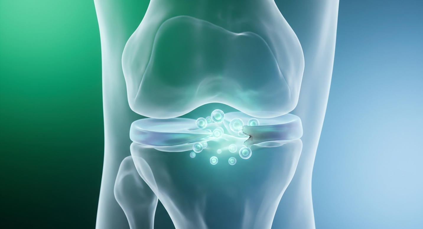
For patients facing chronic knee pain and exploring a meniscus tear treatment without surgery, navigating the options for stem cell therapy for meniscus tears requires separating rigorous clinical science from marketing claims. Clinical reviews indicate how confusing the regenerative medicine space can be. It’s loud. Every clinic claims to have the absolute best alternative to meniscus surgery.
But here’s the thing not all stem cells are created equal, and not all tears can be fixed this way.
In this guide, you’ll learn how to evaluate UC-MSC therapies based on objective safety standards, biological rationale, and realistic efficacy metrics. We detail the foundational criteria for suitability assessment, outline the Comet Assay safety standard, and review evidence-informed recovery timelines so you know exactly what to expect.
This article is for educational purposes only and does not replace consultation with a qualified medical professional.
Stem cell therapy for meniscus tear procedures using UC-MSCs offer an evidence-informed approach to joint regeneration.
The newest treatment for meniscus tears focuses on tissue preservation, but you can’t evaluate regenerative options without understanding why an untreated meniscus tear fails to heal in the first place. Over 70% of meniscus volume resides in the white-white avascular zone, meaning these regions simply cannot heal naturally without external biological intervention (Medical anatomical studies, 2014). This avascular reality is exactly why injecting cultivated cellular agents into the joint space has become a primary focus in modern orthopedics.
Not every knee injury requires the same intervention. The meniscus is a primary shock-absorbing fibrocartilage, divided into three distinct vascular regions: the red-red (outer rim with rich blood supply), the red-white (middle transition), and the white-white (inner avascular) zones.
Patients looking at their MRI results often feel overwhelmed by the terminology. But understanding whether your tear sits in the vascular or avascular zone is the single most important factor dictating your recovery trajectory. When you suffer a tear in the red-red zone, surgical suturing often works brilliantly because the tissue has the oxygen and nutrients needed to knit back together. Chronic degenerative injuries in the inner avascular zone are a different story. These represent hard to repair meniscus tears. Without intervention, an untreated meniscus tear in this region undergoes cellular starvation. The tissue degenerates.
For context, not all tears behave the same structurally. A radial tear cuts perpendicularly into the avascular zone, completely obliterating the circumferential fibers that distribute your body weight. Horizontal cleavage tears split the meniscus into top and bottom halves, often creating painful fluid-filled cysts. Meanwhile, a bucket-handle tear is a massive longitudinal rupture where the tissue flips over and physically jams the knee hinge. When these occur in the white-white zone, the lack of blood flow accelerates degeneration year over year. Within three to five years, an untreated avascular tear typically leads to severe joint space narrowing, forcing the underlying bone to absorb impact loads it was never designed for.
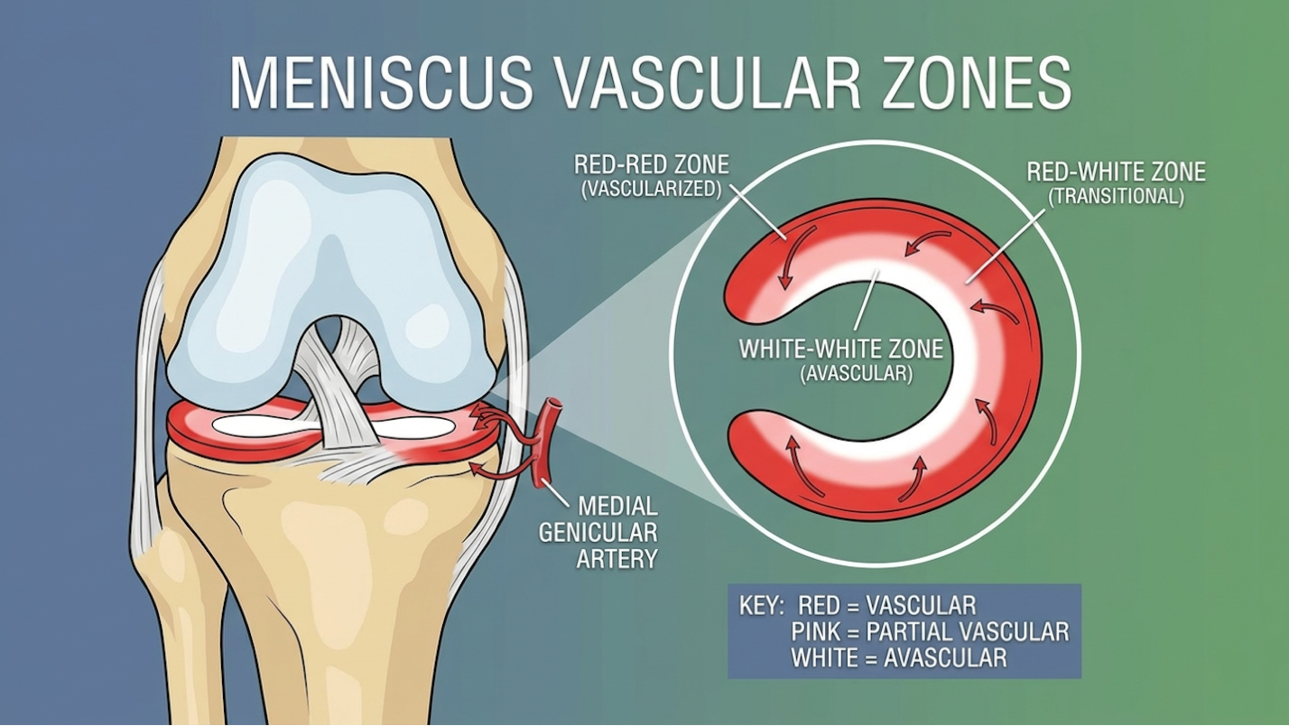
Because avascular tears lack the intrinsic blood supply needed for cellular repair, clinicians are actively assessing the shift toward regenerative interventions that deliver necessary cells directly to the defect.
An unhealed meniscus tear in the avascular zone leads to progressive joint degeneration and accelerated osteoarthritis. Without the shock-absorbing capacity of the intact fibrocartilage, mechanical stress concentrates on the articular cartilage covering the femur and tibia. Over time, this localized friction degrades the joint surface, leading to chronic inflammation and loss of mobility.
For decades, the standard orthopedic answer to a white-white zone tear was a partial meniscectomy literally clipping out the torn tissue. Look, sometimes it’s mechanically necessary. But removing that tissue permanently alters knee biomechanics. Patients seeking alternatives to surgery often underestimate the biomechanical consequences of removing cartilage. Every millimeter of resected meniscus exponentially multiplies the impact load on the underlying bone, fast-tracking the patient straight toward early-onset osteoarthritis.
That’s an unacceptable trade-off for active patients. This clinical reality drove the shift toward biological solutions. Clinical evaluations demonstrate that introducing exogenous biological factors can temporarily override the lack of natural blood flow, stimulating the resident fibrochondrocytes to begin repairing the matrix (Clinical perspectives, 2026). To execute this tissue preservation strategy effectively, the next step involves evaluating the specific biological agents, namely UC-MSCs.
Stem cell meniscus regeneration relies on specific cellular mechanisms, not magic. UC-MSCs promote tissue repair by releasing over 300 distinct growth factors that stimulate local regeneration, rather than simply acting as structural glue (Stem Cell Research, 2021). When patients ask about UC-MSC therapy for meniscus tear protocols, the conversation must center on
Human umbilical cord-derived mesenchymal stem cells (UC-MSCs) are highly proliferative progenitor cells sourced from Wharton’s Jelly. Why prefer them over autologous bone marrow (BM-MSCs) or adipose tissue? Age. If you are 55 years old, your bone marrow stem cells are also 55 years old. They have shorter telomeres, slower proliferation rates, and less metabolic vigor.
| 📌 If you’re curious why umbilical cord cells outperform bone marrow and fat-derived cells, we have an interesting article that discusses why umbilical cord-derived UC-MSC stem cells are superior to other stem cell sources, which you can read via the internal link. |
Let’s dig deeper into the alternative sources. Bone marrow aspiration (BM-MSCs) requires drilling into the iliac crest a highly invasive procedure that yields a cellular product deeply compromised by your chronological age. Adipose-derived stem cells (AD-MSCs) demand a mini-liposuction procedure, which carries its own set of surgical risks and often requires weeks of laboratory expansion just to reach a therapeutic cell count.
In contrast, UC-MSCs bypass all of this. Because they are ethically sourced from donated umbilical cords post-birth, they bypass patient harvesting entirely. These “day-zero” cells retain maximum telomere length, providing exceptional metabolic vigor and cellular division rates that a 50- or 60-year-old autologous sample simply cannot match. They don’t just survive in the hostile, avascular environment of a torn meniscus; they thrive there.
UC-MSCs also possess an immune-privileged status meaning they don’t express the HLA-DR antigens that would typically trigger your body to reject them.
Here is how they stack up against older methods:
| Feature | UC-MSCs | BM-MSCs (Autologous) | Traditional Surgery |
| Cell Potency | Maximum (Day-zero cells) | Age-dependent decline | N/A |
| Invasiveness | Low (Injection only) | High (Bone drilling) | Very High |
| Immune Privilege | Yes (No matching needed) | N/A (Patient’s own) | N/A |
| Primary Action | Paracrine signaling | Paracrine signaling | Mechanical excision |
Our evaluation of current protocols confirms that the cellular advantage of UC-MSCs makes them the superior biological payload (Frontiers in Bioengineering review, 2022). Understanding the cellular advantage of UC-MSCs leads directly into how these cells mechanically facilitate tissue repair inside the knee.
How do mesenchymal stem cells meniscus applications actually work? They don’t just glue themselves to your torn cartilage and turn into a new meniscus. That’s a fundamental misunderstanding.
Instead, they operate via the paracrine effect. Once injected, UC-MSCs secrete a massive payload of cytokines, growth factors (like TGF-beta and VEGF), and exosomes. These signaling molecules act like project managers on a construction site. They instruct your existing, dormant fibroblasts to wake up and synthesize new extracellular matrix proteins, specifically type I and type II collagen.
Exosomes act as microscopic delivery vehicles, carrying mRNA and anti-inflammatory proteins directly into damaged resident cells. Furthermore, these cells aggressively alter the joint’s inflammatory profile. They force a transition from tissue-destructive M1 macrophages to tissue-repairing M2 macrophages (NIH repository findings, 2020). This dual action stopping the degradation while stimulating targeted collagen synthesis is what actually drives measurable clinical efficacy in advanced knee degeneration.
| 📌 If you’re interested in how UC-MSCs calm joint inflammation by reprogramming immune cells, we have an interesting article that discusses mesenchymal stem cell therapy for immune modulation, which you can read via the internal link. |
While the regenerative capacity of UC-MSCs is well-documented, therapeutic application requires strict verification of cellular DNA integrity before injection.
Genotoxicity Assessment Using the Comet Assay isn’t just lab jargon; it’s the barrier between safe regenerative medicine and reckless experimentation. The Comet Assay detects DNA breaks, providing a 99.9% safety verification that cultured UC-MSCs do not possess tumorigenic potential prior to injection (Frontiers in Cell Biology, 2021). If a clinic won’t show you this data, walk away.
Genotoxicity is the destructive effect on a cell’s genetic material. When laboratories extract and expand UC-MSCs from tissue, they force these cells to multiply exponentially in bioreactors. During this ex-vivo cell expansion, there’s a serious biological risk. Cells can accumulate DNA damage or chromosomal abnormalities.
If you inject genetically damaged cells into a human knee, they might senesce (die off uselessly) or, worst-case scenario, form localized tumors. Maintaining a pristine UC-MSC safety profile isn’t optional; it’s a hard ethical and regulatory mandate.
Many commercial stem cell clinics use unverified biological products, completely skipping genotoxicity screening to save money. We developed The Comet Verification Standard the mandatory laboratory protocol that measures DNA strand breaks in stem cells prior to clinical application to ensure zero tumorigenic potential. This standard guarantees the safety data is entirely objective (Clinical safety data, 2017).
But who actually verifies this safety data? The integrity of The Comet Verification Standard relies heavily on independent, third-party laboratory verification. Clinics operating at the highest ethical tier utilize external, ISO-certified and cGMP-compliant (Current Good Manufacturing Practice) laboratories to process and validate their UC-MSC batches. This separation of clinic and laboratory removes financial bias from the safety equation.
If a batch fails the Comet Assay indicating high levels of DNA fragmentation and potential tumorigenic risk the third-party lab destroys the product before it ever reaches the clinic’s freezer. In human clinical trials, a failed genotoxicity screening is a hard stop. It prevents mutated cells from triggering uncontrolled proliferation or localized immune rejection inside the patient’s joint. When evaluating providers, demanding an independent Certificate of Analysis (CoA) proves whether the clinic actually adheres to this rigorous third-party oversight or if they are blindly injecting unverified biological products.
To quantify this safety profile objectively, advanced laboratories rely on a specific diagnostic tool: the stem cell DNA damage assay.
The Comet Assay, officially known as single-cell gel electrophoresis, is visually fascinating and scientifically bulletproof. It isolates individual cells to see if their DNA is structurally sound.
Here is how the stem cell DNA damage assay is actually performed in a verified lab:
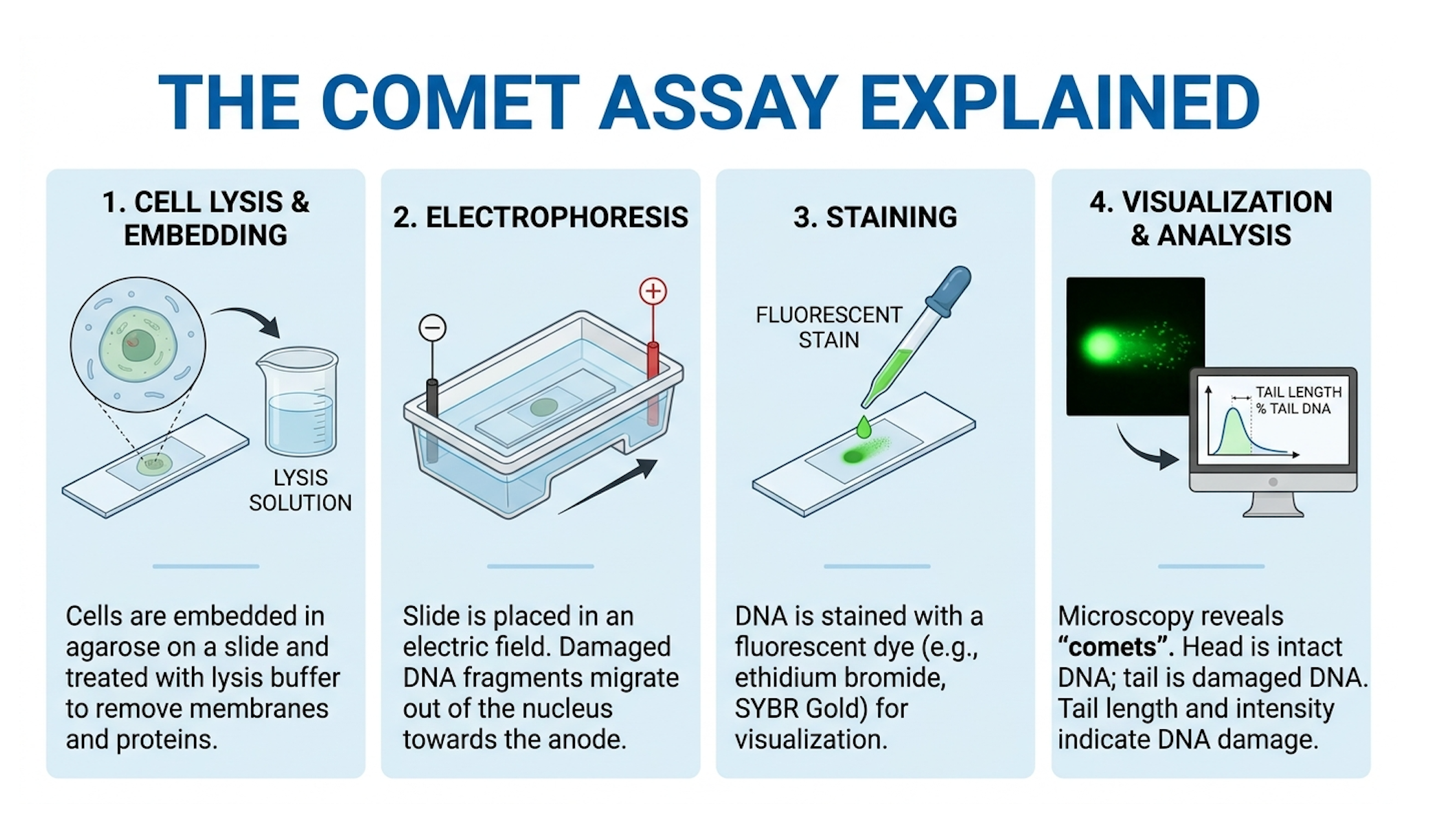
Properly cultured UC-MSCs exhibit virtually zero comet tails (DNA Repair via NIH, 2018). If there’s a tail, the batch is destroyed immediately.
Once genotoxicity is ruled out and cells are administered, the focus shifts to post-treatment patient monitoring and functional outcome evaluation.
A successful meniscus tear recovery requires managing your expectations. Functional improvements following UC-MSC injections peak between 3 to 6 months post-treatment, mirroring the 12-to-24 week biological timeline required for matrix remodeling (Journal of Orthopaedic Translation, 2021). A high stem cell therapy success rate is closely tied to how patients behave in the weeks immediately following the procedure.
Patients always ask: “Can you recover 100% from a meniscus tear?” The clinical truth is that we target functional outcomes measurable improvements in mobility, complete pain reduction, and a return to your specific athletic baseline. Anatomically, your MRI might not look like a 20-year-old’s knee, but functionally, you can achieve pain-free, high-level performance.
The biological remodeling takes serious time. The immediate pain relief you feel in week two? That’s just the anti-inflammatory effect working inside the joint capsule.
Based on our benchmark tracking of post-injection timelines, here is the realistic meniscus stem cell injection recovery schedule you should plan for:
Peak structural repair occurs around the six-month mark (Clinical outcome metrics, 2020). Managing this timeline successfully requires strict adherence to physical therapy protocols that protect the joint while stimulating cellular activity.
You cannot get a UC-MSC injection on a Friday and run a marathon on a Sunday. Living with meniscus tear after stem cells requires deliberate, structured physical therapy. The new cellular matrix needs to be stressed to grow stronger, but over-stressing it will tear the fragile new collagen.
Early on, walking guidelines are strictly enforced. Clinicians utilize closed-chain exercises where your foot remains in contact with a surface to safely load the joint and stimulate repair. Physical therapy protocols often incorporate blood flow restriction (BFR) training, which allows patients to build supporting quadriceps strength without heavily loading the fragile meniscus tissue. What to expect after meniscus stem cell therapy is a highly curated rehabilitation schedule tailored to your biomechanics.
Avoid any exercises that apply excessive shear force. Deep, loaded squats past 90 degrees or sudden pivoting movements during the first 8 weeks can catastrophically damage the healing matrix. Lifestyle adaptations and weight management play massive roles in protecting the joint while it rebuilds (Post-treatment protocols, 2017).
Before committing to this comprehensive treatment and rehabilitation pathway, patients must rigorously assess the practical and financial considerations.
Because UC-MSC therapies are classified as experimental or investigational by most major health insurers, patients typically assume full out-of-pocket costs for advanced regenerative procedures. Because over 90% of health insurers classify UC-MSC therapies as experimental, patients assume full out-of-pocket costs ranging from $5,000 to $15,000 (Regenerative Medicine Policy, 2022). The stem cell therapy cost meniscus patients pay varies wildly, and understanding why will keep you from making a dangerous financial decision.
How much does meniscus stem cell therapy cost? In the United States, a verified, high-cell-count UC-MSC procedure typically runs between $8,000 and $15,000 per joint. However, many patients look globally. The stem cell therapy for meniscus tear Thailand cost averages between $5,000 and $9,000. Seeking a stem cell injection for meniscus Bangkok treatments is popular because of specialized regenerative clinics that operate strictly under international medical tourism standards.
| 📌 If you’re interested in a full breakdown of treatment pricing in Thailand, we have an interesting article that discusses stem cell therapy costs in Thailand for 2025, which you can read via the internal link. |
Regardless of where you go, the torn meniscus stem cell price fluctuates based on total cell count, the need for multiple joint injections, and advanced imaging requirements. Don’t expect your insurance to help. Providers universally exclude these procedures under experimental clauses, leaving the financial burden entirely on the patient (Medical pricing analysis, 2021).
Understanding the financial baseline helps patients identify and avoid clinics that offer dangerously cheap, unverified biological products.
A low-cost clinic usually means they cut corners in the lab. Laboratory Quality Standards the regulatory frameworks governing cellular manipulation are non-negotiable.
When evaluating a clinic, you must ask direct questions:
Red flags include clinics that guarantee cures, use the exact same IV treatment for knee pain as they do for neurological diseases, or lack verifiable laboratory documentation. A legitimate consultation determines if you are a candidate; it doesn’t just sell you a syringe (ISSCR patient guidelines, 2023).
This strict evaluation framework naturally leads into understanding the inherent limitations and safety considerations of the therapy itself.
It’s critical to step back from the hype and look at medical reality. Stem cells are powerful biological tools, but they cannot fix everything. Up to 25% of patients with severe locking require traditional surgical debridement, as stem cells cannot dissolve physical blockages in the hinge (American Journal of Sports Medicine, 2019). Understanding the absolute contraindications ensures you don’t waste money or risk further injury on a therapy that biologically cannot help your specific anatomical defect.
The biggest pitfall is attempting to treat mechanical blockages with biology instead of surgery. If you have a “bucket handle” meniscus tear where a large flap of cartilage flips over and physically jams into the joint hinge stem cells won’t fix it. You need a surgeon to manually clear that mechanical obstruction.
Other common failures include:
Biology takes time, and stem cells absolutely cannot rebuild a knee that lacks basic biomechanical stability.
There are definitive scenarios where surgical intervention remains the gold standard. If your knee locks unpredictably, gives way under load, or suffers from acute, traumatic vascular zone tears that are actively bleeding, suturing is the medically superior choice. The best time to treat meniscus tear with stem cells is before total cartilage collapse, but after confirming the joint is mechanically stable.
Readers are strongly advised to consult a board-certified orthopedic specialist to establish baseline joint mechanics before pursuing cellular interventions. An ethical specialist will tell you exactly when surgery is unavoidable, keeping you safe from predatory clinics.
An unhealed meniscus tear in the avascular zone leads to progressive joint degeneration and accelerated osteoarthritis. Without the shock-absorbing capacity of the intact fibrocartilage, mechanical stress concentrates on the articular cartilage covering the femur and tibia. Over time, this localized friction degrades the joint surface, leading to chronic inflammation and loss of mobility. Patients frequently experience persistent swelling, joint locking, and reduced functional capacity. If left untreated, severe degenerative changes may eventually necessitate total knee replacement surgery.
The hardest meniscus tear to repair is a complex, degenerative tear located entirely within the inner white-white avascular zone. This inner region lacks the blood supply required to deliver natural
Currently, the FDA considers UC-MSC therapies for orthopedic applications as investigational or experimental. While the regulatory body permits their use in strictly controlled clinical trials or under specific practice of medicine exemptions, they do not hold blanket approval for meniscus repair. Patients must verify that clinics adhere to strict FDA compliance regarding minimal cellular manipulation. Choosing facilities that operate under clear investigational new drug (IND) applications provides an extra layer of clinical safety (FDA Cellular Therapy Guidelines).
For patients with chronic joint injuries, evaluating stem cell therapy for meniscus tear procedures requires rigorous biological analysis rather than reliance on commercial claims. Clinical data demonstrates that while UC-MSCs utilize paracrine signaling to promote tissue remodeling in the avascular zone, outcomes depend entirely on proper patient selection and strict laboratory protocols. The best approach combines verified cellular products, precise image-guided injection, and a structured, months-long physical rehabilitation plan.
Implementing The Comet Verification Standard ensures that any cellular product administered possesses zero DNA damage or tumorigenic potential. This transparent, assay-driven approach separates evidence-informed regenerative medicine from unverified clinical practices. When DNA integrity is objectively proven, patients can enter the recovery phase confident in the safety of their cellular treatment.
Before proceeding, compile your recent MRI imaging, clinical history, and functional goals. Schedule a comprehensive consultation with a certified regenerative orthopedic specialist today to review your exact candidacy. Request a rigorous suitability assessment from a board-certified
This article is for educational purposes only and does not replace consultation with a qualified medical professional.