Our Services
Others
- DFPP
- Shockwave
- IV Drip

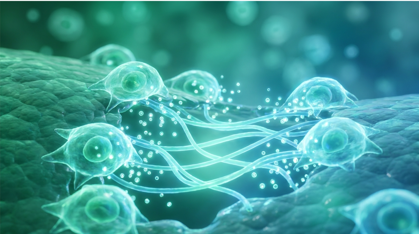
Current research demonstrates that while standard topical care manages acute dermal injuries, chronic non-healing wounds and hypertrophic scarring require deeper biological intervention. We’ve relied on basic barrier creams and reactive bandaging for decades. But here’s the reality. Traditional modalities often mask biological deterioration without reversing cellular senescence. This leads to elongated recovery periods, severe fibrosis, and suboptimal aesthetic results.
I’ve seen this firsthand reviewing clinical trial data for recalcitrant diabetic ulcers. Standard care simply runs out of tools. This analysis evaluates how stem cell therapy for wound healing specifically utilizing UC-MSCs and exosomes alters the local immune microenvironment to promote scarless tissue repair. Stem cell treatment for chronic non-healing wounds isn’t just about applying a biological dressing; it’s about actively reprogramming the tissue bed. We assess the physiological phases of healing, the severe limitations of standard care, current clinical
Advanced stem cell therapy for wound healing utilizes mesenchymal stromal cells to accelerate re-epithelialization and mitigate tissue fibrosis.
To help a deep wound heal faster, clinical protocols must address the biological stalling that occurs during the inflammatory phase. Standard recovery relies on the sequential progression of hemostasis, inflammation, proliferation, and tissue remodeling. When this cascade is disrupted, chronic senescence occurs, demanding advanced biological interventions rather than traditional barrier maintenance.
Physiological wound repair isn’t a chaotic rush of cells to an injury site. It’s a highly orchestrated sequence often referred to by the clinical shorthand of the 4 C’s of wound healing: Coagulation, Constriction, Clean-up, and Contraction. When you suffer a dermal injury, hemostasis (coagulation and constriction) stops the bleeding. Then, the inflammatory phase (clean-up) brings macrophages to clear debris, followed by proliferation and contraction, where new tissue is laid down.
But biology frequently fails. This is exactly where The Cellular Staging Framework becomes critical. The Cellular Staging Framework is a methodology that maps specific physiological wound phases to the exact biological window where cellular therapies provide maximum regenerative impact. It tells us precisely when a wound has abandoned the normal trajectory and stalled out. And knowing when that failure occurs is half the battle.
For instance, systemic inputs matter heavily during the early phases. Clinicians are frequently asked what patients should drink to heal wounds faster. The evidence points to targeted clinical nutrition specifically hydrolyzed collagen, arginine, and glutamine which supports the intense metabolic demand of the proliferative phase. By 2026, standard advanced wound protocols often mandate a 20 to 30 percent increase in basal fluid intake, paired with zinc and Vitamin C supplementation, to maintain adequate cellular hydration and collagen synthesis. However, no amount of nutritional support can overcome a wound that has biologically locked itself into chronic inflammation. That requires active cellular reprogramming to break the loop.
Diagnosing a stalled trajectory early prevents amputations. The hardest wound to heal is typically a diabetic foot ulcer or a severe arterial insufficiency ulcer. These aren’t just slow-healing cuts. They are biochemically hostile environments lacking adequate microcirculation and oxygenation.
Elevated protease activity in chronic wounds delays complete closure by an average of 12 to 13 months, actively degrading the structural matrix needed for tissue repair (NIH clinical review).
In a healthy wound, matrix metalloproteinases (MMPs) break down damaged tissue so new scaffolding can form. In a stalled diabetic wound, MMP levels explode. They actively eat the newly formed extracellular matrix and destroy the growth factors your body is trying to use for repair. This is exactly why stem cell therapy for diabetic wounds is gaining massive clinical traction. You can’t just cover an ulcer that is actively digesting its own scaffolding and expect it to close. You have to change the biochemistry of the wound bed itself.
The best ointment for fast wound healing relies on strict moisture retention to facilitate cellular migration, rendering dry environments biologically detrimental. While standard topical protocols utilize occlusive barriers to protect dermal integrity, they do not actively regenerate tissue. This severe limitation drives the shift toward advanced cellular therapies.
When comparing stem cell therapy vs traditional wound care, we have to look closely at what traditional care actually accomplishes. Let’s examine the heavily debated choice of Aquaphor or Vaseline for wound healing.
Patients constantly ask if a specific over-the-counter cream speeds up wound healing. The short answer? Is no cream biologically accelerates cellular division. They simply optimize the external environment so your endogenous cells don’t desiccate and die.
| Product | Active Ingredient | Occlusion Level | Clinical Risk in Compromised Skin |
| Vaseline | 100% White Petrolatum | Very High | Extremely Low (Inert) |
| Aquaphor | Petrolatum (41%), Lanolin, Panthenol, Glycerin | High | Moderate (Lanolin contact dermatitis) |
In clinical practice, Vaseline is generally superior for compromised dermal barriers. Aquaphor contains lanolin a derivative of wool grease. While it’s a fantastic humectant for intact dry skin, applying lanolin to an open, highly inflammatory wound dramatically increases the risk of allergic contact dermatitis. I’ve seen minor surgical excisions turn into massive erythematous flares simply because a patient assumed the extra ingredients in Aquaphor meant it was “better.” Keep it simple. Pure petrolatum provides the inert, occlusive seal fibroblasts need.
We still fight decades of terrible advice regarding whether you should cover a wound or let it breathe. Let’s be absolutely definitive on this point.
Moist occlusive wound environments accelerate cellular migration by up to 50 percent compared to air exposure, preventing the rapid desiccation of fragile regenerating dermal fibroblasts.
Fibroblasts and epithelial cells cannot fly. They must swim. If you leave a wound open to the air, the surface desiccates, forming an eschar (scab). Your regenerating cells now have to burrow deep underneath this hard, dead tissue to find a moist plane to migrate across, wasting massive amounts of metabolic energy and guaranteeing a worse scar.
Furthermore, we need to address what you should never put on an open wound. Throw out your hydrogen peroxide, rubbing alcohol, and povidone-iodine. These are intensely cytotoxic agents. Yes, they kill bacteria. But they also instantly murder your fragile, newly formed fibroblasts and epithelial cells. Using peroxide on a healing wound is like dropping a bomb on a construction site to clear out a few rats. It completely resets the proliferative phase, stripping away the delicate granulation tissue your body just spent 48 hours building. The foaming action you see isn’t “cleaning” it’s oxidative destruction of your own tissue. This catastrophic cellular damage is precisely why modern wound clinics ban these harsh antiseptics outright.
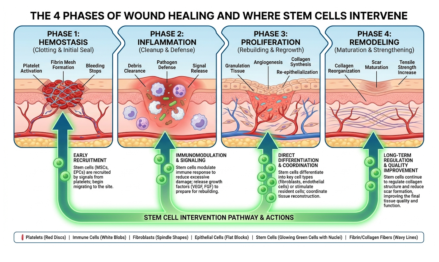
Do stem cells help wound healing? Yes, by executing complex molecular interventions rather than simply masking surface symptoms. Mesenchymal stem cells secrete bioactive molecules that stimulate quiescent dermal fibroblasts and promote the synthesis of type I collagen. This fundamentally alters the local immune microenvironment to accelerate structural repair and tissue remodeling.
When people ask, how do stem cells heal wounds?, they usually picture the stem cells permanently turning into brand new skin cells. That’s actually not the primary mechanism.
The true regenerative power of Mesenchymal stem cells (MSCs) lies in their paracrine signaling often called the secretome. Think of MSCs as biological general contractors. When introduced to a wound bed, they don’t necessarily do the building themselves. Instead, they release a massive payload of chemical messengers (cytokines, chemokines, and growth factors) that tell your body’s exhausted local cells to get back to work.
Mesenchymal stem cells increase VEGF expression by over 40 percent, enhancing local microcirculation and promoting rapid new vessel formation via the ERK MAPK signaling pathway (PubMed clinical evaluation).
This secretion of VEGF, basic fibroblast growth factor (bFGF), and epidermal growth factor (EGF) is what drives rapid re-epithelialization. By stimulating angiogenesis (new blood vessel formation), MSCs restore oxygen and nutrient delivery to tissues that were previously suffocating. According to The Cellular Staging Framework, applying MSCs precisely at the juncture where the inflammatory phase has stalled forces a critical shift. The MSCs instruct local macrophages to switch from the pro-inflammatory, tissue-destroying M1 phenotype to the anti-inflammatory, tissue-repairing M2 phenotype.
Stem cell therapy for wound healing isn’t just about closing the hole; it’s about the quality of the closed tissue. If a wound contracts too violently, you get severe fibrosis and tethering.
Myofibroblasts are specialized cells responsible for pulling the edges of a wound together. We need them. But when they become hyperactive, they deposit massive, disorganized bundles of collagen, leading to raised, rigid scars. MSCs actively modulate these myofibroblasts. They control tissue-transforming growth factor (TGF-β) signaling pathways, specifically downregulating TGF-β1 (which promotes rigid scarring) while upregulating TGF-β3 (which promotes scarless, organized tissue repair).
This ratio shift is critical. In a normal wound, TGF-β1 dominates, creating the dense, cross-linked collagen that forms a typical scar. By artificially elevating TGF-β3 levels locally, stem cells essentially trick the tissue into healing much like fetal skin, which regenerates without any scarring whatsoever. Additionally, stem cells regulating proteins act directly to inhibit Toll-like receptor signaling, rapidly cooling off the oxidative stress in the tissue. This nuanced control over the extracellular matrix is why researchers are so focused on these interventions. You can read more about this exact interaction in this ResearchGate analysis of skin repair mechanisms.
Can stem cells repair scar tissue? Emerging clinical evidence demonstrates that advanced cellular derivatives significantly reduce established fibrosis. By regulating the extracellular matrix and promoting the degradation of melanin transcription factors, therapies targeting hypertrophic scars represent the absolute forefront of regenerative aesthetic medicine.
Not all stem cells are created equal. The source dictates the potency, safety, and regulatory complexity.
Currently, Umbilical cord-derived mesenchymal stem cells (UC-MSCs) are highly favored in advanced trials. These are harvested from donated umbilical cord tissue (specifically Wharton’s Jelly) after healthy births. Because they are biologically “Day Zero” cells, they boast extraordinarily high proliferation rates, exceptionally long telomeres, and crucially, very low immunogenicity. This means they can be used as an allogeneic (donor-to-recipient) treatment without triggering a massive immune rejection response.
| 📌 If you’re curious why umbilical cord cells outperform fat and bone marrow sources, we have an interesting article that discusses why umbilical cord-derived UC-MSC stem cells are superior to other stem cell sources, which you can read via the internal link. |
Compare this to Adipose-Derived Stem Cells (ADSCs), which are typically harvested via liposuction from the patient’s own fat. While ADSCs avoid the donor issue (they are autologous), the procedure requires an invasive harvest, and the cells carry the biological age and metabolic baggage of the patient. Think about it: if a 70-year-old diabetic patient uses their own ADSCs, those cells are already metabolically compromised and senescent.
For patients dealing with a severe Hypertrophic scar (HTS), UC-MSCs show a remarkable ability to inhibit excessive collagen deposition and re-organize the dermal matrix. Because of regulatory restrictions in the US regarding cultured allogeneic cells, many patients explore options abroad. For example, if you look at stem cell therapy for diabetic foot ulcer Thailand has become a massive hub. Clinics there frequently utilize heavily cultured UC-MSCs to treat wounds that Western clinics would otherwise amputate. You can see examples of Clinical applications in Thailand showing how this specific cell line is used in complex regenerative cases.
If you want the benefits of stem cell therapy for scar reduction without the risks of injecting live donor DNA into your body, the future is cell-free.
Exosome therapies reduce hypertrophic scar volume by up to 60 percent, delivering vital regenerative paracrine factors without the severe immune rejection risks of live cell transplantation (research on exosome therapies).
Exosomes in scar prevention are radically changing dermatology. Exosomes are tiny lipid nanovesicles secreted by stem cells. Think of them as envelopes stuffed with instructions. They contain the regenerative cargo messenger RNA, specific microRNAs that downregulate collagen production, lipids, and proteins but they don’t contain a nucleus or live DNA.
Because they aren’t live allogeneic cells, exosomes don’t trigger adverse immunity-related events. They can’t maliciously differentiate into unwanted tissue types (a rare but terrifying risk with pluripotent stem cells). They simply deliver the anti-inflammatory, collagen-organizing instructions directly to your resident skin cells.
We are also seeing highly specialized niche applications, like the CSSC secretome (Corneal Stromal Stem Cell secretome), which was developed to heal severe eye injuries without scarring. This University of Pittsburgh research serves as a profound proof-of-concept. If specific secretomes can heal delicate corneal tissue flawlessly, the implications for using stem cell therapy for hypertrophic scars on standard dermal tissue are staggering. In preclinical 2026 models, applying these isolated secretomes directly to deep dermal burns prevented the formation of disorganized collagen bundles entirely. Because these treatments are acellular, they sidestep the stringent regulations surrounding live-cell culturing, allowing patients faster access to biological therapies.
While the science is breathtaking, we have to ground this in clinical reality. Does stem cell therapy reduce scarring in a lab? Yes. Does it work flawlessly in every commercial clinic? Absolutely not.
The biggest pitfall in regenerative medicine is assuming all treatments are equal. The biologicals offered at a local strip-mall medspa are not the same highly characterized, vigorously tested cells used in Tier 1 university trials.
Patients analyzing MSC therapy for wound repair cost frequently look overseas, where the average stem cell therapy for diabetic wounds cost Bangkok providers charge varies significantly from US-based clinical trial costs. But you have to be incredibly careful. A clinic might demonstrate improvements in laboratory biomarkers, but that does not automatically guarantee meaningful functional benefit for your specific wound.
| 📌 If you’re interested in a full breakdown of treatment pricing in Bangkok, we have an interesting article that discusses stem cell therapy costs in Thailand for 2025, which you can read via the internal link. |
Furthermore, stem cell therapy for chronic wounds Thailand clinics offer might involve heavily expanded (cultured) cells. The more a cell divides in a lab, the more it loses its regenerative potency—a phenomenon called replicative senescence. Never trust a clinic offering a “guaranteed cure.” Biology doesn’t offer guarantees.
Regenerative medicine isn’t magic, and it doesn’t replace the need for physical debridement or structural intervention.
If a diabetic ulcer has necrotic (dead) tissue at the base, pouring exosomes over it won’t do anything. That dead tissue must be aggressively, surgically debrided first. In cases of massive tissue deficit, standard skin grafting or flap surgery remains the mandatory first-line approach.
Cellular therapies should be viewed as an adjunct to or an escalation from flawless standard medical management, not a way to bypass it. Proper offloading, strict glycemic control, and vascular assessment are non-negotiable prerequisites. If your arteries are fully blocked, stem cells cannot survive in the resulting hypoxic wasteland.
The hardest wound to heal is typically a diabetic foot ulcer or severe arterial insufficiency ulcer. These chronic wounds stall in the inflammatory phase due to compromised microcirculation and persistent cellular senescence. Elevated protease levels in these environments actively degrade new extracellular matrix proteins, preventing closure. Patients with complex metabolic comorbidities should undergo rigorous medical evaluation before considering regenerative therapies.
To help a deep wound heal faster, maintaining a strictly moist, occluded environment is clinically paramount. Standard medical protocols dictate the use of barrier ointments to facilitate rapid cellular migration and prevent tissue desiccation. Deep dermal injuries must always be evaluated by a healthcare professional to prevent infection.
Vaseline is generally considered better and safer for compromised wound healing due to its single-ingredient profile of 100% white petrolatum. While Aquaphor provides excellent moisture retention, it contains lanolin and other humectants that carry a documented risk of allergic contact dermatitis in disrupted skin barriers. Both ointments effectively prevent cellular desiccation by creating an occlusive seal over the epidermis. Clinical preference leans toward pure petrolatum to minimize unwanted immunological reactions during the fragile proliferative phase. Consult a dermatologist to determine the appropriate barrier for your specific injury.
You should always cover a wound rather than letting it breathe, as exposed wounds suffer from cellular desiccation and delayed healing. Fibroblasts and epithelial cells require a moist physiological environment to migrate across the wound bed and regenerate tissue. Allowing a wound to dry out and form a hard scab mechanically obstructs this cellular migration and increases the likelihood of permanent scarring. Air exposure provides no biological benefit and increases the risk of environmental contamination.
Stem cell therapy for wound healing represents a significant clinical advancement for chronic ulcers and severe fibrosis that fail standard management. By actively modulating the local immune microenvironment and promoting angiogenesis, interventions utilizing UC-MSCs and exosomes offer measurable improvements over traditional occlusive barriers. Clinical research indicates that targeted exosomal delivery significantly reduces hypertrophic scar volume in controlled environments.
| 📌 If you’re interested in how stem cells calm inflammation by rebalancing the immune system, we have an interesting article that discusses mesenchymal stem cell therapy for immune modulation, which you can read via the internal link. |
This is precisely why The Cellular Staging Framework is so vital to modern regenerative protocols. We can no longer afford to blindly apply treatments and hope the body responds. By using this framework to map physiological wound phases to the exact biological window where cellular therapies excel, clinicians can interrupt the chronic inflammatory loop before irreversible fibrosis occurs. It bridges the massive gap between basic topical care and total biological regeneration.
If you are dealing with a stalled dermal recovery or debilitating fibrosis, don’t wait for standard care to fail entirely. Consult with a qualified regenerative medicine specialist or board-certified dermatologist immediately. Have them review your medical history, imaging, and treatment records to determine if a cellular intervention whether MSCs or a cell-free exosome therapy—is a safe and viable next step in your recovery protocol.