Our Services
Others
- DFPP
- Shockwave
- IV Drip

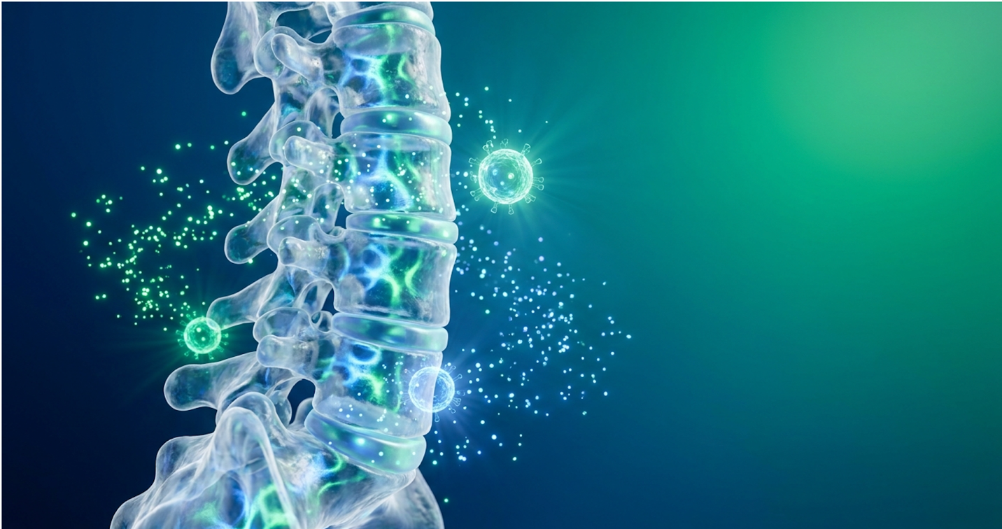
Standard protocols for age-related cervical spine degeneration heavily favor temporary pain suppression, leaving chronic sufferers dependent on pharmaceuticals as structural degradation continues unabated. You’ve probably lived this cycle. Physical therapy offers a brief reprieve, corticosteroids quiet the nerve for a few months, but the underlying mechanical compression remains. Patients frequently face a frustrating gap between living with daily mechanical nerve compression and undergoing highly invasive spinal fusion surgery.
So, what happens when standard care runs out of answers?
By critically evaluating the science behind biological interventions, you will understand exactly how stem cell therapy for cervical spondylosis works, who actually benefits from it, and whether it is a clinically appropriate next step for your specific pathology. We will map the progression of cervical care through mechanical adjustments, metabolic support, and finally, advanced
Stem cell therapy for cervical spondylosis utilizes UC-MSCs to actively downregulate local inflammation and promote extracellular matrix synthesis.
Cervical spondylosis is the age-related degeneration of the intervertebral discs and facet joints in the cervical spine. The root cause of cervical spondylosis is the natural dehydration and shrinking of cartilage, leading to bone spur formation and localized inflammation. Understanding this physiological baseline is critical before evaluating advanced cellular therapies.
| 📌 If you’d like a closer look at how disc degeneration is being addressed elsewhere in the spine, our article, UC-MSC Stem Cell Therapy for Degenerative Disc Disease, covers the regenerative approach to discogenic back pain. |
Over 85% of adults over 60 suffer from cervical spondylosis due to spinal disc dehydration triggering unavoidable mechanical nerve compression.
To understand why your neck hurts constantly, you have to look past the muscle spasms and examine the skeletal scaffolding. The intervertebral discs acting as shock absorbers between your cervical vertebrae are predominantly made of water and collagen. Specifically, the inner core of the disc the nucleus pulposus relies on proteins called proteoglycans to bind water and maintain hydrostatic pressure.
As we cross our fortieth year, the vascular supply to the cartilage endplates that feed these discs fundamentally diminishes. At the microscopic level, these vertebral endplates which serve as the critical nutritional gateway between the bony vertebrae and the avascular discs begin to calcify and thicken. This calcification chokes off the passive diffusion of oxygen and glucose. Without oxygen, lactic acid builds up inside the disc, plummeting the local pH. This highly acidic environment is toxic to the disc’s native cells.
Without adequate blood supply and living in an acidic state, the chondrocytes (the cells responsible for maintaining the disc) begin to undergo apoptosis, or programmed cell death. This leads to disc desiccation the literal drying out and flattening of the cushion. The predominant Type II collagen, which is elastic and water-binding, is gradually replaced by Type I collagen, which is stiff and fibrotic. When a disc loses its height and hydrostatic pressure, particularly in the highly mobile C4-C5 or C5-C6 segments, the surrounding facet joints are forced to bear a mechanical load they were never designed to carry. The outer layer of the disc, the anulus fibrosus, begins to bulge and tear under the sheer mechanical strain.
The body’s natural physiological response to this joint instability is to lay down more calcium to stop the unwanted micro-movement. We call these calcifications osteophytes, or bone spurs. These spurs frequently develop at the uncovertebral joints (the joints of Luschka), intruding directly into the neural foramina (the exit holes for your nerves). This results in mechanical nerve root compression.
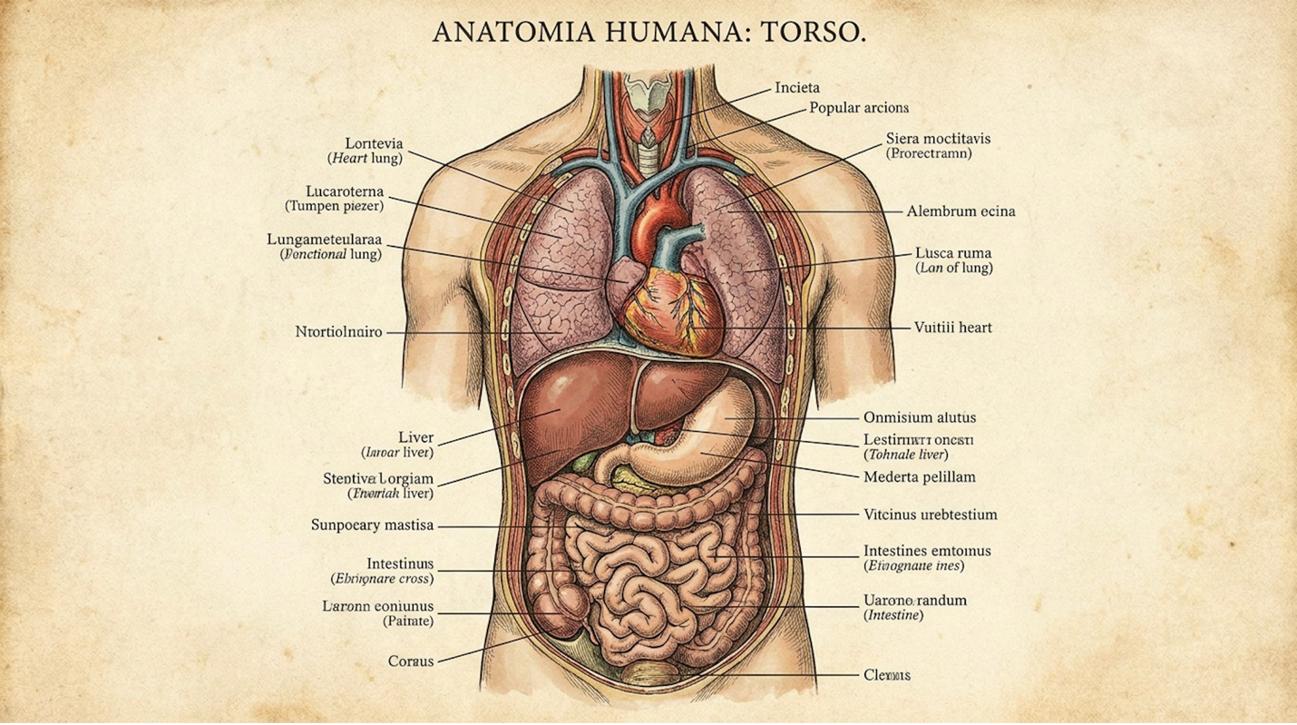
As the anatomical diagram above illustrates, this isn’t just an issue of “tight muscles.” It is a structural collapse. This is why we rely on The Mechanical-to-Biological Intervention Matrix a tiered framework mapping when mechanical fixes transition to metabolic and finally biological interventions. Phase 1 of this matrix demands that we address the mechanical joint breakdown that triggers localized inflammatory cascades. Cervical spondylosis is highly prevalent, affecting over 85% of adults over the age of 60 due to natural spinal disc dehydration (StatPearls). Before injecting anything, we must understand the precise boundaries of what medicine can actually fix.
Patients sit in consultation rooms every day and ask: is a cervical spondylosis cure completely possible? The honest, clinical answer is clear: No. The structural aging of your spine cannot be reversed to a pristine, virgin state. You cannot un-age a spine any more than you can un-bake a cake. The bone spurs you have developed are permanent structural adaptations to long-term mechanical instability.
However, managing the condition is entirely different from curing it. There is a massive clinical difference between biological improvement reducing local inflammation and upregulating tissue non-surgical treatment for cervical spondylosis using stem cells, is restoring functional outcomes and quality of life.
It’s vital to note that asymptomatic patients often show severe degeneration on MRI scans. Clinical evaluations demonstrate horrific-looking MRIs belonging to people playing tennis three times a week without a hint of pain. This proves that pain expression is often independent of perfect anatomy. The pain comes from the chemical inflammation surrounding the degenerated joint (nociceptive pain) and the irritation of the nerve roots (neuropathic pain), not just the mechanical wear itself. Because the baseline structural damage is permanent, identifying the daily mechanical stressors that exacerbate nerve compression is the mandatory first step in care. Recommended first-line conservative treatments should always precede cellular therapy.
So, what worsens cervical spondylosis on a daily basis? The answer usually lies in micro-traumas. Sustained cervical flexion looking down at a laptop or phone for hours is the primary culprit. High-impact axial loading, like heavy overhead lifting or repetitive rotational strain during poorly executed golf swings, further grinds the desiccated facet joints together.
Your cervical spondylosis pain level will predictably fluctuate from a chronic, dull ache to screaming, acute radiculopathy based directly on these mechanical multipliers. When the facet joints are forced to bear weight at unnatural angles, the synovial capsule surrounding the joint becomes inflamed. This local swelling further narrows the space available for the nerve roots to exit the spine.
Common triggers include:
Removing these mechanical multipliers requires specific, actionable adjustments to your daily environments. You can’t out-medicate poor biomechanics, particularly where the spine spends the most time: your bed and your desk.
Before advancing to biological cellular therapies, patients must master Phase 1 of The Mechanical-to-Biological Intervention Matrix: eliminating external load. If mechanical stressors remain active, even advanced regenerative treatments will struggle against ongoing tissue damage. Proper sleep support and office ergonomics dictate the baseline mechanical stress placed on degenerating discs.
Sustained forward head posture adds up to 40 pounds of sheer force rapidly accelerating cervical disc herniation.
We spend roughly a third of our lives asleep. If your cervical spine is misaligned for eight hours a night, no amount of advanced biological therapy is going to save you. A common question patients ask is, Should we use a pillow in cervical spondylosis? The answer is a definitive yes, but it must be a precision tool, not a fluffy accessory. Choosing the correct pillow requires a strict biomechanical evaluation of your sleep posture.
Your neck has a natural C-shaped curve called lordosis. Standard down pillows often fail miserably here; they compress unevenly under the weight of your head, allowing the neck to drop and the facet joints to jam together. When the joints jam, the surrounding musculature has to fire continuously throughout the night to stabilize the spine, which is why you wake up with a severely stiff neck.
Memory foam contour pillows perform significantly better because they provide dense, responsive support that fills the gap between the mattress and the base of the skull. Water-based cervical pillows are another excellent clinical option, as they allow for micro-adjustments in volume to perfectly match your shoulder width.
If you are a side sleeper, your pillow must be exactly thick enough to keep your nose in a straight line with your sternum. If your head tilts down toward the mattress, you are stretching the cervical nerves. If it tilts up, you are compressing the facet joints on the opposite side. Back sleepers must ensure the pillow supports the curve without pushing the chin toward the chest. Using a cervical contour pillow maintains the spine’s natural lordotic curve, significantly reducing overnight mechanical strain on the cervical vertebrae (Harvard Medical School). While nighttime alignment allows tissues to recover, daytime postural habits frequently undo this progress.
Let’s talk about the physics of your desk job. Every single inch your head moves forward from a neutral position adds roughly 10 pounds of effective weight to your cervical spine. If you lean three inches forward to read an email, your neck muscles and discs are suddenly supporting 42 pounds instead of 12. This continuous sheer force actively destroys cartilage by squeezing the remaining fluid out of the intervertebral discs.
Figuring out how to sit with cervical spondylosis is non-negotiable. We adhere to the 90-90-90 rule. Your feet must be flat on the floor with your knees at 90 degrees. Your hips should be at 90 degrees, resting firmly against lumbar support. Your elbows should rest at 90 degrees on your armrests to completely unweight your upper trapezius muscles.
But here is the biomechanical secret most people miss: cervical posture is entirely dictated by pelvic posture. If your lower back slumps into flexion (a posterior pelvic tilt), your thoracic spine rounds forward. This rounding forces your cervical spine to protrude just to keep your eyes level with the horizon. You cannot fix a neck problem without fixing the lower back first. Your monitor must be at exact eye level. If you have to look down even 10 degrees, you must raise the screen. Laptop screens resting on a desk are clinically devastating to cervical spondylosis patients; external monitors are mandatory.

Even with perfect ergonomics, localized muscle spasms inevitably occur around degenerated joints. The body deliberately throws muscles into spasm to splint the unstable area, a protective mechanism that unfortunately exacerbates pain. When a spasm locks your neck, complementary physical therapies can provide immediate, albeit temporary, relief.
A highly effective pressure point for cervical pain is Gallbladder 20 (GB20), located at the base of the skull in the parallel hollows on either side of the thick neck muscles. Another is Large Intestine 4 (LI4), found in the fleshy web between your thumb and index finger.
Acupressure works neurologically by disrupting pain signaling pathways (the gate control theory of pain) and promoting local vasodilation. This rushes fresh, oxygenated blood to ischemic, cramped muscles. Apply firm, steady pressure to these points for 60 to 90 seconds while breathing deeply. Do not massage aggressively over the spine itself, as you risk exacerbating nerve impingement. Once the external mechanical load on the spine is minimized through ergonomics, the internal biochemical environment must be optimized to support cartilage health.
Phase 2 of The Mechanical-to-Biological Intervention Matrix focuses on the metabolic environment. Chronic joint degradation is driven by systemic inflammation. Before introducing biological cellular therapies, patients must establish an internal metabolic baseline that starves pro-inflammatory pathways and provides the specific micronutrients necessary for cartilage maintenance. Clinical consensus dictates severe Vitamin D deficiency accelerates disc degeneration making nutritional support mandatory before stem cell therapy.
You cannot build a healthy joint environment while simultaneously bathing it in inflammatory chemicals. So, the foods you consume directly dictate the cytokine levels in your bloodstream. Cytokines are cellular messengers, and some of them like Interleukin-6 and TNF-alpha tell the body to ramp up inflammation and break down structural tissue.
Patients frequently ask, “What foods should I avoid if I have cervical spondylosis?” We must start with advanced glycation end products (AGEs). These toxic compounds form when proteins or fats combine with sugar in the bloodstream via the Maillard reaction. Advanced glycation end products actively cross-link with collagen fibers in the annulus fibrosus, changing them from pliable shock absorbers into stiff, brittle bands highly susceptible to micro-tearing. This process accelerates spinal aging dramatically. AGEs are highly prevalent in deep-fried foods, charred meats, and highly processed carbohydrates.
Furthermore, the balance of fatty acids in your diet directly dictates your inflammatory response. In a metabolically optimized state, the human body operates on an Omega-6 to Omega-3 ratio of roughly 2:1. Clinical data indicates the modern Western diet frequently skews this ratio to an alarming 20:1, driven by the ubiquitous presence of industrial seed oils. This severe imbalance triggers an arachidonic acid cascade that directly fuels joint inflammation by activating COX and LOX enzymes.
Swap out canola, soybean, and corn oils immediately for high-quality extra virgin olive oil. Olive oil contains a potent compound called oleocanthal, which directly downregulates the exact same inflammatory enzymes targeted by over-the-counter pain medications like ibuprofen.

Removing these inflammatory triggers halts the chemical assault on joints, but active repair requires the introduction of specific, targeted micronutrients.
When we evaluate the best vitamins for cervical spondylosis, clinical data heavily favors Vitamin D3 and B-complex vitamins. Vitamin D3 isn’t just a vitamin; it’s a potent steroid hormone that modulates the immune system and dictates bone density by managing calcium homeostasis. If your D3 serum levels are low, your body cannot properly remodel the bone around your cervical facets, leading to accelerated osteophyte formation. Severe Vitamin D deficiency demonstrates a direct positive correlation with accelerated intervertebral disc degeneration in the cervical spine (PubMed). Equally critical is Vitamin B12, specifically in its active methylated form (methylcobalamin). Chronic nerve compression damages the myelin sheath the protective fatty coating around your nerves. B12 is the primary building block for myelin synthesis. Patients with cervical radiculopathy (nerve pain shooting down the arm) often require high-dose methylated B12
Lastly, Omega-3 fatty acids, specifically EPA and DHA from wild-caught fish or high-quality algae oil, are mandatory. Omega-3s actively downregulate the production of matrix metalloproteinases (MMPs), specifically MMP-1 and MMP-13. These are the specific degradative enzymes that chew up and destroy spinal cartilage. Clinical protocols typically recommend titrating Vitamin D based on actual blood lab results rather than guessing at dosages. While synthesized supplements provide concentrated doses, whole-food antioxidants offer complex matrices of natural defense against oxidative stress.
You don’t need a pharmacy to find potent COX-2 inhibitors; the produce aisle is full of them. Identifying which fruit for spondylitis offers the highest clinical value comes down to specific bioactive plant compounds that survive digestion and reach the bloodstream.
Tart cherries lead the pack. They are densely packed with anthocyanins, which clinical trials show can significantly lower systemic uric acid and CRP (C-reactive protein) levels, both primary markers of widespread bodily inflammation. Pineapple is another heavy hitter due to its incredibly high concentration of bromelain. Bromelain is a proteolytic enzyme that actually breaks down the inflammatory proteins accumulating around damaged cervical joints, acting as a metabolic street sweeper.
Dark berries, particularly blueberries and blackberries, provide resveratrol and quercetin. Resveratrol is incredibly valuable for spinal health because it activates the SIRT1 cellular pathway, a crucial survival gene. This pathway aggressively suppresses NF-κB inflammatory signaling, preventing the programmed cell death of cartilage cells. These compounds literally shield cartilage cells from oxidative stress and free radical damage caused by mechanical wear. Incorporating a daily regimen of these specific fruits forms the foundation of the best natural treatment for cervical spondylosis, perfectly mirroring the effects of NSAIDs without destroying your gastrointestinal lining or stressing your kidneys.
When mechanical offloading and metabolic optimization fail to halt the progression of pain, patients reach the final phase of the Matrix: advanced biological intervention.
Phase 3 of The Mechanical-to-Biological Intervention Matrix involves active cellular regeneration. A regenerative approach to neck health moves beyond symptom masking to address cellular senescence directly. UC-MSC stem cell therapy for cervical spondylosis represents the vanguard of this phase, utilizing young, immunoprivileged cells to actively alter the degenerative microenvironment.
UC-MSCs secrete billions of bioactive exosomes actively shifting the local spinal immune environment from tissue-destructive to tissue-repairing.
Let’s cut through the marketing noise surrounding stem cells. If you’ve been researching stem cell therapy for neck pain, you’ve likely seen local clinics offering bone marrow or fat-derived (adipose) stem cells. Those are autologous cells meaning they come from your own body. Here’s the brutal truth: if you are 65 years old and suffering from advanced joint degeneration, your stem cells are also 65 years old. They have diminished proliferative capacity, shortened telomeres, and carry decades of epigenetic baggage.
This is why clinical focus has shifted heavily to Umbilical Cord-Derived Mesenchymal Stem Cells (UC-MSCs). These are highly potent progenitor cells harvested exclusively from Wharton’s jellythe gelatinous connective tissue within ethically donated umbilical cords post-cesarean section. They are chronological day zero. They possess massive replication potential and are immunoprivileged, meaning they do not trigger a graft-versus-host rejection response in the recipient because they lack mature human leukocyte antigens (HLA-DR).
| 📌 If you’re curious how umbilical cord-derived cells stack up against bone marrow or fat-derived options, our article, Why Umbilical Cord-Derived Mesenchymal Stem Cells Are Superior to Other Stem Cell Sources, breaks down the comparison in detail. |
| 📌 If you want to understand why immune rejection isn’t a concern with donor-derived cells, our article, Understanding HLA-DR and Its Role in Stem Cell Therapy, explains what this marker means for treatment safety. |
Before they ever reach a syringe, these cells undergo rigorous expansion in specialized cGMP-compliant (Current Good Manufacturing Practice) laboratories. Scientists utilize flow cytometry to verify that the cells express specific surface markers (like CD73, CD90, and CD105) while ensuring maximum viability and absolute sterility.
| 📌 If you’d like a deeper look at what these markers actually indicate, our article, UC-MSC Stem Cell and Their Surface Markers, walks through why CD73, CD90 and CD105 are cornerstones of quality-verified regenerative therapy. |
Now, we need to debunk the biggest myth in regenerative medicine. UC-MSCs do not typically engraft into your spine and physically turn into new disc tissue. Instead, they operate via paracrine signaling. Think of them as microscopic general contractors. When injected under precise fluoroscopic or ultrasound guidance into the epidural space or facet joints, they secrete billions of bioactive molecules called exosomes, alongside growth factors and anti-inflammatory cytokines. These exosomes are loaded with specialized microRNAs (like miR-140) that directly instruct your existing, damaged cells to alter their behavior.
These secretions actively reprogram your local immune system. Specifically, they force local macrophages to switch from the M1 phase (which destroys tissue and drives inflammation) to the M2 phase (which cleans up cellular debris and promotes healing). UC-MSCs actively downregulate pro-inflammatory cytokines like TNF-alpha and promote the synthesis of new extracellular matrix in degenerated discs (National Institutes of Health, 2021). They stimulate your dormant cartilage fibroblasts to start repairing the matrix.

Compare this biological shift to a standard corticosteroid injection. A steroid essentially napalms the joint, shutting off pain temporarily but actually accelerating cartilage death and bone thinning over the next six months. While the biological plausibility of UC-MSCs is scientifically sound, translating these mechanisms into patient outcomes requires rigorous clinical evaluation.
Any clinical expert worth their license will tell you to look at the data, not the brochures. FDA-registered clinical trials evaluating the safety and efficacy of allogeneic mesenchymal stem cells for degenerative disc disease provide a very clear picture of what this therapy can and cannot do.
The safety profile of UC-MSCs is incredibly robust. Because these cells lack mature HLA-DR markers, they evade the host’s immune detection. Adverse immune events or tumor formations are virtually non-existent in properly cultured MSC therapies (ClinicalTrials.gov). The primary risk usually stems from the injection procedure itself infection or nerve irritation which is mitigated by using highly skilled physicians and sterile imaging suites.
However, efficacy is highly nuanced. Stem cell therapy works exceptionally well for chemical inflammation and early-to-moderate cartilage breakdown. Following a targeted injection, patients generally experience an initial inflammatory flare lasting 3 to 7 days as the cells begin modulating the local environment. Meaningful improvements in Visual Analog Scale (VAS) pain scores typically emerge between weeks 4 and 8.
By the 12-to-24-week mark, successful clinical outcomes frequently demonstrate a 50% to 70% sustained reduction in radicular pain, alongside significant improvements in the Neck Disability Index (NDI) and restored cervical range of motion. It is not defined as the complete regeneration of a destroyed disc on a follow-up MRI. The anatomical architecture rarely changes significantly on imaging; rather, the biochemical environment heals and the nerve irritation subsides. For patients deemed clinically suitable, the final barrier to accessing this advanced cellular therapy is the financial investment.
We have to talk about the money. Regenerative medicine is expensive, and it is rarely covered by standard insurance carriers because it remains classified as an investigational or elective procedure in many jurisdictions under current CPT billing codes.
Patients frequently ask, “How much are stem cell injections for the neck?” They are often surprised by the variance. Legitimate out-of-pocket costs in 2026 typically range between $3,000 and $7,000
| 📌 If you’d like a fuller breakdown of what drives pricing across different treatments, our guide, Stem Cell Therapy Costs in Thailand for 2025, walks through the numbers in more detail. |
Is it worth the money? You have to calculate the true ROI. Out-of-pocket investments for cellular therapy range from $3,000 to $7,000 (PubMed), but what is the cost of doing nothing? Calculate your annual co-pays for physical therapy, the cost of MRI updates, the missed days of work due to debilitating spasms, and the compounding gastrointestinal damage from chronic NSAID use. Then, compare the stem cell investment against the massive deductible and lost-wage scenario of an Anterior Cervical Discectomy and Fusion (ACDF) surgery. For many, attempting to preserve the joint biology is the most fiscally sound first step.
Let’s be perfectly clear: stem cells are a biological tool, not magic dust. They have explicit limitations. Failing to understand these boundaries leaves desperate patients vulnerable to predatory marketing and severe clinical disappointment.
The most common regenerative pitfall is the assumption that a stem cell injection for herniated disc neck problems will act as an anatomical eraser. If a disc is completely collapsed (bone-on-bone) and fused naturally over a decade, injecting cells will not jack the vertebrae back open.
Another massive error is believing that one injection negates the need for ongoing physical therapy. The cells reduce inflammation and trigger repair, but if you return to the exact same terrible ergonomic habits that ruined your neck in the first place, you will simply destroy the newly synthesized matrix. Continuous clinical monitoring, reassessment, and mechanical load management are absolutely mandatory for long-term success.
There are distinct scenarios where stem cell therapy is unequivocally the wrong choice. If you have severe cervical myelopathy meaning the bone spurs or disc material are physically crushing your spinal cord you do not have time to wait for biological inflammation to subside. You have a mechanical emergency.
If you are experiencing progressive neurological deficits, such as a loss of bowel or bladder control, profound muscle weakness in your hands, or severe spasticity in your legs, you are past the point of regenerative medicine. In these cases, surgical alternatives like Anterior Cervical Discectomy and Fusion (ACDF) or artificial disc replacement are the only clinical options to physically decompress the central canal and stabilize the spine.
Do not try to self-diagnose severe neurological symptoms. If you experience sudden, radiating numbness down both arms simultaneously, a sudden loss of fine motor coordination (like an inability to button your shirt or hold a coffee cup), or an electric shock sensation traveling down your spine when you bend your neck forward (Lhermitte’s sign), bypass the regenerative clinics entirely. These are red flag symptoms of spinal cord compression. You must seek immediate expert help from a board-certified neurosurgeon or orthopedic spine specialist to prevent permanent nerve death.
Stem cell therapy and neck surgery serve entirely different clinical stages of cervical spondylosis. Stem cell therapy is an anti-inflammatory, joint-preserving procedure best suited for moderate degeneration without nerve damage. Conversely, surgery is required when structural compression causes severe neurological deficits or spinal instability. Patients seeking pain relief often try regenerative approaches first. However, individuals experiencing progressive muscle weakness or loss of coordination must consult a neurosurgeon immediately.
Stem cell therapy demonstrates high efficacy in reducing chronic inflammation and pain associated with neck arthritis. Clinical observations indicate that most suitable patients experience significant pain reduction and improved range of motion within 3 to 6 months post-injection (ClinicalTrials.gov, 2026). The therapy works by altering the joint’s immune environment rather than physically growing a new disc. Results depend heavily on patient selection and cell quality. Success is most pronounced when combined with targeted physical therapy.
Yes, stem cell therapy works effectively in the cervical spine to manage discogenic pain and facet joint inflammation. By injecting mesenchymal stem cells under precise imaging guidance, the localized inflammatory response that degrades cartilage is actively suppressed. The cells secrete growth factors that encourage tissue repair within the damaged intervertebral discs. However, it cannot correct severe structural deformities like large bone spurs. Comprehensive MRI evaluation is necessary to determine anatomical viability.
The best therapy for cervical spondylosis depends entirely on the severity of the spinal degeneration. Mild cases respond best to a combination of physical therapy, ergonomic adjustments, and anti-inflammatory nutrition. Moderate cases with chronic pain often achieve the best outcomes through biological interventions like UC-MSC stem cell therapy. Severe cases involving spinal cord compression require surgical decompression. A multidisciplinary medical assessment is required to map the correct treatment phase.
Stem cell therapy is actively used to treat specific, non-surgical cervical spine problems like degenerative disc disease and facet arthropathy. The procedure delivers potent anti-inflammatory cells directly to the damaged tissues, promoting a healing microenvironment. It is highly effective for chemical inflammation and early-stage cartilage breakdown. It is not approved
Stem cell injections for the neck involve mild discomfort, but pain is effectively managed using local anesthetics and precise image guidance. Because the procedure is performed using fluoroscopy or ultrasound, the physician can avoid sensitive nerve roots, significantly minimizing procedural pain. Following the injection, patients commonly experience a temporary inflammatory flare lasting three to seven days. This post-injection soreness is a normal biological response and is easily managed with prescribed non-NSAID pain relievers.
For patients suffering from chronic cervical spondylosis, UC-MSC stem cell therapy delivers advanced biological pain relief by directly modulating spinal inflammation. Clinical evidence shows these cells downregulate joint inflammation and promote tissue synthesis (National Institutes of Health, 2021). The best approach combines these regenerative treatments with strict ergonomic corrections and a metabolically supportive anti-inflammatory diet.
This approach succeeds precisely because it follows The Mechanical-to-Biological Intervention Matrix. Biological treatments are most effective when mechanical loads are actively minimized and metabolic environments are optimized for healing. You can’t just inject cells and hope for the best; you have to stop the mechanical trauma causing the damage in the first place. This framework resolves the frustration of temporary pharmaceutical fixes by shifting the focus to genuine tissue restoration and sustained inflammatory control.
Stop guessing about your prognosis and start acquiring objective clinical data. Request a formal suitability assessment with a qualified regenerative medicine clinic that prioritizes transparency and image-guided delivery. Prepare your recent MRI imaging (ideally less than six months old) and compile a detailed history of your previous conservative treatments. A comprehensive, expert evaluation is the only way to determine if you are a viable candidate for advanced cellular therapy.