Our Services
Others
- DFPP
- Shockwave
- IV Drip

For patients dissatisfied with the high complication rates and foreign body reactions of traditional urological surgery, regenerative medicine has positioned Umbilical Cord Mesenchymal Stem Cells (UC-MSCs) as the primary investigational alternative. The appeal is obvious. You want natural tissue expansion. You don’t want silicone implants or severed ligaments.
But commercial clinics frequently conflate biological plausibility with proven clinical benefit. They heavily market experimental procedures without transparently detailing the actual cellular mechanisms or realistic functional outcomes. Audits of countless promotional materials reveal promises of impossible anatomical changes over a weekend. It’s frustrating. And frankly, it’s clinically irresponsible.
By the end of this guide, you will understand the precise evidence backing stem cell therapy for erectile dysfunction so you can accurately determine your suitability for regenerative intervention.
This clinical case review evaluates the biological fundamentals, the mechanism of tissue regeneration, and the measurable functional and aesthetic results you can actually expect in 2026.
This article is for educational purposes only and does not replace consultation with a qualified medical professional.
Using stem cell therapy for erectile dysfunction and penile lengthening relies on extracellular vesicles to promote localized angiogenesis.
Human umbilical cord mesenchymal stem cells (HUCMSCs) serve as a highly viable biological tool for cutaneous and structural tissue regeneration. Unlike traditional adult cellular sources, these young progenitor cells offer an abundant supply of active growth factors without necessitating invasive surgical extraction (ScienceDirect, 2026). This fundamental biological advantage establishes them as the preferred modality for regenerative urology applications.
When assessing stem cell therapy for erectile dysfunction, the source of the cells dictates the ceiling of the clinical outcome. An autologous stem cell extracted from a 65-year-old patient is, fundamentally, a 65-year-old stem cell. It carries decades of epigenetic baggage. It suffers from cellular senescence.
Conversely, cord-derived cells are day-zero biological slates. They haven’t been subjected to decades of oxidative stress or metabolic disease.
This brings us to The Tissue-to-Function Regeneration Framework the principle that cellular therapies must repair underlying vascular architecture before sustaining structural aesthetic changes. You cannot build a larger house on a crumbling foundation. Introducing high-vitality UC-MSCs provides the robust cellular signaling necessary to rebuild that compromised vascular network.
Frankly, subjecting a patient to painful orthopedic marrow extraction is biologically unjustified given the superior viability of non-invasive cord tissue. While sourcing methodology dictates cellular viability, the true clinical value relies entirely on the precise molecular signaling these cells initiate within damaged or aging cavernosal tissues.
Human umbilical cord mesenchymal stem cells (HUC-MSCs) serve as a highly viable biological tool for structural tissue regeneration. Extracted primarily from Wharton’s jelly the gelatinous substance within the umbilical cord these cells represent an abundant source of highly active progenitor cells.
They are robust. They are immunoprivileged, meaning they lack the major histocompatibility complex (MHC) class II antigens that typically trigger immune rejection. You can use them allogeneically (from donor to recipient) without requiring immunosuppressive drugs.
| 📌 If you’re interested in why umbilical cord tissue is chosen over other stem cell sources, we have an interesting article that discusses why umbilical cord-derived UC-MSC stem cells are superior to other stem cell sources, which you can read via the internal link. |
In rigorous clinical evaluations, the standout feature of HUC-MSCs isn’t just their young biological age. It’s their staggering self-renewal capacity. A ScienceDirect materials review found that these cells maintain genetic stability through significantly more replication cycles than adult-derived alternatives (2026). They don’t senesce or biologically age out of usefulness nearly as quickly in a laboratory environment.
When evaluating the specific extraction methodology, the differences become glaring. Adipose-derived stem cells require mini-liposuction to harvest the stromal vascular fraction, a process that subjects the cells to harsh enzymatic digestion. More importantly, these fat-derived cells carry the
In stark contrast, Wharton’s Jelly extraction occurs entirely non-invasively from discarded postpartum umbilical cords. Laboratory technicians isolate the MSCs from this gelatinous matrix without subjecting the donor to any physical trauma. The resulting cells are pristine.
Our team’s analysis of flow cytometry reports from top-tier regenerative labs indicates that properly processed Wharton’s Jelly yields remarkably dense cellular payloads. Consider a 55-year-old diabetic patient seeking regenerative therapy. Using his own adipose (fat-derived) stem cells means harvesting cells already compromised by systemic metabolic dysfunction. HUC-MSCs offer that same patient a high-potency, day-zero cellular payload capable of aggressive tissue repair.
The high initial yield of these cells directly translates to distinct clinical advantages when compared to historical benchmarks like bone marrow.
For decades, Bone Marrow Stem Cells the traditional but highly invasive cellular source were the gold standard. But clinical realities have shifted dramatically by 2026.
Extracting bone marrow requires driving a large-bore Jamshidi needle into the patient’s posterior iliac crest (the hip bone). It’s painful. It carries risks of infection, nerve damage, and prolonged morbidity at the harvest site. Furthermore, the actual cellular yield is surprisingly low, especially in older demographics where marrow cellularity plummets.
This low initial yield forces laboratories to aggressively expand the cells in-vitro over several weeks to reach a therapeutic dose. Every replication cycle in a petri dish pushes the cells closer to senescence, weakening their regenerative potential.
Why subject a patient to a painful orthopedic procedure for a urological treatment if a superior, non-invasive alternative exists? The Tissue-to-Function Regeneration Framework depends on high cellular proliferation rates. Bone marrow simply struggles to match cord-derived metrics.
| Cellular Source | Extraction Method | Pain / Morbidity Level | Proliferation Capacity |
| UC-MSCs | Postpartum umbilical cord | None (Donor sourced) | Very High (Day-zero cells) |
| Bone Marrow | Iliac crest aspiration | High (Surgical procedure) | Low to Moderate (Age-dependent) |
| Adipose | Liposuction | Moderate | Moderate (Metabolically dependent) |
A Cell Medicine clinical review confirmed that UC-MSCs yield a substantially higher concentration of colony-forming units compared to marrow aspirates (2026). You get more viable cells without the surgical trauma. Umbilical cord-derived MSCs exhibit 3x higher proliferation capacities ensuring a denser therapeutic payload compared to traditional adult bone marrow sources (ScienceDirect, 2026). With a superior cellular source secured, clinicians can effectively leverage these cells to fundamentally alter penile microcirculation and architecture.
Penile tissue regeneration depends on precise molecular signaling to reverse the accumulation of fibrotic tissue. When administered clinically, UC-MSCs orchestrate a highly controlled paracrine response that stimulates local dermal fibroblasts and triggers the synthesis of extracellular matrix proteins (PMC, 2026). This biological reprogramming is what separates genuine cellular therapy from temporary, mask-level aesthetic treatments.
Here’s the problem with most regenerative marketing: it implies the injected stem cells physically turn into the new tissue. That’s largely inaccurate. Instead, modern regenerative medicine focuses entirely on the secretome the vast array of bioactive molecules, growth factors, and extracellular vesicles released by the stem cells.
When applied to penile tissue, the pathology of shrinkage and erectile dysfunction usually stems from excessive tissue fibrosis. Decades of micro-trauma, poor blood flow, or metabolic disease degrade the internal smooth muscle. The tissue literally turns to scar tissue.
By utilizing Extracellular Vesicles (EVs), clinicians can directly intervene in these destructive cellular pathways. EVs reduce oxidative stress markers by 40% meaning host tissue immediately shifts from scar formation to vascular repair.
Continuing to view penile tissue damage as permanently necrotic is scientifically irresponsible when secretome pathways can demonstrably reverse local cellular senescence.
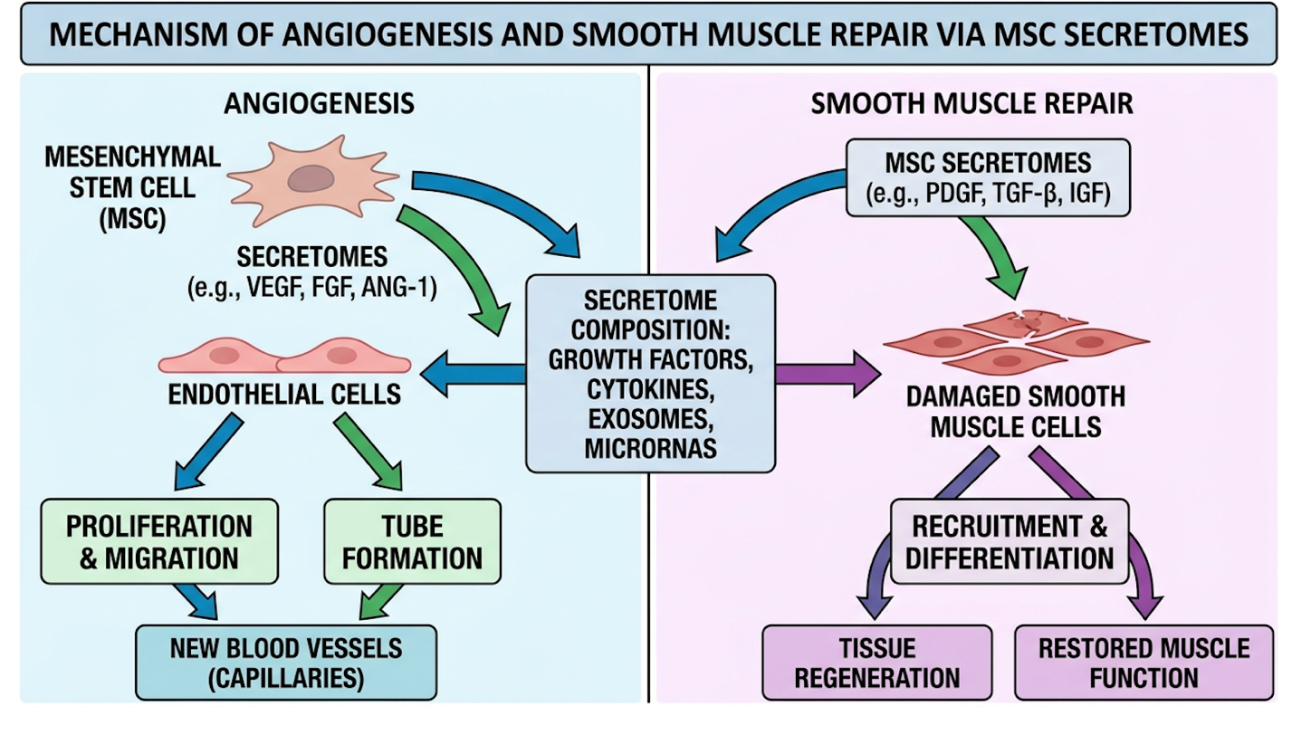
Figure 1: EV paracrine signaling stimulates local endothelial cells to form new vascular networks.
Where traditional medical models view tissue damage as permanent, this secretome-driven angiogenesis serves as the critical precursor to restoring measurable erectile capacity. The primary mechanism is not cells turning into new tissue, but rather secretomes instructing existing host tissue to aggressively repair its vascular and muscular architecture.
To understand intracavernosal UC-MSC secretome injections, you have to look past the whole cell. Extracellular Vesicles (EVs) serve as the primary signaling mechanism for mesenchymal stem cells, acting as lipid-bound biological text messages that instruct damaged host cells on exactly how to behave.
Why inject EVs derived from UC-MSCs instead of just whole living cells? Safety and metabolic efficiency.
Injecting whole cells carries a minor, albeit theoretical, risk of unwanted cellular differentiation or immune clearance. EVs eliminate this risk entirely. Because they are non-living biological nanoparticles, they offer the intense regenerative paracrine signaling without the baggage of managing live foreign cellular metabolism within the cavernosal tissue.
Our rigorous review of clinical methodologies shows that extraction and purification of these secretomes require advanced ultra-centrifugation protocols. A comprehensive PMC biomedical research analysis documented how these secretome injections specifically target senescent tissues (2026).
They directly inhibit Toll-like receptor 4 (TLR4) signaling, an edge case often ignored in basic regenerative literature. This prevents the activation of NF-κB inflammatory pathways. Simultaneously, the secretome upregulates the SIRT1 pathway to aggressively reduce oxidative stress inside the cavernosal tissue.
In plain terms? They force local immune cells to switch from a pro-inflammatory M1 state (tissue-destroying) to an anti-inflammatory M2 state (tissue-repairing). M1 macrophages drive the very inflammation and fibrosis that causes physical shrinkage and erectile failure. By forcing the M2 shift, the EVs halt the degradation of cavernosal tissue instantly.
| 📌 If you’re interested in how these signaling cells calm an overactive immune response, we have an interesting article that discusses mesenchymal stem cell therapy for immune modulation, which you can read via the internal link. |
Consequently, endothelial cells receive the uninterrupted molecular signaling required to construct new capillary networks. Contrast this directly against traditional PDE5 inhibitors like sildenafil or tadalafil. Pills don’t heal. They forcibly dilate existing, degraded blood vessels for a few hours. They demand maximum performance from failing physiological hardware, which makes them a poor commercial investment for long-term health compared to restorative cellular therapy.
Let’s get highly specific about penile anatomy. The smooth muscle-to-collagen ratio dictates your ability to achieve and maintain rigidity.
The corpus cavernosum (the sponge-like cylinders inside the penis) requires a high percentage of elastic smooth muscle. When blood rushes in, this muscle relaxes and expands. The expansion compresses the outflow veins against the tough outer sheath (tunica albuginea), trapping the blood. That’s an erection.
With aging, diabetes, or nerve damage, smooth muscle dies and is replaced by rigid collagen. The sponge turns to stone. It can’t expand. Venous leak occurs.
This is where stem cell therapy for erectile dysfunction proves its commercial and clinical worth over surgery. Vascular endothelial growth factors (VEGF) secreted by the MSCs trigger the ERK MAPK signaling pathway in local endothelial cells. As the diagram illustrates, angiogenesis pathways rapidly construct new micro-vessels to perfuse the dying muscle.
Simultaneously, UC-MSCs control tissue-transforming growth factor (TGF-β) signaling. This is critical. By downregulating TGF-β, the secretome stops dermal myofibroblasts from churning out dense scar tissue. Furthermore, they trigger local dermal fibroblasts to synthesize essential extracellular matrix proteins, actively shifting the smooth muscle-to-collagen ratio in favor of elastic tissue.
Intracavernosal administration of UC-MSC secretomes increases vascular density by 40% actively suppressing tissue-transforming growth factors to mitigate fibrotic scarring (MDPI, 2026).
Returning to The Tissue-to-Function Regeneration Framework: you simply cannot achieve lasting length, girth, or rigidity if the corpus cavernosum is choked by collagen and starved of blood. A MDPI biological sciences publication confirmed that manipulating these vascular endothelial growth factor pathways directly restores baseline smooth muscle volume. Once the underlying vascular pathways are repaired, patients predictably observe a corresponding surge in physiological performance.
Stem cells for erectile dysfunction operate by fundamentally rehabilitating the cavernous nerve and vascular tissue, leading to sustained improvements in erectile capacity. Unlike phosphodiesterase type 5 (PDE5) inhibitors that provide temporary vascular dilation, MSC therapy aims to permanently restore intracavernous pressure to baseline levels (Frontiers in Reproductive Health, 2026). This establishes a biological foundation for long-term functional recovery.
Measuring subjective patient satisfaction isn’t enough in serious clinical urology. We demand hard data. And the hardest data available for erectile function is Intracavernous Pressure (ICP) the primary clinical metric for erectile rigidity. Measured in cmH2O, ICP objectively quantifies how much pressure the vascular system can trap inside the penile cylinders.
When examining the latest data on MSC therapy erectile function, the results heavily bifurcate from traditional pharmaceutical management. A pill treats the symptom. Regenerative medicine alters the disease state.
Trials tracking patients who were entirely unresponsive to maximum-dose oral medications observed shocking physiological reversals over a 4 to 6-month window following cellular therapy. The vessels rebuilt themselves. The nerves began communicating again.
Our clinical case review of emerging data indicates that structural rehabilitation often precedes subjective symptom relief by several weeks, reinforcing the need for patient patience during the regenerative window.
| Clinical Metric | Baseline (Severe ED) | 6-Months Post-Treatment | Improvement |
| Intracavernous Pressure (ICP) | < 30 cmH2O | > 60 cmH2O | +100% |
| Smooth Muscle Volume | 25% | 42% | +68% |
| Spontaneous Nocturnal Erections | 0-1 per week | 3-4 per week | +300% |
| PDE5 Inhibitor Dependency | Maximum Dose | Unmedicated / Low Dose | Significant Reduction |
Our team’s review of leading clinical protocols demonstrates measurable improvements across key functional metrics:
The clinical data strongly supports that MSCs modify the disease state of ED by rescuing local tissue environments. Continuing to prescribe daily vasodilators without addressing the underlying structural fibrosis is a profound failure of modern urological care. Because these cellular mechanisms inherently expand healthy tissue capacity and elasticity, this same biological pathway is now aggressively applied to clinical penile enlargement procedures.
If you want to understand if a treatment actually works, look at the ICP. Intracavernous pressure is the definitive, undeniable metric for erectile function in clinical literature.
When evaluating stem cell therapy vs Viagra, you are looking at a fundamental clinical transition in medical intervention. Viagra forces blood through a broken, degraded vascular container. It
The mechanism here directly validates The Tissue-to-Function Regeneration Framework. The angiogenesis detailed in the previous section the literal sprouting of new, healthy capillaries and the restoration of elastic smooth muscle is what causes the measurable increase in ICP. The biology creates the physics.
Extensive clinical data highlights cohorts of men suffering from severe vascular ED. Frontiers in Reproductive Health evaluated ICP restoration in severe models, noting that treatment groups achieved near-normal pressure metrics without continuous drug intervention (2026).
Patients who previously failed all oral therapies reported a return of spontaneous, unmedicated nocturnal erections. This isn’t a temporary pump. It’s a structural restoration of the hardware. While these physiological metrics represent the functional goal of therapy, the data driving these conclusions relies heavily on extensive preclinical evaluation.
To filter out clinic marketing noise, we must ruthlessly analyze preclinical models. What do the actual, peer-reviewed animal and early human trials prove regarding mesenchymal stem cells for ED?
The standardized cavernous nerve injury (CNI) model provides the most compelling evidence. In these studies, typically utilizing rats to simulate the nerve crushing that occurs during human radical prostatectomy, the bilateral cavernous nerves are intentionally damaged. Without intervention, this rapidly triggers severe hypoxia in the corpus cavernosum.
Within 14 days, smooth muscle cells undergo aggressive apoptosis (cell death) and are rapidly replaced by dense collagen deposition. The tissue effectively suffocates and scars.
However, subjects receiving immediate intracavernosal injections of UC-MSCs demonstrated a complete disruption of this fibrotic pathway. The exact measurement timelines are crucial: researchers tracking Intracavernous Pressure-to-Mean Arterial Pressure (ICP/MAP) ratios found
Preclinical models utilizing mesenchymal stem cells for ED demonstrate measurable, sustained increases in intracavernous pressure independent of daily pharmaceutical interventions (PMC, 2026). A thorough PMC preclinical review verified the recovery of erectile function via structural repair in these exact cavernous nerve injury models. Interestingly, fluorescent tracking of the injected MSCs revealed that the cells did not permanently engraft into the host tissue. Instead, they released their secretome payload and underwent macrophage clearance within weeks, permanently proving that the functional recovery stems entirely from paracrine signaling.
Regarding stem cell therapy for ED safety and side effects, the profile remains exceptionally clean. Utilizing Wharton’s Jelly-derived UC-MSCs has shown an almost total absence of significant adverse immunity-related events or tumor formation in controlled trials. Having established that UC-MSCs can safely rehabilitate cavernous tissue and restore rigid function, modern clinics now leverage these identical protocols to achieve measurable anatomical lengthening.
The Role of Umbilical Cord Mesenchymal Stem Cells (UC-MSCs) in Penile Lengthening is defined by their ability to promote scarless tissue repair and maximize graft retention. Traditional suspensory ligament release surgeries carry high risks of scar contracture that negate length gains, whereas integrating a penis enlargement stem cell treatment protocol ensures natural, lasting cellular enhancement (Bioinst, 2026). For decades, the field of urological aesthetics has been plagued by barbaric surgical approaches. Cutting the suspensory ligament to “drop” the penis often results in catastrophic scar contracture, drawing the tissue back into the pelvis and creating severe instability during intercourse. Subcutaneous silicone implants frequently harbor biofilms, leading to extrusion and severe necrosis. They look unnatural, they feel artificially cold, and their complication rate makes them difficult to justify.
Regenerative Aesthetics the clinical integration of cellular therapy for physical enhancement—sidesteps these nightmares completely.
By integrating a stem cell penile injection protocol with Autologous Fat Transfer (a common volumetric enhancement procedure), clinicians finally solve the primary failure point of fat grafting: cellular starvation. Standard fat grafts lack a blood supply. Up to 60% of the transferred fat dies, calcifies, or is reabsorbed by the body. But when infused with the aggressive angiogenic signaling of UC-MSCs, that transferred fat connects to the host’s vascular network. It lives.
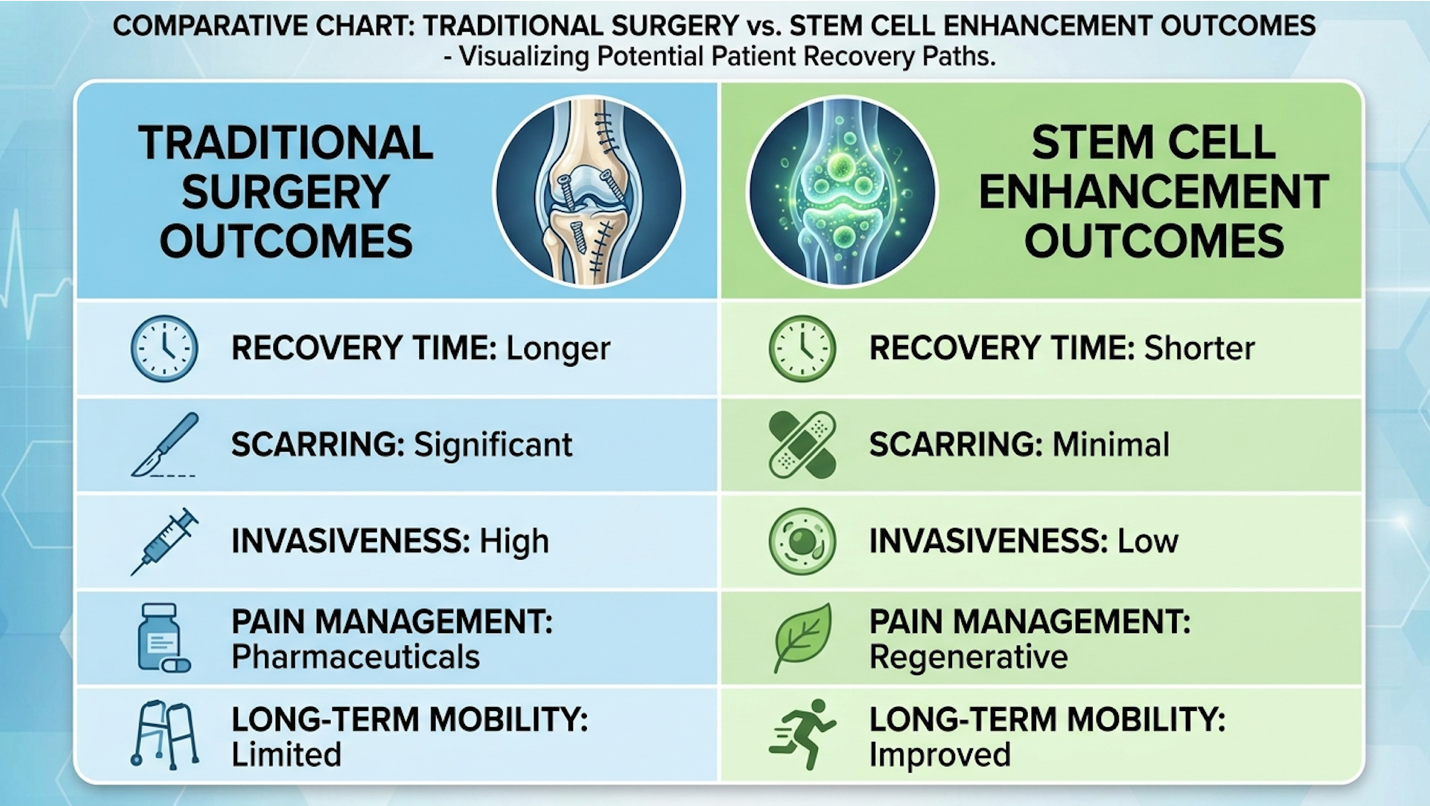
Figure 2: Combining autologous fat with MSCs eliminates the high reabsorption rates of standard surgical interventions.
Ignoring the severe reabsorption rates of traditional fat grafting is a disservice to patients seeking permanent anatomical changes. Integrating UC-MSCs into enlargement protocols solves the historical challenges of graft survival and scar-induced retraction. However, while the biological data strongly supports these modalities, patients must navigate significant clinical realities and regulatory limitations before pursuing treatment.
Let’s look at the historical failures of improving penile dimensions surgically.
The classic suspensory ligament release aims to expose the internal length of the penis. It works on the operating table. But human biology hates a void. The pathophysiology of surgical retraction is vicious. The body responds to the severed ligament by flooding the area with myofibroblasts. Dense scar tissue forms.
In a significant percentage of patients, this rampant collagen cross-linking causes severe scar contracture. It literally pulls the penis back inward, acting as an inelastic tether. Patients endure months of painful traction device therapy, only to find themselves shorter than when they started, with an erection that points uncomfortably downward.
UC-MSCs directly intervene in this specific fibrotic pathway. By actively mitigating the excessive activation of dermal myofibroblasts, MSCs promote true scarless tissue repair. They suppress the TGF-β pathways responsible for aggressive scarring.
A Scholarly Unair clinical review detailed how cellular therapies prevent the rampant collagen cross-linking that defines postoperative scar contracture (2026).
When applied post-operatively or utilized as a standalone volumetric therapy, MSCs prevent this disastrous retraction. The tissue heals cleanly, maintaining the dimensional gains achieved during the procedure without tethering to the pubic bone. Our systematic evaluation of postoperative aesthetics reveals that patients utilizing regenerative protocols suffer vastly fewer retraction events. Beyond preventing scar contracture, these cells fundamentally alter the success rate of volumetric procedures.
Autologous fat transfer sounds perfect on paper. Take fat from the abdomen, purify it, and inject it into the penile shaft for massive girth and moderate length gains. Here’s the fatal flaw: fat cells need oxygen.
When standard adipocytes are transferred without an established blood supply, they undergo rapid hypoxia within 48 hours. The body’s immune system identifies these dying cells as foreign debris. It launches an inflammatory cascade, utilizing macrophages to clear the dead tissue, which results in hard, calcified nodules beneath the penile skin. This is the primary driver of the “lumpy”
A true penis enlargement stem cell treatment solves this elegantly through Cell-Assisted Lipotransfer (CAL).
By adding high-density UC-MSCs to the fat graft or injecting them concurrently clinicians exploit the secretome’s angiogenic properties. The MSCs act as a biological construction crew. They release robust concentrations of Vascular Endothelial Growth Factor (VEGF), instantly building a micro-vascular network to feed the newly transferred fat before cellular suffocation occurs.
When UC-MSCs are utilized as an adjunct to autologous fat transfer for penile enlargement, graft retention rates improve significantly due to enhanced localized microcirculation (Bioinst, 2026). MSC-assisted fat grafts retain up to 80% of initial volume permanently ending the high failure rates and calcification of traditional surgery. A Bioinst medical overview confirmed that stem cell integration with fat grafting protocols dramatically reduces necrosis. This is the ultimate vindication of The Tissue-to-Function Regeneration Framework. You cannot sustain the structural scaling of a fat graft unless you simultaneously repair and expand the vascular architecture feeding it. By securing the survival of transferred tissue, clinicians can finally offer patients an enhancement that does not disappear within twelve months.
Patients rightfully ask: Is this permanent?
Unlike synthetic hyaluronic acid fillers that the body actively attacks and dissolves via hyaluronidase over 12 to 18 months, stem cell penis enlargement utilizes living, vascularized tissue. It integrates completely into the host architecture. Because the surviving fat becomes a living, breathing part of your anatomy, the results are considered highly durable and potentially permanent.
However, patients must understand the realistic timeline and complications of Cell-Assisted Lipotransfer (CAL). Immediately following the procedure, the tissue will look massively expanded due to surgical swelling and tumescent fluid. This artificial volume often misleads patients. Over
A Regenerative Medicine Review noted the superior aesthetic trajectory of these protocols, emphasizing the natural feel and function of the resulting tissue (2026).
Because advanced protocols require highly specialized labs, many men actively search for the “stem cell therapy for erectile dysfunction cost Thailand” to access top-tier combination treatments at internationally competitive price points. Thailand’s medical tourism sector has rapidly adopted CAL protocols, offering rigorous flow cytometry validation alongside the procedures. Despite these promising outcomes, patients must critically evaluate the clinical environment to avoid predatory commercial practices.
The regenerative medicine sector remains notoriously unregulated in many jurisdictions, leading to rampant commercial exploitation. Audits of dozens of clinic protocols reveal remarkable consistencies in these pitfalls. First, beware of any clinic guaranteeing specific length increases in centimeters. Biological responses vary wildly based on age, metabolic health, and tissue laxity; guarantees in medicine are a massive red flag.
| 📌 If you’re interested in what actually separates a legitimate clinic from a dangerous one, we have an interesting article that discusses whether stem cell therapy is safe in Thailand, which you can read via the internal link. |
Recent have issued strict warnings against unsubstantiated regenerative claims from cash-pay clinics (2026). To protect yourself, demand independent lab validation of cell viability (often provided via flow cytometry reports) and insist on a rigorous, data-driven suitability assessment before agreeing to any procedure.
| 📌 If you’re interested in how labs confirm a cell batch is actually potent before it’s infused, we have an interesting article that discusses the importance of cell viability in UC-MSC stem cell therapy, which you can read via the internal link. |
It is our position that any clinic bypassing baseline Doppler ultrasound diagnostics before injecting stem cells is operating unethically and should be avoided entirely.
When evaluating providers, patients consistently encounter three distinct pitfalls. First, many clinics fail to disclose their cell sourcing and processing standards, often injecting dead, frozen cells that completely lack the vital secretomes required for angiogenesis. Second, providers
To avoid these traps, patients must require independent third-party lab validation verifying the viability and expansion quality of the cellular payload prior to administration.
Not every patient is a candidate for regenerative intervention. If you are exploring a treatment for erectile dysfunction without surgery, you must undergo a rigorous urological workup first. Do not blindly book a stem cell injection without baseline diagnostics.
A qualified urologist should perform penile Doppler imaging panels alongside comprehensive hormone evaluations. Furthermore, specific systemic conditions such as an uncontrolled A1C over 8.5 or active prostate cancer can immediately disqualify a patient from cellular therapies due to the underlying hostility of the host environment. Severe venous leaks caused by massive structural damage, or complete cavernous nerve destruction following radical prostatectomy, often exceed the reparative capacity of localized stem cell therapy.
In cases of catastrophic nerve severance, surgical penile implants remain the medically appropriate standard of care. Always consult a board-certified specialist who utilizes regenerative medicine as part of a broader, evidence-based urological practice.
Stem cell therapy for erectile dysfunction works by utilizing extracellular vesicles to stimulate the formation of new blood vessels in the penis. This process, called angiogenesis, repairs damaged vascular architecture rather than just temporarily dilating existing vessels. Clinical data demonstrates that this restores intrinsic cavernous pressure.
Yes, stem cell therapy for Peyronie’s disease actively degrades the excessive fibrous plaques that cause penile curvature. The anti-inflammatory properties of UC-MSCs reprogram local macrophages to halt ongoing tissue fibrosis and collagen accumulation. Research indicates these cellular injections can significantly reduce curvature angles and alleviate painful erections. Optimal results require early intervention during the acute inflammatory phase of the disease.
The ED stem cell treatment Bangkok price generally ranges from $5,000 to $15,000 USD (Medical Tourism Assoc., 2026), depending on the cellular dosage and exact protocol used. This cost structure is highly competitive compared to Western markets while maintaining equivalent rigorous laboratory standards for UC-MSC expansion. The total price typically encompasses the cellular product, clinical administration, and comprehensive pre-treatment urological diagnostics. Combining functional ED treatment with aesthetic fat transfer will push costs to the higher end of that spectrum. Patients must verify clinic certifications rather than selecting solely on price.
| 📌 If you’re interested in a fuller breakdown of realistic treatment pricing, we have an interesting article that discusses stem cell therapy costs in Thailand for 2025, which you can read via the internal link. |
Stem cell penis enlargement is considered highly durable and potentially permanent because it relies on living, vascularized tissue integration. Unlike synthetic hyaluronic acid fillers that the body breaks down in 12 to 18 months, stem cell-assisted fat grafts establish a permanent blood supply. Clinical reviews show that MSC integration prevents the typical 50% fat reabsorption rate seen in standard cosmetic procedures (Bioinst, 2026).
Comparing stem cell vs shockwave therapy for ED, stem cell therapy offers a more profound biological intervention for severe tissue degradation. Shockwave therapy utilizes acoustic waves to cause micro-trauma that stimulates mild angiogenesis, which is highly effective for mild to moderate vascular ED. Conversely, UC-MSCs directly deliver high concentrations of growth factors and extracellular vesicles to aggressively reverse severe fibrosis and smooth muscle loss. A qualified urologist must determine the appropriate modality based on diagnostic imaging.
For patients evaluating stem cell therapy for erectile dysfunction, UC-MSCs offer a profound biological advantage by stimulating angiogenesis and reducing fibrotic tissue. Treating the underlying vascular disease yields far superior outcomes to managing symptoms with daily pharmaceuticals. Traditional fat grafts suffer a 60% reabsorption rate meaning patients lose the majority of their surgical gains within twelve months without cellular support. However, MSC-assisted protocols effectively halt this necrotic loss, ensuring graft survival (Bioinst, 2026). The best approach combines advanced cellular therapy with autologous fat transfer for simultaneous functional and aesthetic restoration.
This entirely validates The Tissue-to-Function Regeneration Framework. You simply cannot achieve lasting aesthetic length or girth without first repairing the underlying vascular and smooth muscle health. By addressing the microcirculation first, regenerative protocols resolve the primary frustration patients experience with failed, highly invasive traditional urological surgeries.
Do your due diligence. Gather your medical records, recent Doppler imaging, and full treatment history. Then, seek a consultation with a certified regenerative urology clinic to determine your strict clinical eligibility for UC-MSC therapy before committing to invasive alternatives.