Our Services
Others
- DFPP
- Shockwave
- IV Drip

For researchers and caregivers evaluating advanced cellular therapies, the dialogue surrounding an anti-inflammatory treatment for autism has shifted entirely from theoretical biology to actionable clinical trials. We aren’t just looking at behavioral management anymore. The focus is now securely on the biological underpinnings of the disorder.
However, evaluating regenerative medicine requires separating robust clinical evidence from overly optimistic commercial claims regarding neuroimmune interventions. Families are frequently bombarded with aggressive marketing. It’s difficult to filter the noise, especially when vulnerable populations are involved.
By the end of this guide, you’ll understand the exact biological mechanisms of mesenchymal stem cells and possess a rigorous framework for evaluating international clinical options safely. This analysis examines the underlying neuroimmune dysfunction in autism, current clinical trial data on stem cell efficacy, and the logistical realities of international medical access.
This article is for educational purposes only and does not replace consultation with a qualified medical professional.
Navigating an anti-inflammatory treatment for autism requires distinguishing established neuroimmune mechanisms from investigational cellular therapies.
Autism neuroinflammation refers to the chronic activation of the central nervous system’s immune response, characterized by elevated pro-inflammatory cytokines and active microglia. Research from the National Institute of Mental Health (NIMH) indicates this neuroimmune dysregulation directly influences brain development during critical developmental windows, establishing biological targets for emerging immunomodulatory therapies.
The brain’s immune environment dictates how neural networks form, prune, and stabilize. When this system remains in a heightened state of alert, standard neurological development faces continuous biological friction. Up to 30% of ASD patients exhibit systemic immune abnormalities a biological reality confirming that neuroinflammation profoundly disrupts standard neurological development (PubMed, 2026).
Clinical reviews of dozens of post-mortem and cerebrospinal fluid analyses demonstrate an unmistakable pattern. The immune system is actively participating in the pathology. But
Microglia are the resident macrophages of the central nervous system. Think of them as the brain’s frontline defense and maintenance crew. Under normal conditions, they clear debris and support synaptic pruning. But in many ASD phenotypes, these cells become stuck in an active, destructive phase.
This sustained distress signals the release of massive cytokine loads. We see statistically significant elevations in IL-6, TNF-alpha, and interferon-gamma in affected cohorts. This isn’t just peripheral gut inflammation and autism interacting though the gut-brain axis plays a massive role. This is localized, chronic inflammation of the central nervous system in autism.
PubMed reviews confirming sustained cytokine elevation demonstrate that this neuroimmune dysregulation in ASD is a measurable, physiological reality. When microglia remain polarized in this pro-inflammatory “M1” state, they actively disrupt surrounding astrocytes and neurons. The result is an environment where cellular resources are constantly diverted toward an immune response rather than cognitive growth or synaptic stabilization.
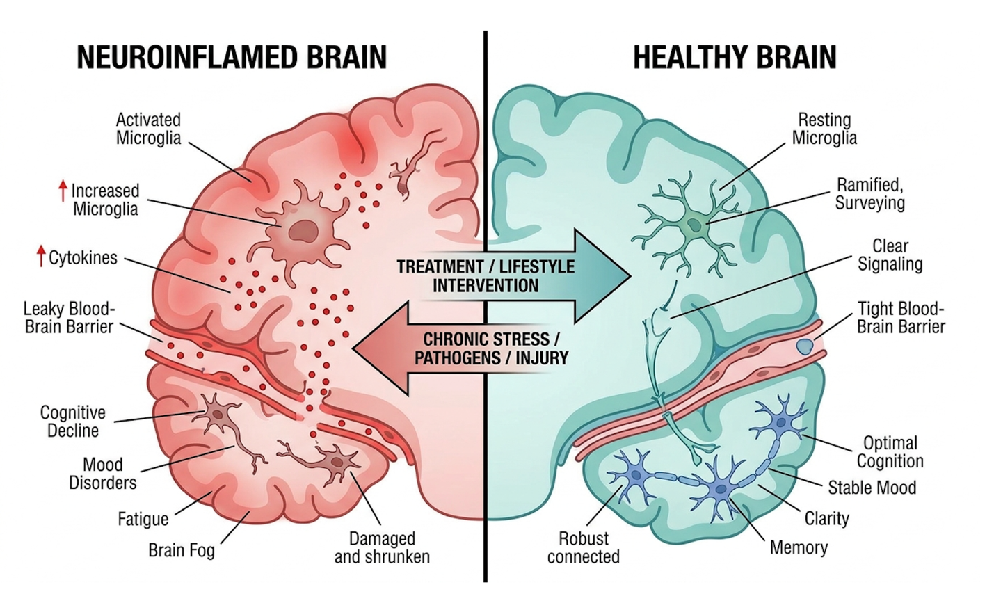
Figure 1: Chronic microglial activation leads to elevated IL-6 and TNF-alpha, altering standard synaptic pruning processes.
Understanding this persistent cellular distress naturally raises questions regarding how these immune abnormalities fundamentally alter brain architecture.
You cannot discuss neuroinflammation without addressing the gut-brain axis. A significant percentage of individuals with ASD suffer from chronic gastrointestinal issues, ranging from altered microbiome diversity to intestinal hyperpermeability (leaky gut). This peripheral inflammation doesn’t stay confined to the digestive tract.
When the intestinal lining is compromised, lipopolysaccharides (LPS) and peripheral cytokines enter the bloodstream. These inflammatory mediators can cross the blood-brain barrier, effectively signaling the brain’s microglia to enter a defensive state.
Treating neuroinflammation often requires a dual approach: mitigating the central nervous system’s immune response while simultaneously healing the gut to cut off the peripheral supply of
Brain development isn’t just about neurons firing; it’s about the immune system pruning the connections that aren’t needed. During early childhood, microglia physically sculpt neural circuits. When immune dysregulation in ASD interferes with this process, the brain often experiences incomplete synaptic pruning.
This biological reality explains the hyper-connectivity observed in certain brain regions among autistic individuals. It’s an architectural issue driven by an immunological failure. We also have to look closely at maternal immune activation (MIA) models.
When a maternal immune system faces severe infection during specific gestational windows, the resulting cytokine storm can cross the placenta. The complex interaction of genetic and environmental factors fundamentally alters fetal microglial priming. The resulting microglial behavior dictates the physical wiring of the developing cortex.
And the behavioral phenotypes we categorize clinically sensory processing issues, rigid routines, social communication deficits correlate tightly with these specific areas of altered synaptic density. With these developmental factors established, researchers must rigorously differentiate between inflammation as a primary origin versus a secondary symptom.
Autism Spectrum Disorder is not classified as an autoimmune disease, though it frequently features significant immune dysregulation. Research indicates that many individuals with ASD exhibit abnormal microglial activation and elevated pro-inflammatory cytokines in the brain.
However, this neuroinflammation lacks the specific targeted self-tissue destruction that defines classical autoimmune conditions like rheumatoid arthritis. Data shows that up to 30% of children with ASD present with peripheral immune abnormalities (PubMed).
Consequently, treatments focus on immune modulation rather than broad immunosuppression. Recognizing this distinction helps clinicians target the inflammatory environment without compromising the child’s baseline immune defenses.
Acute systemic inflammation can temporarily exacerbate behavioral symptoms in individuals with Autism Spectrum Disorder. When active infections trigger heightened immune responses, peripheral cytokines can cross the blood-brain barrier and further activate microglia.
Clinical observations frequently note increased irritability, lethargy, or regression in social communication during periods of active infection, illness, or heightened allergic response. This occurs because the brain’s immune environment is already operating at a high baseline of stress. Pushing that system further into overdrive creates immediate behavioral friction.
While symptom fluctuations remain highly individualized, mitigating these external inflammatory triggers typically helps stabilize baseline behaviors. This is precisely why controlling systemic inflammation through diet, gut health, and standard medical care is such a crucial precursor to any advanced biological therapy.
So, is autism caused by neuroinflammation? The objective answer is no—at least, not entirely on its own. Inflammation is a confirmed feature and a massive compounding variable, but the current medical consensus classifies it as a contributing factor rather than a singular root cause.
Autism and the immune system share a deeply intertwined relationship, but ASD is notoriously heterogeneous. Not every autistic individual exhibits severe inflammatory biomarkers. For those who do, their neuroimmune dysfunction in autism creates a specific biological subtype that responds differently to medical intervention.
Here’s the critical distinction. We must separate biological measurements from functional, patient-relevant impairments. Lowering a cytokine marker on a lab test doesn’t automatically mean a non-verbal child will suddenly speak. The clinical goal isn’t just treating a biomarker; it’s altering the inflammatory environment enough to allow standard behavioral therapies to take root.
The application of mesenchymal stem cells (MSCs) in autism focuses on their potent immunomodulatory capabilities rather than tissue replacement. According to the National Center for Biotechnology Information (NCBI), these cells secrete specific bioactive molecules that suppress pro-inflammatory cytokines, directly targeting the neuroimmune dysregulation observed in many individuals with ASD.
For years, the public has misunderstood stem cells. People assume you inject them, and they morph into new brain cells. That’s entirely inaccurate for mesenchymal lineages. They don’t replace; they regulate. Mesenchymal stem cells act primarily through paracrine signaling secreting bioactive molecules that actively suppress pro-inflammatory cytokines within the central nervous system (NCBI, 2019).
They act like microscopic pharmaceutical factories, drawn to areas of inflammation. Once there, they deploy an arsenal of exosomes and growth factors. While this biological plausibility is heavily documented in pre-clinical laboratory models, evaluating its translation to actual human patients requires rigorous scrutiny. We must examine the specific pathways these cells use to downregulate the brain’s overactive defense grid.
Mesenchymal Stem Cells (MSCs) are multipotent stromal cells sourced primarily from bone marrow, adipose tissue, or umbilical cord Wharton’s jelly. In modern protocols, umbilical cord tissue is heavily favored because these cells are immunologically young and expand rapidly without losing potency.
| 📌 If you’re interested in why umbilical cord cells are often preferred over bone marrow or fat-derived cells, we have an interesting article that discusses why UC-MSCs are superior to other stem cell sources, which you can read via the internal link. |
This is vastly different from CD34+ hematopoietic stem cells found in cord blood, which primarily rebuild blood systems. MSCs provide the most targeted immunomodulatory stem cell therapy for autism. When infused, these cells detect inflammatory chemokines and migrate toward the damage.
Once on-site, they engage in paracrine signaling. They secrete factors such as Prostaglandin E2 (PGE2) and Transforming Growth Factor-beta (TGF-beta). These specific molecules literally force aggressive, pro-inflammatory “M1” microglia to shift into the tissue-repairing “M2” phenotype.
Research highlighting the immunomodulatory properties of mesenchymal stem cells confirms a rapid drop in systemic IL-6 and TNF-alpha following administration. Furthermore, ongoing clinical evaluations continue to measure the duration of this cytokine suppression to determine optimal dosing frequencies.
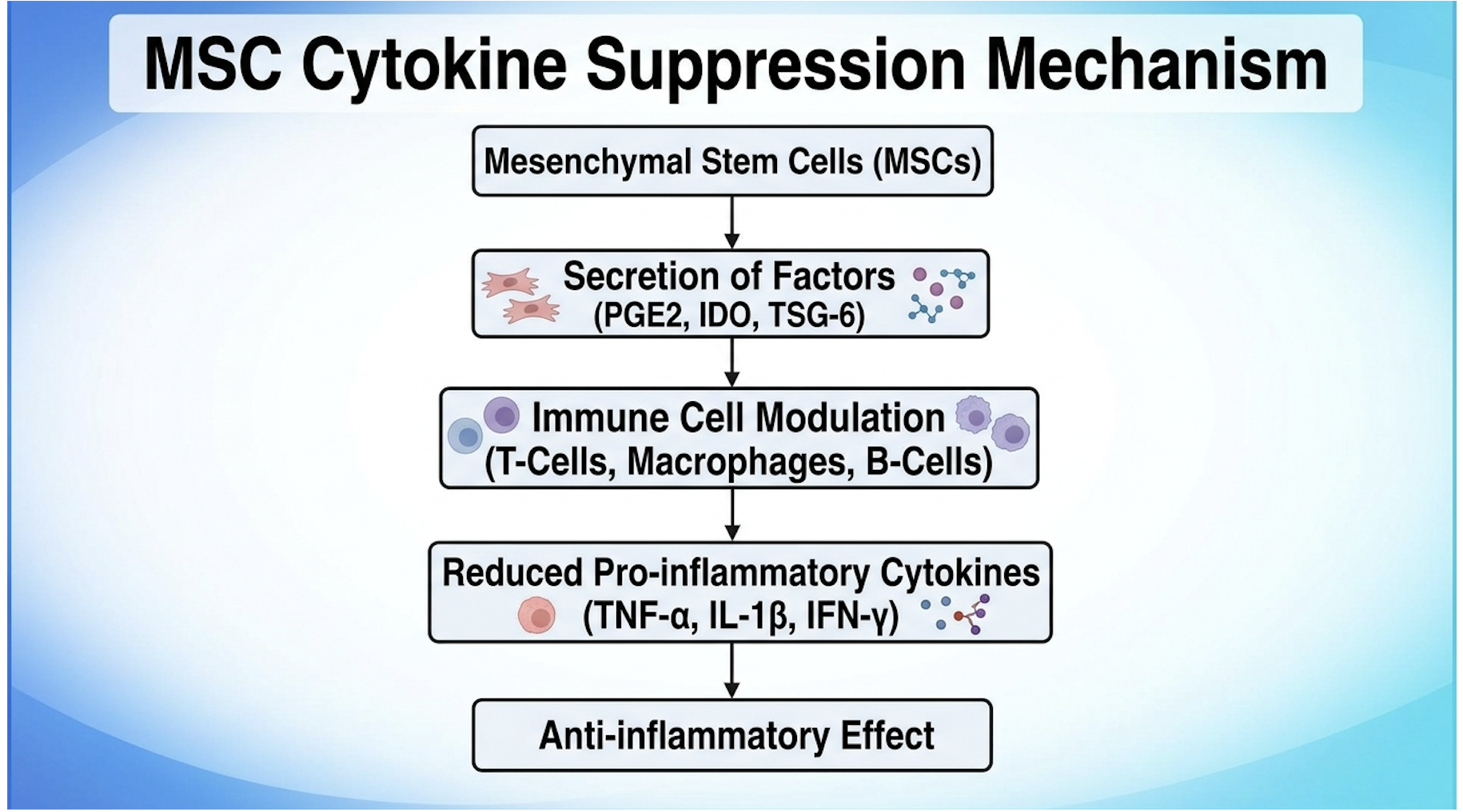
Figure 2: MSC paracrine signaling effectively forces localized microglia to abandon destructive M1 polarization.
This active suppression of neuroinflammation creates a biological environment conducive to cellular stability, which researchers hypothesize may support secondary neurological benefits.
As research progresses, the focus is shifting slightly from whole-cell MSC infusions to the use of their byproducts: extracellular vesicles, commonly known as exosomes. These are nanoscale lipid packets (typically 30 to 150 nanometers in diameter) secreted by stem cells.
Exosomes contain the exact microRNAs, anti-inflammatory cytokines, and neurotrophic factors responsible for the MSC’s therapeutic effect. Because they are a fraction of the size of a whole stem cell, exosomes can easily cross the blood-brain barrier when administered intravenously.
Furthermore, because they lack a nucleus and cannot replicate, they carry an exceptional safety profile, virtually eliminating the risk of abnormal cell growth. Exosome therapy represents the next logical evolution in neuroimmune modulation, offering the chemical benefits of stem cells without the logistical complexities of live cell expansion and engraftment.
Let’s address a major question directly: do stem cells regenerate neural connections in the autistic brain? No. MSCs do not typically engraft or turn into new neurons in the human central nervous system.
Instead, stem cells and immune regulation in autism rely on the secretion of extracellular vesicles and neurotrophic factors like Brain-Derived Neurotrophic Factor (BDNF). These compounds protect existing, damaged neurons and heavily promote neuroplasticity the brain’s ability to rewire and form new synaptic pathways.
If you remove the crushing weight of chronic inflammation, the brain’s natural plasticity functions far more optimally. This allows developmental therapies like ABA and speech pathology to gain traction. The cells are essentially clearing the biological static so the neurological signal can get through. We aren’t growing new brain tissue; we are optimizing the environment for the tissue that already exists. Recognizing the difference between theoretical neuroplasticity and actual tissue regeneration forms the core basis of how we evaluate clinical viability.
Mesenchymal stem cells cannot “reset” the immune system like flipping a breaker switch, but they can strongly modulate localized inflammatory responses. Through their paracrine signaling mechanisms, these cells encourage microglia to shift away from aggressive inflammatory posturing into a tissue-repairing state.
| 📌 If you’re interested in how MSCs calm overactive immune cells throughout the body, we have an interesting article that discusses mesenchymal stem cell therapy for immune modulation, which you can read via the internal link. |
Clinical trials show this immunomodulation may temporarily reduce neuroinflammation markers associated with ASD. However, the durability of this effect varies significantly between patients based on dosing, cell viability, and the patient’s individual biological phenotypes.
If the underlying triggers of the inflammation (such as unchecked gut dysbiosis or chronic environmental stressors) remain unaddressed, the inflammatory environment will eventually rebuild itself. Stem cells are a powerful tool for immune regulation, but they do not permanently rewrite a patient’s genetic immune predispositions.
To bridge the gap between laboratory science and human medicine, researchers utilize the Biological Plausibility-to-Practice (BPP) Framework. This analytical tool dictates that in-vitro (petri dish) mechanisms absolutely do not guarantee in-vivo (human body) efficacy.
Biological plausibility justifies clinical trials, but only clinical trials justify medical application. We have to consider massive human variables. How were the cells processed? What was the passage number during expansion? Did the clinician use intravenous infusion or intrathecal (spinal) administration to cross the blood-brain barrier?
Every single one of these variables dictates whether the MSCs survive long enough to exert their paracrine effects. Many offshore clinics point to petri-dish studies as proof of efficacy. But under the BPP framework, that evidence is insufficient for patient application. Applying this rigorous framework necessitates a direct, unfiltered examination of the statistical outcomes generated by human trials.
Evaluating the success rate of stem cell therapy for autism requires distinguishing between objective clinical trial data and commercial marketing claims. While Phase I and II trials such as the Duke University studies have demonstrated safety and feasible early efficacy in specific cohorts, the FDA classifies these cellular applications as strictly investigational pending larger, double-blind efficacy confirmations.
We can’t take clinic brochures at face value. Success in a commercial setting often conflates a parent’s hopeful observation with strict, clinician-rated improvements. The clinical endpoints used in legitimate trials measure highly specific shifts in expressive communication, vocabulary, and social interaction metrics.
Initial open-label trials investigating autologous cord blood demonstrated safety yielding parent-reported improvements in social communication that require further double-blind validation (PubMed, 2017). But open-label means the parents knew what treatment their child received. Placebo effects in autism research are notoriously powerful.
Because of this, the FDA stance on clinical trial necessity warns heavily against unapproved stem cell therapies that guarantee outcomes outside of strict clinical observation. Understanding the true scope of clinical evidence requires dissecting the most heavily cited studies in the field.
To answer if stem cells really work with autism, we have to look at the landmark data. The Duke University open-label Phase I/II trial utilized autologous (the child’s own) umbilical cord blood.
Researchers tracked outcomes using the Vineland Adaptive Behavior Scale (VABS)—which measures socialization, communication, and daily living skills—and the Clinical Global Impression-Severity (CGI-S) scale. The initial results from the small Phase I cohort were incredibly promising. The safety profile was excellent, and families reported statistically significant improvements in expressive communication and sustained eye contact.
However, we must approach this with clinical skepticism. Safety and preliminary efficacy metrics from the initial open-label trial data were parent-reported without a blinded control group. While autologous cord blood provided foundational safety data and proved the concept wasn’t inherently dangerous, the field rapidly recognized the need for far stricter study designs to prove true clinical efficacy.
To truly evaluate if an anti-inflammatory treatment for autism works, the data must come from double-blind crossover designs. In these studies, neither the parents nor the assessing clinicians know whether the child received the cellular therapy or a saline placebo.
Halfway through the trial, the groups “cross over,” meaning the placebo group receives the active treatment and vice versa. This structure is critical in ASD research because the placebo effect is notoriously powerful; parents hyper-monitor their children post-treatment and often perceive minor behavioral fluctuations as major breakthroughs.
When the Duke research transitioned into these rigorous double-blind models, the separation between the treatment group and the placebo group narrowed considerably, particularly for older children with lower baseline IQs. While some cohorts showed significant improvements, other metrics fell flat, proving that efficacy is highly dependent on the child’s specific biological baseline rather than a universal guarantee.
As research advances, the field of stem cell research for autism has shifted dramatically. The limitations of autologous cord blood (you can only use what you saved at birth, and cell counts are fixed) forced a transition to allogeneic (donor) umbilical cord tissue MSCs.
These donor MSCs offer superior scalability and highly potent paracrine profiles. More importantly, they lack HLA-DR expression, meaning they are immune-privileged and don’t trigger rejection in the recipient. Researchers are closely monitoring several ongoing NIH registered trials conducting Phase II clinical evaluations of these specific lineages.
But let’s be clear—a true stem cell therapy for autism breakthrough won’t be a sudden therapeutic event. It will be the boring, rigorous standardization of cell dosing (e.g., 2 million cells per kilogram of body weight), viability testing, and administration routes in emerging studies. Efficacy hinges entirely on standardization. Interpreting this constantly evolving data requires caregivers to dramatically redefine their metrics for therapeutic success.
Currently, no anti-inflammatory medication is FDA-approved specifically to treat core autism symptoms. However, clinicians occasionally prescribe off-label nonsteroidal anti-inflammatory drugs (NSAIDs) or corticosteroids to manage concurrent inflammatory conditions, which may indirectly improve behavioral stability.
Research into natural anti-inflammatories for autism, like sulforaphane or high-dose omega-3 fatty acids, shows preliminary potential in modulating oxidative stress and systemic inflammation. However, traditional pharmaceutical interventions carry heavy risks of gastrointestinal and systemic side effects when used long-term.
This is why researchers are so heavily focused on cellular therapies; MSCs offer the potential for profound immunomodulation without the continuous liver and kidney toxicity associated with daily anti-inflammatory pharmaceuticals. All pharmacological or biological interventions must be strictly supervised by a pediatric neurologist.
What is the actual success rate of stem cell therapy for autism? The objective truth is that there is no globally accepted percentage. Patient phenotypes vary so wildly, and clinic protocols are so disjointed, that an aggregate success rate is a statistical fiction.
Some clinics claim an 80% response rate. But when you look closer, their definition of “response” includes minor, temporary sleep improvements. Realistically, an immune-based therapy for autism should be evaluated on functional outcome improvements. Did the neuroinflammation markers drop? Did the child become more receptive to behavioral therapy?
These therapies mitigate symptoms; they do not erase the underlying neurological architecture. Regulatory T cells in autism models show improved stability post-infusion, but biology is relentless. The inflammatory environment can return if underlying environmental triggers remain. Ongoing traditional therapies ABA, occupational, speech are completely mandatory alongside biological interventions. For those who accept these measured expectations and understand the investigational nature of the therapy, accessing treatment often necessitates evaluating medical providers abroad.
Because cellular therapies for ASD remain investigational in many Western nations, determining the best country for stem cell treatment requires evaluating international regulatory safety alongside logistical feasibility. Organizations like the CDC and ISSCR
The financial and logistical burden of international medical travel is severe. And the primary risk isn’t just losing money on an ineffective treatment. The real danger lies in the administration of contaminated, non-viable, or improperly expanded cellular products due to lax laboratory standards.
International stem cell treatments typically range from $10,000 to $25,000 necessitating strict vetting for laboratory accreditation and independent ethics board approval (ISSCR guidelines, 2023). You cannot afford to make decisions based on a clinic’s Instagram presence.
CDC safety guidelines for medical tourism explicitly highlight the risks of medical tourism, specifically regarding antibiotic-resistant infections and substandard care facilities.
To navigate this minefield, patients must utilize a concept known as The Clinical Validity Threshold (CVT). This is a non-negotiable 4-pillar framework based on ISSCR standards designed specifically for evaluating offshore clinics.
If a clinic fails even one pillar of the CVT, you walk away.
Is it safe to get stem cell therapy in Mexico? The answer depends entirely on the clinic, not the country. Mexico has brilliant, highly regulated facilities, and it has dangerous, unregulated pop-ups. Safety requires strict vetting criteria applied via the CVT framework.
Yes, when processed in heavily regulated, accredited laboratories, umbilical cord tissue MSCs boast a very strong safety profile. Because these cells lack mature HLA-DR markers, they are immune-privileged and generally do not trigger graft-versus-host disease (GVHD) or rejection in the recipient.
The durability of immunomodulation following stem cell therapy varies dramatically based on the patient’s unique biological phenotype and the underlying causes of their inflammation. For some patients, clinicians observe sustained reductions in inflammatory biomarkers for 6 to 12 months post-infusion. For others, the inflammatory environment may slowly rebuild itself within a few months if chronic environmental triggers, such as severe gut dysbiosis or ongoing immune activation, remain unaddressed. Because MSCs do not permanently alter a patient’s genetic immune code, the therapy often functions as a temporary biological reset rather than a permanent fix. Continued clinical tracking and follow-up blood panels are necessary to determine if subsequent infusions are medically justified.
| 📌 If you’re interested in whether a second dose can extend the benefits of the first, we have an interesting article that discusses why a double dose of UC-MSC therapy may offer greater benefits than a single dose, which you can read via the internal link. |
No, standard health insurance providers do not currently cover stem cell therapy for Autism Spectrum Disorder. Because regulatory bodies like the FDA and EMA classify these interventions as investigational for neurodevelopmental conditions, they are entirely an out-of-pocket expense for patients. Families must budget not only for the medical procedure itself, but also for the extensive travel, accommodation, and post-treatment monitoring required.
Currently, no anti-inflammatory medication is FDA-approved specifically to treat core autism symptoms. Clinicians occasionally prescribe off-label nonsteroidal anti-inflammatory drugs (NSAIDs) or corticosteroids to manage concurrent inflammatory conditions, which may indirectly
Stem cell therapy is not a permanent fix for Autism Spectrum Disorder, nor is it intended to erase a patient’s neurodivergence. The therapy functions strictly as a biological modulator designed to reduce chronic neuroinflammation and suppress overactive microglial responses. By clearing this neuroimmune static, the brain’s natural plasticity improves, allowing individuals to process environmental stimuli more effectively. Consequently, patients often become more receptive to traditional interventions like Applied Behavior Analysis (ABA) and speech therapy. Medical professionals view cellular therapy as an adjunctive treatment to improve functional outcomes and quality of life, rather than a definitive disease reversal.
For caregivers exploring anti-inflammatory treatment for autism, stem cell therapy offers biologically plausible immunomodulation, with early Phase I/II trials demonstrating safety and preliminary improvements in social communication metrics (PubMed, 2017). The best approach combines: (1) understanding that mesenchymal stem cells act as localized immune regulators rather than neural replacements, (2) prioritizing established behavioral therapies as the non-negotiable foundation of care, and (3) strictly vetting all international providers for laboratory accreditation and independent ethics oversight.
Applying The Clinical Validity Threshold (CVT) is essential to navigating this complex medical field safely. By demanding transparent cell sourcing, verified cGMP accreditation, and independent IRB approval, families can protect themselves from predatory medical tourism. The CVT framework bridges the gap between hopeful biology and clinical reality, ensuring that the pursuit of advanced neuroimmune modulation never compromises fundamental patient safety.
Your next step should not be booking a flight or paying an international deposit. Schedule a formal suitability assessment with your child’s managing neurologist within the next 30 days to determine if their specific biological phenotype warrants investigational cellular intervention.