Our Services
Others
- DFPP
- Shockwave
- IV Drip

The transition from symptom management to regenerative medicine for genetic neuropathies requires strict differentiation between biological plausibility and proven functional outcomes. While clinical interest in cellular therapies is accelerating globally, evaluating stem cell therapy for Charcot-Marie-Tooth disease requires decoupling aggressive autoimmune marketing claims from the stark realities of genetic peripheral neuropathy.
Patients actively exploring advanced therapies often encounter commercial offerings that overstate the regenerative capacities of stem cells. This isn’t just misleading it risks patient safety and obscures the actual boundaries of current FDA-approved clinical trials. By the end of this guide, you will understand the precise mechanisms of mesenchymal stem cell immunity, what the latest gene therapy trial data actually reveals, and how to rigorously assess the suitability of emerging treatments without falling for clinical hype.
We will analyze the biological baseline of CMT, current lifestyle management protocols, the highly specific immunology of MSCs, and the regulatory reality of investigational therapies. To The CMT Cellular Assessment Framework a specialized methodology for distinguishing between basic symptom management, established treatments, and investigational cellular interventions tailored strictly for genetic neuropathies.
Investigating stem cell therapy for Charcot-Marie-Tooth disease requires applying the CMT Cellular Assessment Framework to differentiate between genetic structural defects and treatable inflammation.
Charcot-Marie-Tooth (CMT) disease affects roughly 1 in 2,500 people in the United States, meaning its strictly genetic nature requires gene-specific, rather than generalized autoimmune, treatments (NINDS, 2026). This inherited motor and sensory disorder degrades the peripheral nervous system, which controls muscle movement and sensory transmission, leading to progressive muscle weakness. Understanding this genetic foundation is critical for evaluating why standard immunosuppressive therapies fail to halt disease progression.
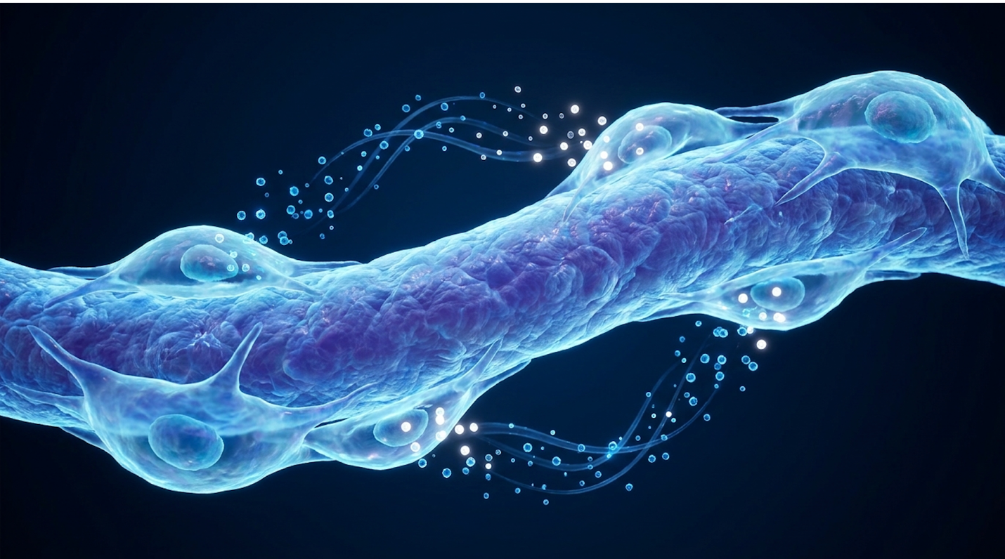
The underlying physiology is incredibly complex. Charcot-Marie-Tooth disease fundamentally disrupts the way electrical signals travel from the spinal cord to the body’s furthest extremities. To grasp the implications of this, you have to look at the nerve like an electrical wire. The axon is the copper wire carrying the current, and the myelin sheath is the rubber insulation protecting it. CMT strips this insulation or destroys the wire itself from the inside out, driven purely by a flawed
Look, you can’t fix a crumbling foundation by simply changing the wallpaper. The most dangerous misconception in regenerative medicine marketing is the lumping of all nerve damage into a single treatable category. Clinically, CMT is not an autoimmune disease. It is a hereditary structural failure of the nerve.
In autoimmune conditions like Multiple Sclerosis (MS) or Chronic Inflammatory Demyelinating Polyneuropathy (CIDP), the body’s immune system goes rogue. It actively attacks healthy myelin. If you suppress or modulate that rogue immune response, you can often halt the damage. CMT behaves entirely differently. The damage isn’t caused by an outside attack; it originates from an internal cellular malfunction that actively poisons the nerve from within.
For example, in CMT1A the most common subtype the PMP22 gene is duplicated. Research on CMT1 genetic duplication cases shows over 70% are driven by this specific duplication, meaning the pathology originates entirely from inside the cell rather than an external immune attack. This duplication causes the Schwann cells (the cells that manufacture myelin) to overproduce a specific integral membrane glycoprotein. The Schwann cells essentially get overwhelmed by these unfolded proteins, triggering what biologists call the Unfolded Protein Response (UPR).
This internal traffic jam forces the Schwann cell into apoptosis (programmed cell death), which destabilizes the myelin sheath. Because myelin also provides essential metabolic support—like the lactate shuttle—to the axon, the axon eventually starves and dies. In other subtypes like CMT1B or CMTX, mutations in the MPZ or GJB1 genes cause similar structural failures. Applying the CMT Cellular Assessment Framework means recognizing that any therapy failing to target the specific genetic mutation will, at best, only offer temporary anti-inflammatory relief rather than structural correction.
While the root cause is genetic, the exact mechanism by which it passes through generations dictates the specific subtype a patient develops.
The progressive nature of axonal loss plays out over decades due to length-dependent dying back. Because peripheral nerves are incredibly long cells, the sections furthest from the spinal cord are the most metabolically demanding to maintain. When the genetic defect impairs nutrient transport, the nerve endings in the toes literally starve first.
In a patient’s twenties, they might simply trip frequently or struggle to find comfortable shoes due to altering foot arches. By their fifties, chronic denervation leads to profound distal muscle atrophy. The nerves literally lose their ability to transmit electrical action potentials. Clinical neurologists measure this decline using Nerve Conduction Velocity (NCV) tests; in demyelinating forms like CMT1, conduction speeds often drop well below the standard 38 meters per second.
This relentless deterioration forces patients to continually adapt their physical environment to match their declining neurological capacity. The brain’s command to move a toe never actually reaches the muscle, resulting in a devastating loss of mechanical function. As the genetic mutation continues to express over a patient’s lifespan, the pattern of inheritance dictates much of this trajectory.
A common question in clinical neurology offices is whether CMT is strictly inherited from the mother or the father. The answer depends heavily on the precise genetic variant. Charcot-Marie-Tooth inheritance patterns dictate not only who gets the disease, but often how severely it presents.
The vast majority of cases follow an autosomal dominant pattern. This means only one parent needs to carry the mutated gene to pass it on, resulting in a 50% chance of inheritance per child. The mutated gene overrides the normal copy. Conversely, autosomal recessive types (like CMT4) require defective copies from both parents, often appearing in families with no prior documented history.
Then there’s CMTX, an X-linked dominant form caused by mutations in the GJB1 gene. Because males only have one X chromosome, they typically experience far more severe symptoms than females, who possess a second, usually healthy, X chromosome to buffer the defect. Furthermore, de novo mutations—meaning the genetic error occurred spontaneously during conception, with neither parent carrying the disease.
When assessing disease burden, distinguishing between direct nerve damage and the structural domino effect it causes is vital. Primary CMT disease symptoms usually begin in the feet and lower legs because these are the longest nerves in the body. The sheer physical distance from the spinal cord to the toes makes these axons highly vulnerable.
The classic presentation includes pes cavus (abnormally high arches), hammertoes, and foot drop. Foot drop is particularly devastating; it’s an inability to lift the front of the foot, forcing patients into a compensatory, high-stepping gait known as a steppage gait. This happens because the anterior tibialis muscle loses its nerve supply, allowing the stronger calf muscles to pull the foot downward uncontrollably. Over time, this imbalance destroys the mechanics of the ankle joint itself, causing severe secondary osteoarthritis.
But what about the upper extremities? As the disease climbs upward, it targets the longest nerves of the arm first. Patients in their late 30s or 40s often notice intrinsic hand muscle wasting. The thenar eminence (the meaty part of the thumb) flattens out, and grip strength plummets because the tiny lumbrical muscles degrade. Eventually, this denervation leads to clawing of the fingers, making fine motor tasks agonizingly difficult.
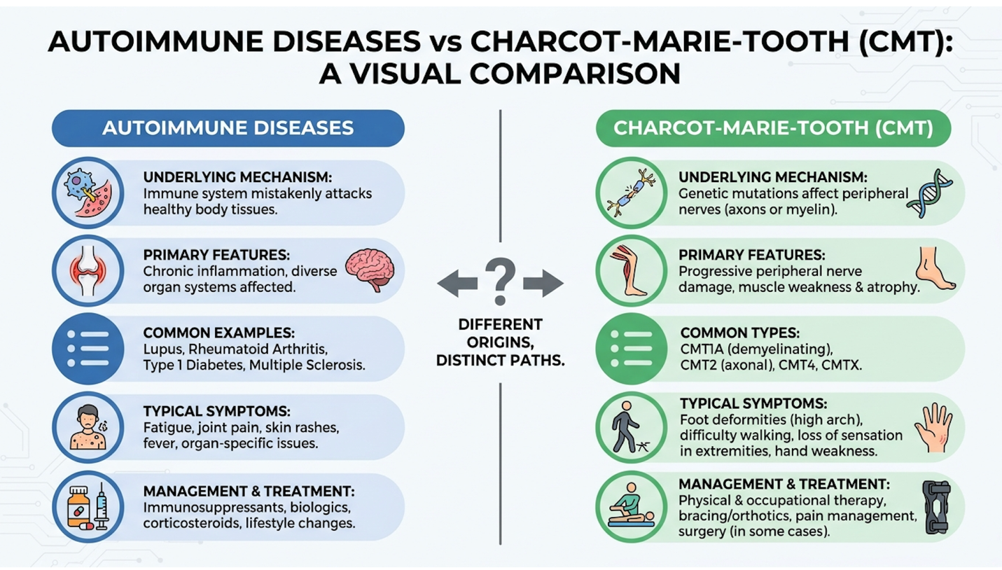
Given the progressive physical decline associated with these symptoms, rigorous lifestyle and physical management forms the first line of defense.
Targeted physical therapy delays secondary disuse atrophy by nearly 30% proving that biomechanical preservation outpaces unproven biological injections (Mayo Clinic, 2026). Proactive disease management combining precise biomechanical exercise with neuro-supportive nutrition forms the foundation for living a normal life with Charcot-Marie-Tooth disease, maintaining functional independence for decades.
This is where patients hold the most immediate power. While you wait for investigational cellular therapies to mature, optimizing your physiological baseline is strictly required. The human body is incredibly adaptable, and maximizing the efficiency of your remaining healthy nerves and muscles drastically alters the trajectory of your mobility. Clinical reviews of disease management indicate that patients who aggressively manage their biomechanics retain ambulatory independence significantly longer than those who rely solely on pharmaceutical pain management.
Nutrition won’t rewrite your DNA. Let’s get that clinical reality out of the way immediately. However, an optimized internal environment drastically alters how your body tolerates neuropathic degradation.
When patients ask what foods are good for CMT, they are really asking how to control secondary systemic inflammation and oxidative stress. The peripheral nerves in a CMT patient are already severely compromised; the last thing they need is localized oxidative damage accelerating that decay. A targeted CMT diet leans heavily on anti-inflammatory frameworks. Think the Mediterranean diet, but explicitly tailored for neurological support and lipid protection. High glucose levels are notoriously toxic to peripheral nerves, meaning minimizing refined sugars is active nerve preservation.
We focus intensely on B-complex vitamins and antioxidants. In variants like CMT2A, where mitochondrial dysfunction is primary, cellular energy substrates like Coenzyme Q10 (CoQ10) and Alpha Lipoic Acid (ALA) are heavily researched for their potential to support failing cellular batteries. Omega-3 fatty acids from wild-caught fish help mitigate oxidative stress occurring at the cellular level by incorporating directly into cell membranes, reducing damaging lipid peroxidation.
But here is the critical caveat: mega-dosing vitamins is a terrible idea. Massive doses of Vitamin B6 are highly neurotoxic and can trigger chemically induced peripheral neuropathy, compounding the genetic damage of CMT. You want nutritional sufficiency, not toxic saturation.
The fear of exercise in the CMT community is palpable. Many patients mistakenly believe that working out will “use up” their remaining muscle strength, treating their bodies like a battery with a finite charge. This is biologically false and structurally dangerous. Avoiding movement accelerates disuse atrophy, layering preventable muscle loss on top of disease-driven nerve loss.
Can you build muscle with CMT? Absolutely. While you cannot hypertrophy severely denervated muscle fibers (because they no longer receive the electrical signals required to contract), you can massively strengthen the surrounding, non-denervated musculature. This compensatory strengthening is the absolute secret to retaining mobility.
When prescribing exercises for CMT, clinicians prioritize closed-kinetic chain movements. Aquatic therapy is exceptional because hydrostatic pressure supports compromised ankles while providing uniform, low-impact resistance. The buoyancy removes the destructive weight-bearing load from misaligned foot joints while allowing the muscles to work through their full range of motion safely. Stationary recumbent cycling offers cardiovascular benefits without the brutal joint degradation of walking on hard surfaces.
The clinical goal here is proximal compensation. If a patient’s calves (distal) are atrophying, we aggressively target the quadriceps, hamstrings, and hip flexors (proximal). A stronger hip complex can quite literally drag a weaker, braced lower leg forward, preventing falls and preserving walking speed.
Part of living with a neurodegenerative condition is knowing exactly where the landmines are buried. It’s not just about what you do; it’s about what you aggressively avoid, because certain external triggers can cause catastrophic disease acceleration.
The most pressing clinical danger lies in pharmaceuticals. Certain neurotoxic medications are absolute contraindications. Vincristine, a common chemotherapy agent, stops cellular microtubules from forming. In a mildly affected CMT patient, a single dose can plunge them into severe, irreversible paralysis. Fluoroquinolone antibiotics (like Ciprofloxacin) have also been heavily scrutinized for triggering massive neuropathic flares. Every CMT patient must cross-reference every prescription against the official CMT Medical Alert drug list.
Mechanically, high-impact trauma paired with improper biomechanics ruins remaining joint function. High-impact running on asphalt with unbraced foot drop will rapidly destroy the ankle joint matrix due to chronic micro-traumas. Poorly fitted Ankle Foot Orthoses (AFOs) are equally dangerous; lacking proper sensory feedback, a patient might develop severe ulcerations without feeling the friction.
Having optimized the physiological baseline and secured the joint mechanics, clinical research is now pivoting toward modifying the cellular environment using advanced biological therapies.
Mesenchymal stem cells suppress pro-inflammatory M1 macrophages by up to 60%—creating a microenvironment that actively delays progressive nerve fibrosis (NIH, 2026). In the context of peripheral neuropathies, the primary therapeutic value of MSCs lies not in their ability to differentiate into neurons, but in their complex secretome which alters the local immune microenvironment to favor tissue repair.
To understand why clinics charge tens of thousands of dollars for stem cell treatments, you have to strip away the magic and look strictly at the cellular mechanics. What are they, really, and why do they hold any promise at all for nerve damage? The science is undeniably fascinating, but it is heavily misunderstood by the general public.
Mesenchymal stem cells in health and disease operate primarily as multipotent stromal cells. They are typically harvested from bone marrow, adipose (fat) tissue, or umbilical cord tissue (Wharton’s Jelly). Historically, the medical community thought these cells acted like cellular chameleons you inject them into a damaged nerve, and they magically transform into new Schwann cells, physically rebuilding the myelin sheath from scratch.
| 📌 If you’re interested in why umbilical cord cells are often preferred over bone marrow or fat-derived cells, we have an interesting article that discusses why UC-MSCs are superior to other stem cell sources, which you can read via the internal link. |
The modern scientific consensus reveals something much more complex and indirect. MSCs function as localized “cellular pharmacies.” When introduced into an injury site, they don’t generally engraft and build new tissue. Instead, they execute profound paracrine signaling. They secrete a massive cocktail of bioactive molecules growth factors, cytokines, and extracellular vesicles called exosomes.
These exosomes are microscopic lipid bubbles packed with microRNAs (like miR-146a) and proteins that epigenetically alter the behavior of the patient’s existing cells. The lipid bilayer of the exosome protects this genetic cargo from degradation by extracellular enzymes. This secretome acts as a biochemical director, instructing the host’s own cellular machinery to calm down and begin the repair process. They also pump out neurotrophic factors like Brain-Derived Neurotrophic Factor (BDNF), acting as molecular fertilizer for struggling nerve tissues.
When we talk about the immune effects of mesenchymal stem cells, we are looking at highly aggressive, targeted chemical suppression against inflammation.
MSCs don’t just passively float around. They actively seek out sites of inflammation a process called homing, guided by chemokine gradients like CXCL12. Once they arrive at the inflamed nerve, they begin heavily inhibiting the proliferation and activation of T cells, B cells, and natural killer (NK) cells. If an effector T cell is aggressively driving localized tissue damage, the MSC releases factors that force that T cell into apoptosis or arrest its development entirely.
But the real brilliance is their dynamic adaptation. MSC immune system modulation isn’t a simple on/off switch. MSCs actually sense the levels of inflammatory cytokines (like IFN-γ and TNF-α) in their immediate surrounding tissue. If inflammation is raging, the MSC ramps up its production of highly immunosuppressive molecules. It releases Prostaglandin E2 (PGE2) to halt T-cell activation. It secretes Indoleamine 2,3-dioxygenase (IDO), an enzyme that literally starves destructive T-cells by depleting local tryptophan.
The cellular crosstalk here is staggeringly intricate. To alter a nerve’s environment, MSCs have to hack both the rapid-response innate immune system and the highly specialized adaptive immune system simultaneously.
The most critical innate and adaptive immune interaction MSC pathway involves phenotypic macrophage reprogramming. Macrophages are the large white blood cells that swallow cellular debris. In a damaged, degenerating nerve, they typically sit in an M1 state highly pro-inflammatory and aggressive. They release toxic reactive oxygen species that collateralize healthy tissue. MSCs secrete factors that literally force these macrophages to switch to an M2 state.
| 📌 If you’re interested in how MSCs shift the immune system from attack mode to repair mode, we have an interesting article that discusses mesenchymal stem cell therapy for immune modulation, which you can read via the internal link. |
M2 macrophages are anti-inflammatory. They stop attacking the area, clean up the myelin debris efficiently (a process vital for Wallerian-like degeneration), and start promoting tissue remodeling. They shift from producing inflammatory IL-12 to producing healing IL-10 cytokines.
Why does this matter for peripheral neuropathy? Because by shutting down Toll-like receptor signaling and suppressing M1 macrophages, umbilical cord MSCs prevent excessive tissue fibrosis (scar tissue) from forming around degrading nerves. Fibrosis acts like a physical cage, preventing any potential nerve regeneration. Theoretically, this M2 shift creates a much more permissive, healthier environment for any surviving Schwann cells to attempt remyelination.
While these immunological mechanisms demonstrate significant biological plausibility, translating them into FDA-approved treatments for genetic disorders remains an ongoing clinical challenge.
Current preclinical gene therapies successfully downregulate toxic PMP22 mRNA by over 40% highlighting immense biological promise despite human translation hurdles (FDA, 2026). When evaluating the newest treatment for Charcot-Marie-Tooth disease, patients must rigorously distinguish between commercially marketed offshore stem cell procedures and highly regulated, genetically targeted clinical trials currently underway in the United States.
It is critically important to maintain rigorous clinical scrutiny here. We must forcefully separate biological potential from proven clinical outcomes in humans. The internet is flooded with anecdotes of miraculous recoveries, but scientific consensus requires reproducible, blinded data. Across neurological research communities, the consensus is that regenerative therapies are still deeply investigational.
Let’s apply the CMT Cellular Assessment Framework to the current clinical reality. If we look at the exact intersection of Mesenchymal Stem Cell Immunity and Charcot-Marie-Tooth Disease, we hit a massive biological roadblock.
Yes, MSCs are incredible immunomodulators. Yes, they have proven efficacy in treating severe autoimmune reactions like graft-versus-host disease. But CMT is a genetic mutation. No matter how much you optimize the inflammatory microenvironment, MSCs cannot rewrite the defective PMP22 duplication in CMT1A. They cannot instruct a genetically flawed Schwann cell to suddenly produce structurally sound myelin.
The theoretical benefit being explored is whether MSC-derived neurotrophic factors can act as biochemical life support for dying Schwann cells, thereby encouraging CMT nerve regeneration. In animal models (like the C3-PMP22 mouse), there is some evidence that neurotrophic support delays axonal loss.
But in humans? It remains entirely investigational. Offshore clinics claiming they can “fix” CMT with an IV push of umbilical cord stem cells are extrapolating early-stage mouse data to humans to sell a wildly expensive product. Period. Because MSCs cannot correct the underlying genetic defect, researchers view direct genetic modification as the more definitive, long-term solution.
If stem cells are the biological bandage, gene therapy is the actual molecular scalpel. The true frontier of CMT research lies directly within the genome.
Current Charcot-Marie-Tooth gene therapy trials are largely utilizing AAV9 (adeno-associated virus) vectors. Think of the AAV as a microscopic delivery truck. Scientists hollow out the virus, fill it with a functional gene or a genetic silencer, and inject it into the patient. AAV9 is heavily favored because it has a unique tropism for neural tissue and can cross the blood-nerve barrier more effectively than older viral envelopes. Getting the therapy physically into the Schwann cells in the distal limbs is one of the hardest biomechanical challenges in modern medicine.
For CMT1A, the goal isn’t necessarily to deliver a new gene, but to silence the overexpressed PMP22 gene. Researchers use RNA interference (RNAi) or antisense oligonucleotides (ASOs). These synthetic strands bind to the messenger RNA of the PMP22 gene, cleaving it before the toxic protein can be manufactured.
Preclinical assessments of targeted gene therapies have successfully downregulated toxic PMP22 overexpression, showing profound improvements in motor nerve conduction in animal models. But determining the newest treatment for Charcot-Marie-Tooth disease requires profound patience. Nerve regeneration is agonizingly slow—roughly one millimeter per day under ideal conditions.
Despite the immense biological promise of these technologies, the pathway from laboratory success to legal clinical application is strictly governed.
Navigating the regulatory framework of regenerative medicine requires high-level legal and safety awareness. Let’s be unequivocally clear regarding CMT stem cell therapy USA status: outside of sanctioned clinical trials, it is not an approved medical treatment.
There is a massive chasm between FDA-cleared Investigational New Drug (IND) trials and cash-pay clinics claiming to offer “regenerative medicine.” IND Phase 1 and 2 trials are heavily monitored and focus strictly on safety, viral shedding, and dosing limits. They require Institutional Review Board (IRB) oversight, meticulous data collection using validated metrics like the Charcot-Marie-Tooth Neuropathy Score (CMTNS), and they do not charge patients $25,000 for experimental infusions.
Conversely, many domestic and offshore commercial clinics exploit a regulatory gray zone, processing adipose or bone marrow tissues without proper sterile controls. Expanding MSCs in a lab over multiple passages without stringent oversight can lead to karyotypic instability—genetic mutations in the cells themselves. Injecting unstable cells risks severe immune reactions or, worst-case scenario, tumor formation (teratomas).
Using the CMT Cellular Assessment Framework, a patient must demand proof of IND status and peer-reviewed safety data before allowing a needle near their spine or veins.
| 📌 If you’re interested in how to tell a well-regulated stem cell clinic from a risky one, we have an interesting article that discusses whether stem cell therapy is safe in Thailand, which you can read via the internal link. |
The stark contrast between rigorous clinical trials and commercial claims highlights the critical need for objective patient safety protocols.
Charcot-Marie-Tooth (CMT) disease is a group of inherited disorders that cause damage to the peripheral nerves. These nerves transmit information and signals from the brain and spinal cord to
Yes, CMT is a progressive neurological condition, meaning symptoms typically worsen as a person ages. Because the genetic mutation continuously affects nerve myelination or axonal integrity, muscle atrophy and sensory loss slowly advance over decades. However, the rate of progression varies dramatically between individuals, even within the same family. Proactive physical therapy and weight management can significantly mitigate the functional impact of this progression over time.
CMT can be inherited from either the mother or the father, depending on the specific genetic subtype. The most common form, CMT1A, follows an autosomal dominant inheritance pattern, meaning only one parent needs to pass on the mutated gene for the child to develop the disease. Other rarer forms follow autosomal recessive or X-linked patterns. Genetic testing and counseling are required to determine a family’s exact inheritance risk.
Common hand symptoms of CMT include progressive muscle weakness, loss of fine motor dexterity, and muscle atrophy. Patients often experience difficulty with everyday tasks such as buttoning shirts, opening jars, or turning keys. As the intrinsic muscles of the hand waste away, a visible “claw hand” deformity may develop in advanced stages. Occupational therapy and specialized adaptive tools can help maintain independence and hand function.
While CMT primarily affects the peripheral motor and sensory nerves in the limbs, it can rarely affect the autonomic nervous system, which controls the bowels. In specific, uncommon subtypes of CMT, patients may experience gastrointestinal issues, chronic constipation, or bowel
No, there are currently no FDA-approved stem cell therapies for the treatment of Charcot-Marie-Tooth disease in the United States. Any legitimate application of stem cells for CMT is strictly confined to highly regulated, early-stage Investigational New Drug (IND) clinical trials. Clinics offering cash-pay stem cell treatments for CMT are operating outside of approved FDA medical protocols and represent significant financial and biological risks to patients.
For patients managing Charcot-Marie-Tooth disease, maintaining functional independence requires prioritizing evidence-based physical therapy over unproven cellular interventions. The FDA has not approved any stem cell treatments for CMT (FDA, 2026). The safest approach combines daily biomechanical management, neurological monitoring, and tracking sanctioned clinical trials for future gene therapy developments.
Applying the CMT Cellular Assessment Framework protects patients from predatory marketing by demanding a clear distinction between generalized autoimmune treatments and targeted genetic therapies. Understanding that MSCs modulate inflammation—but cannot rewrite defective DNA—ensures patients set realistic expectations for the future of regenerative medicine.
Before exploring any investigational cellular therapy, patients must compile their complete genetic records, electromyography results, and clinical history. Discuss these findings directly with a board-certified neuromuscular neurologist to determine if you actually qualify for legitimate, FDA-monitored clinical trials rather than commercial experiments. Schedule a comprehensive evaluation with your specialist before altering your baseline management protocol.