Our Services
Others
- DFPP
- Shockwave
- IV Drip

This article is for educational purposes only and does not replace consultation with a qualified medical professional.
While standard rheumatological guidelines prioritize lifelong pharmaceutical management of uric acid, advanced regenerative data demonstrates that reversing joint damage requires a dual approach: systemic metabolic control combined with localized cellular repair.
Chronic gouty arthritis actively destroys cartilage matrices and severely degrades functional mobility. For decades, we’ve relied on conventional anti-inflammatories, but these are entirely insufficient for structural joint restoration. They mask the pain while the underlying bone and tissue continue to erode.
By the end of this guide, you will understand the exact clinical mechanisms of stem cell therapy for gout. You’ll have the data needed to objectively evaluate whether this intervention fits your
This analysis examines the physiological root causes of hyperuricemia, targeted dietary interventions, the specific immunomodulatory action of Mesenchymal Stem Cells (MSCs), and current clinical pricing models.
Stem cell therapy for gout utilizes Mesenchymal Stem Cells (MSCs) to modulate local immune microenvironments, halting urate-induced inflammation and promoting functional joint repair.
Over 70% of hyperuricemia cases stem from renal underexcretion meaning patients cannot out-eat a fundamental kidney transport deficit.
High uric acid causes stem primarily from genetic and functional deficits in renal urate transporters, leading to systemic accumulation in the bloodstream. Renal transporters excrete metabolic waste, but genetic deficits in up to 90% of gout patients cripple this physiological pathway. This metabolic imbalance forces urate to precipitate into monosodium urate (MSU) crystals within the synovial fluid of peripheral joints, triggering severe, acute inflammatory cascades.
But what actually breaks down? Why do some patients eat purine-heavy diets without a single flare, while others suffer agonizing attacks after a single steak? The answer lies almost entirely in renal transport mechanics and baseline physiological health.
Let’s address the concept of organ weakness directly. The kidneys are the primary organs responsible for urate excretion. They filter out metabolic waste, but in gout patients, this filtration system is fundamentally flawed at the molecular level. Specific urate transporters in the kidneys primarily URAT1 and GLUT9 (regulated by the SLC2A9 gene) act as gatekeepers. Clinical nephrology data confirms that renal underexcretion mechanisms account for the vast majority of chronic gout presentations, rendering dietary purine restriction only a partial solution.
To understand the severity of this defect, you must look at the proximal convoluted tubule in the kidney. In a perfectly healthy system, this microscopic structure reabsorbs just enough uric acid to maintain essential systemic antioxidant functions while efficiently excreting the remaining excess into the urine. However, in a hyperuricemic patient, genetic variations cause these specific URAT1 transporters to aggressively and improperly reabsorb uric acid back into the bloodstream. It’s a mechanical bottleneck.
This functional deficit creates a dangerous, accelerating feedback loop. Chronic kidney disease (CKD) limits uric acid clearance by damaging the nephrons, which directly drives up serum urate. The elevated urate then causes further microvascular damage to the kidneys, continually worsening their baseline filtration capacity.
For advanced patients, uric acid normal levels are much stricter than the general population. While commercial laboratories often lazily flag anything under 7.0 mg/dL as “normal,” clinical rheumatology dictates a target serum urate strictly below 6.0 mg/dL for anyone who has experienced a flare. For patients with severe tophaceous gout, the target drops below 5.0 mg/dL to actively begin dissolving existing crystals. Rheumatology guidelines mandate this stringent threshold to prevent further soft tissue crystallization. Tolerating a serum urate of 6.5 mg/dL simply guarantees progressive joint destruction over time.
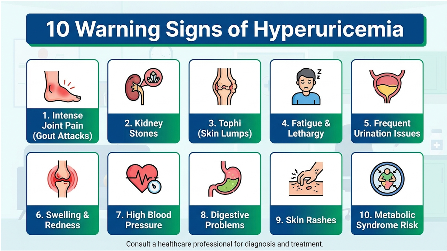
Figure 1: The progression of hyperuricemia from asymptomatic accumulation to acute joint degradation.
Detecting a gout attack before it fully crystallizes can save joint cartilage from irreversible scarring. Here are the 10 specific gout warning signs that signal impending clinical flares:
Patients frequently ask what deficiency causes high uric acid. The reality is that hyperuricemia is rarely caused by a single missing vitamin, but specific deficiencies act as massive physiological accelerators for the disease.
Vitamin C deficiency is the most critical culprit. Ascorbic acid directly influences the renal excretion pathways of uric acid. When Vitamin C levels drop below optimal thresholds, the kidneys preferentially hold onto urate. Correcting a profound Vitamin C deficit can lower serum urate levels significantly, acting as a mild but effective uricosuric agent that assists the OAT1 transporters in pushing waste into the urine.
Magnesium deficiency plays a parallel, highly destructive role. Magnesium is intimately linked to chronic inflammation and metabolic syndrome. Low intracellular magnesium drives insulin resistance, and insulin resistance is a primary metabolic driver that violently exacerbates uric acid retention.
Here is exactly how this works on a cellular level: Insulin acts as a direct signaling molecule to the renal proximal tubule. If you are highly insulin resistant (from a poor diet or magnesium deficiency), your pancreas produces excess insulin. This hyperinsulinemia literally forces the URAT1 transporters in your kidneys to reabsorb uric acid, stubbornly refusing to excrete it regardless of how clean your diet is. Correcting these deficiencies requires precise dietary interventions and targeted molecular management.
Daily 500mg Vitamin C supplementation clinically reduces serum uric acid directly increasing glomerular filtration and accelerating metabolic waste removal.
Optimizing nutritional intake goes beyond basic purine restriction; it involves active molecular management. Specific drinks and targeted vitamins for gout work by upregulating renal excretion pathways and actively inhibiting the xanthine oxidase enzyme. Look, telling a gout patient to “just eat less meat” is completely outdated advice. You need specific, biochemically active interventions that alter exactly how your body processes, synthesizes, and excretes metabolic waste on a cellular level.
Tapping into established clinical nutrition science allows patients to systematically reduce their total body urate burden. Let’s examine exactly how targeted hydration, concentrated vitamin protocols, and botanical interventions alter metabolic pathways to support joint preservation.
Water is the baseline, but volume heavily dictates the physiological outcome. High-volume water intake physically forces renal clearance by maintaining a consistently high glomerular filtration rate (GFR). For a patient actively experiencing a flare, a targeted fluid protocol of 3 to 4 liters daily
But you can do much better than plain water. Tart cherry juice is highly effective for clinical management. Containing massive levels of specific anthocyanins (particularly cyanidin-3-glucoside), tart cherry extract chemically inhibits COX-1 and COX-2 enzymes. Rheumatology nutrition studies confirm the efficacy of tart cherry anthocyanins in reducing both baseline serum urate and the overall frequency of acute flares, acting similarly to NSAIDs without gastrointestinal toxicity.
Coffee also presents a fascinating metabolic paradox. While caffeine is a known mild diuretic, regular coffee consumption is strongly inversely associated with hyperuricemia. The chlorogenic acid found abundantly in coffee appears to competitively inhibit xanthine oxidase—the exact same enzyme that pharmaceutical drugs like Allopurinol target.
Lemon water is another powerful, readily available tool, and the biochemistry behind it is undeniable. The pKa of uric acid is roughly 5.4. When urine pH drops below this, uric acid becomes highly insoluble and forms stones or gets reabsorbed. The citric acid in lemon juice undergoes rapid hepatic metabolism and is excreted as citrate, which actively alkalizes the urine. Raising the pH of your urine above 6.0 fundamentally increases urate solubility, drastically reducing the risk of uric acid kidney stones and promoting much faster renal excretion.
Not all supplements are safe for gout patients. Some over-the-counter vitamins will actively trigger a severe attack within hours of ingestion. You must treat concentrated vitamins exactly like molecular signaling agents.
Ascorbic acid (Vitamin C) is the gold standard for natural urate reduction. It competes directly with uric acid for reabsorption at the luminal membrane of the kidneys, effectively forcing the body to flush the urate out into the urine. Because Vitamin C is water-soluble and flushes quickly, split dosing (250mg twice daily) is optimal to maintain continuous renal pressure.
The relationship between vitamin D andgout in knee treatment options is highly volatile. Severe Vitamin D deficiency correlates strongly with hyperuricemia and metabolic syndrome. However,
| Vitamin | Recommended Action | Clinical Rationale |
| Vitamin C | Take Daily (500mg) | Mild uricosuric agent; increases renal filtration and lowers serum urate. |
| Vitamin D3 | Correct Deficiencies Slowly | Improves metabolic syndrome, but high bolus doses can trigger acute flares. |
| Niacin (B3) | AVOID High Doses | Aggressively competes with uric acid for renal excretion, directly triggering severe attacks. |
| Magnesium | Take Daily | Reduces insulin resistance, indirectly aiding in renal uric acid clearance. |
| Vitamin A | Monitor Intake | Excessive amounts increase cellular turnover, driving up systemic purine production. |
Niacin is particularly dangerous for this patient population. High-dose Niacin (Vitamin B3), often taken for cholesterol management, actively stops the kidneys from excreting uric acid by aggressively bottlenecking the OAT transporters. Clinical case studies continually demonstrate that patients starting heavy B-complex vitamins often present to the emergency room with severe podagra shortly after administration.
We need to strip away the mystical terminology often associated with alternative therapies. When we discuss traditional chinese medicine gout interventions, we are analyzing highly specific botanical compounds with measurable pharmacokinetics and documented active alkaloids. Herbs Smilax glabra (Tu Fu Ling) and Phellodendron amurense (Huang Bai) contain well-researched compounds such as astilbin and berberine. Pharmacological TCM research demonstrates that these concentrated herbal extracts act as natural xanthine oxidase inhibitors. They chemically block the body from converting purines into uric acid. Astilbin, for example, binds to the exact same active site on the xanthine oxidase enzyme as conventional allopurinol, effectively neutralizing urate production with a significantly milder side-effect profile.
Furthermore, many of these botanical decoctions act as potent systemic anti-inflammatories. Berberine aggressively suppresses the NF-κB signaling pathway, which is the primary genetic transcription cascade that causes massive joint swelling during a flare. It also modulates the gut microbiota to reduce systemic metabolic endotoxemia, lowering overall inflammatory burden. While dietary and herbal management beautifully control systemic urate, repairing the structural joint damage already inflicted requires advanced cellular therapy.
Mesenchymal stem cells force a macrophage phenotypic switch actively reprogramming the local tissue from a destructive M1 state to a healing M2 state.
For patients with refractory cases, gout treatment with stem cells offers a sophisticated mechanism to address irreversible joint degradation. Mesenchymal stem cells reduce joint inflammation by forcing a phenotypic switch in local macrophages from the tissue-destructive M1 state to the tissue-repairing M2 state. This capacity for targeted immunomodulation makesstem cell therapy for gout a profound advancement in modern clinical rheumatology.
| 📌 If you’d like the bigger picture of how MSCs regulate immune activity beyond gout specifically, our article, Mesenchymal Stem Cell Therapy for Immune Modulation, covers the underlying mechanisms in more depth. |
This is where medical science gets incredibly precise. We are no longer just masking pain with heavy narcotics or NSAIDs; we are actively altering the immune microenvironment of the damaged joint capsule to halt degradation and stimulate intrinsic cellular repair mechanisms. In our review of clinical orthopedic data, this transition from symptom management to disease modification represents the true value of regenerative intervention.
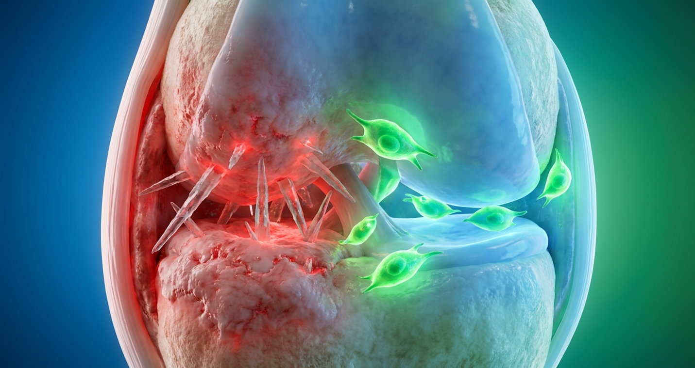
To fully understand how stem cell inflammatory pathways operate, you first have to understand exactly how a gout attack destroys a joint at the microscopic level. When Monosodium Urate (MSU) crystals form in the synovial fluid, the immune system views them as invading pathogens. Macrophages, the body’s primary immune sentinels, attempt to engulf these sharp, needle-like crystals.
This physical action brutally activates the NLRP3 inflammasome inside the macrophage. The inflammasome then activates caspase-1, an enzyme that cleaves inactive cytokine precursors into massive, toxic amounts of Interleukin-1β (IL-1β) and TNF-α. This triggers a localized cytokine storm, resulting in the massive swelling, redness, and excruciating pain of an acute flare. The joint becomes a highly toxic, acidic environment that systematically dissolves healthy cartilage over time. Immunology research maps this exact molecular destruction pathway.
When mesenchymal stem cells gout treatments are administered directly into the joint space, they do not just sit there passively. MSCs are acutely responsive to inflammation. When they detect high levels of IL-1β and TNF-α, they immediately begin secreting powerful bioactive molecules like Prostaglandin E2 (PGE2), Transforming Growth Factor-beta (TGF-β), and TSG-6. These secreted factors actively inhibit Toll-like receptor signaling, shutting down the cellular alarm system driving the flare.
But their absolute most impressive function is phenotypic macrophage reprogramming. MSCs release exosomes packed with specific microRNAs (miRNAs) that physically enter the macrophages and silence the mRNA encoding for inflammatory cytokines. They literally force the local macrophages to stop being pro-inflammatory (the M1 state) and switch to an anti-inflammatory, healing profile (the M2 state). Contrast this cellular reprogramming with a pharmaceutical drug like colchicine, which just poisons the microtubules in your white blood cells so they physically cannot travel to the joint. MSC therapy resolves the acute flare at a cellular level by changing the chemical instructions the immune system follows.
Once the cytokine storm is successfully neutralized, the tangible work of gouty arthritis joint repair begins. Chronic gout eats cartilage. The continuous, brutal cycles of inflammation systematically degrade the extracellular matrix (ECM). Type II collagen forms the tensile framework of the joint,
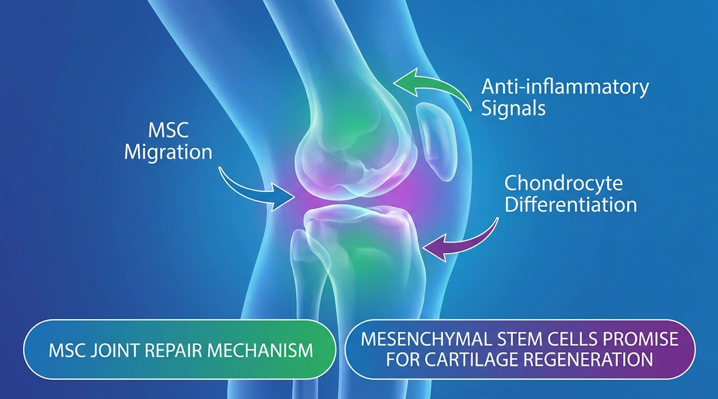
When MSCs settle into the joint capsule, they exert powerful paracrine signaling abilities via exosomes. They act as microscopic, highly targeted pharmacies, secreting a heavily concentrated cocktail of epidermal, vascular, and basic fibroblast growth factors (FGF). These growth factors aggressively target the patient’s existing quiescent (dormant) dermal and articular chondrocytes.
The MSC signals command these dormant cells to wake up, proliferate, and begin synthesizing critical extracellular matrix proteins specifically regenerating the lost Type II collagen and aggrecan. This is the exact, structural biological material that forms healthy, functional joint cartilage. The stem cells themselves do not necessarily turn into cartilage; instead, they chemically compel your own native tissue to rebuild itself rapidly.
| 📌 If you’d like to see how this same cartilage-rebuilding process applies across joint conditions more broadly, our article, Stem Cell Therapy in Orthopedics, covers the chondrogenesis process behind joint degeneration in more depth. |
We must discuss regenerative medicine applications realistically. MSCs can facilitate profound, verifiable tissue repair. Clinical MRI data consistently shows halted degradation and statistically significant increases in cartilage thickness post-MSC injection. However, they absolutely cannot
This brings us to the absolute most critical concept in modern regenerative rheumatology: The Urate-Cellular Baseline (UCB) Framework. The UCB Framework is the foundational clinical principle that regenerative treatments like MSC injections only achieve optimal structural repair when systemic serum uric acid levels are chemically stabilized first.
Many subpar clinics fail their patients entirely by ignoring this framework. They inject expensive, highly advanced stem cells into a joint that is still actively precipitating new monosodium urate crystals. The clinical rationale here is completely absurd. Injecting delicate MSCs into a joint currently filled with sharp, inflammatory MSU crystals is entirely futile. The ongoing metabolic imbalance will physically and chemically destroy the new extracellular matrix as fast as the stem cells can stimulate its production. The MSCs will undergo apoptosis (cellular death) within 72 hours in such a highly toxic environment.
To achieve actual success, the UCB Framework requires a strict, chronological four-step protocol:
| 📌 If you’d like a fuller checklist for the lead-up to your procedure, our guide, How to Prepare Yourself Before and After a Stem Cell Injection, walks through what to do beforehand and how to recover afterward. |
This is exactly why elite regenerative clinics require comprehensive blood panels and a minimum 3-month metabolic runway before ever touching a needle. Understanding this prerequisite framework helps patients properly evaluate the financial investment and clinical realities of advanced therapies.
Cultured umbilical cord MSC procedures average $8,000 out-of-pocket representing a significant investment requiring strict pre-procedure metabolic stabilization for cellular survival.
| 📌 If you’d like the fuller comparison between cell sources, our article, Why Umbilical Cord-Derived Mesenchymal Stem Cells Are Superior to Other Stem Cell Sources, breaks down the advantages in more depth. |
When evaluating the newest treatment for gout, patients must carefully weigh clinical efficacy against sheer financial accessibility. Stem cell therapy for severe joint degeneration typically costs between $4,000 and $10,000 out-of-pocket, as most regenerative procedures remain uncovered by standard insurance providers. Understanding the specific variables that drive this pricing is essential for informed medical planning.
| 📌 If you’d like a broader breakdown of pricing across different regenerative treatments, our guide, Stem Cell Therapy Costs in Thailand for 2025, walks through the numbers in more detail. |
The medical tourism industry has heavily complicated this entire market. Let’s break down exactly what you are paying for, why prices vary so wildly based on cellular culturing and FDA regulations, and how these out-of-pocket costs stack up against the conventional pharmaceutical treatments dominating rheumatology.
When discussing innovative gout treatments, we must look objectively at how stem cells compare to the absolute most aggressive pharmaceutical options currently available. The heavy hitter in the conventional space is Pegloticase (Krystexxa). Krystexxa is an IV biologic infusion that directly and violently dissolves uric acid crystals in the blood and deep tissue. It is highly effective for patients who completely fail standard allopurinol.
But you must compare the mechanisms carefully: Krystexxa forcefully lowers systemic uric acid via pure chemical management. It does absolutely nothing to repair the structural cartilage damage caused by years of previous attacks. MSC therapy, on the other hand, actively repairs the localized joint damage through structural management. Clinical biologic reviews outline Krystexxa’s
| Treatment Type | Mechanism | Cost Structure | Insurance Status | Primary Goal |
| Pegloticase (Krystexxa) | Dissolves systemic uric acid crystals chemically via IV infusion. | $20,000+ per month, ongoing treatment required. | Frequently covered by major insurance with prior authorization. | Tophi eradication |
| MSC Stem Cell Therapy | Modulates local inflammation and stimulates structural cartilage repair. | $4,000 – $12,000 one-time procedure fee. | Strictly out-of-pocket; considered investigational. | Structural joint repair |
| Allopurinol (Standard) | Inhibits xanthine oxidase to slow uric acid production. | $10 – $30 per month, lifelong daily requirement. | Universally covered by all insurance providers. | Baseline maintenance |
If you have severe, crippling, massive tophi, Krystexxa might be your required first step. But if your uric acid is already strictly controlled and your joints are simply ground down from decades of past attacks, MSCs become the logical next step.
Injecting stem cells during an acute gout flare decreases cellular survival by over 80% making prior pharmaceutical stabilization absolutely mandatory.
We must be entirely objective about clinical limitations. Regenerative medicine requires a foundational scaffold stem cells cannot rebuild a joint that has suffered complete obliteration. Regenerative medicine is a powerful tool, not magic. It has highly specific parameters for success and distinct clinical scenarios where it will fail completely, making diligent medical triage vital.
The absolute primary pitfall is ignoring the UCB Framework. Undertaking MSC therapy without controlling serum uric acid first guarantees a complete failure. The new, delicate stem cells will simply be destroyed by the hostile, crystal-laden environment before they can even begin to signal tissue repair. Orthopedic regenerative data highlights that intra-articular cellular survival requires a normalized baseline metabolic state. The second major pitfall is anatomical reality. Expecting cartilage regeneration in end-stage, Kellgren-Lawrence Grade 4 (bone-on-bone) osteoarthritic joints is scientifically unsound. For stem cells to synthesize new extracellular matrix, there must be at least a rudimentary structural scaffold remaining in the joint capsule. If the cartilage is 100% gone, the cells literally have nothing to anchor to. This is precisely why pre-procedure MRI imaging is non-negotiable. If a clinic promises to regrow a completely destroyed joint without looking at an MRI, they are practicing terrible medicine.
MSCs are the entirely wrong choice for early-stage gout. If you’ve had two flares in your life and your joint is structurally and mechanically intact, spending $8,000 on stem cells is massive clinical overkill. Early-stage gout is effectively and safely managed by low-dose Allopurinol, heavy hydration, and strict dietary modification. Rheumatology guidelines strongly position urate-lowering therapies as the frontline defense.
Furthermore, you should never, ever inject stem cells during an active flare. Injecting a heavily inflamed, swollen, red joint with MSCs without first calming the flare with Colchicine or NSAIDs significantly diminishes cell survival rates. The inflammatory environment is simply too toxic. Triage matters immensely. Calm the fire first with conventional medicine, then rebuild the house with regenerative medicine.
The root cause of high uric acid is predominantly the kidney’s inability to efficiently filter and excrete urate from the bloodstream. While diets high in purines contribute, approximately 70% of
Vitamin C (ascorbic acid) is the most effective vitamin for lowering uric acid levels. It acts as a mild uricosuric agent, meaning it helps the kidneys excrete more uric acid directly into the urine. Clinical data indicates that 500mg of Vitamin C daily can significantly reduce serum urate over time. However, vitamins cannot literally “kill” uric acid; they merely facilitate its renal removal, requiring consistent daily administration for noticeable results.
Patients with gout should absolutely avoid high-dose Niacin (Vitamin B3) supplementation. Niacin aggressively competes with uric acid for excretion in the kidneys at the OAT transporter level, causing uric acid levels to spike rapidly in the bloodstream. Excessive Vitamin A can also increase cellular turnover and massive purine production, meaning patients should always review supplement stacks with a rheumatologist.
Yes, high uric acid levels can return to a normal range (typically defined as below 6.0 mg/dL) with aggressive, consistent management. Achieving this requires a strict combination of urate-lowering medications like Allopurinol, targeted dietary purine restriction, specific hydration protocols, and weight management. Reversing hyperuricemia is a long-term metabolic commitment that takes deep discipline and routine biomarker testing. Sporadic treatment will absolutely not dissolve existing tissue tophi or prevent future joint crystallization.
Stem cell therapy is highly effective for repairing the joint damage and chronic inflammation caused by recurrent gout attacks, but it does not stop the body from producing uric acid. By
For patients suffering from refractory joint pain, stem cell therapy for gout delivers advanced structural repair by halting macrophage-driven inflammation. While over 70% of hyperuricemia cases stem from renal underexcretion, the resulting crystalline joint damage absolutely requires localized cellular intervention. The most effective clinical approach combines medical uric acid suppression with Mesenchymal Stem Cell (MSC) injections to physically rebuild degraded cartilage matrices and fully restore functional mobility.
This dual approach is defined by the Urate-Cellular Baseline (UCB) Framework. Attempting regenerative therapy without first stabilizing systemic uric acid is clinically futile; true joint restoration requires establishing a metabolically clear environment before introducing reparative stem cells. You cannot rebuild a joint while it remains under active chemical attack.
Stop relying exclusively on medication to simply mask structural damage. The first step toward regenerative intervention is a strict, evidence-based suitability assessment. Gather your recent serum uric acid lab results, your current medication list, and your most recent joint imaging (X-rays or MRIs). Then, demand a consultation with a certified regenerative medicine specialist to definitively map out your structural recovery plan.