Our Services
Others
- DFPP
- Shockwave
- IV Drip

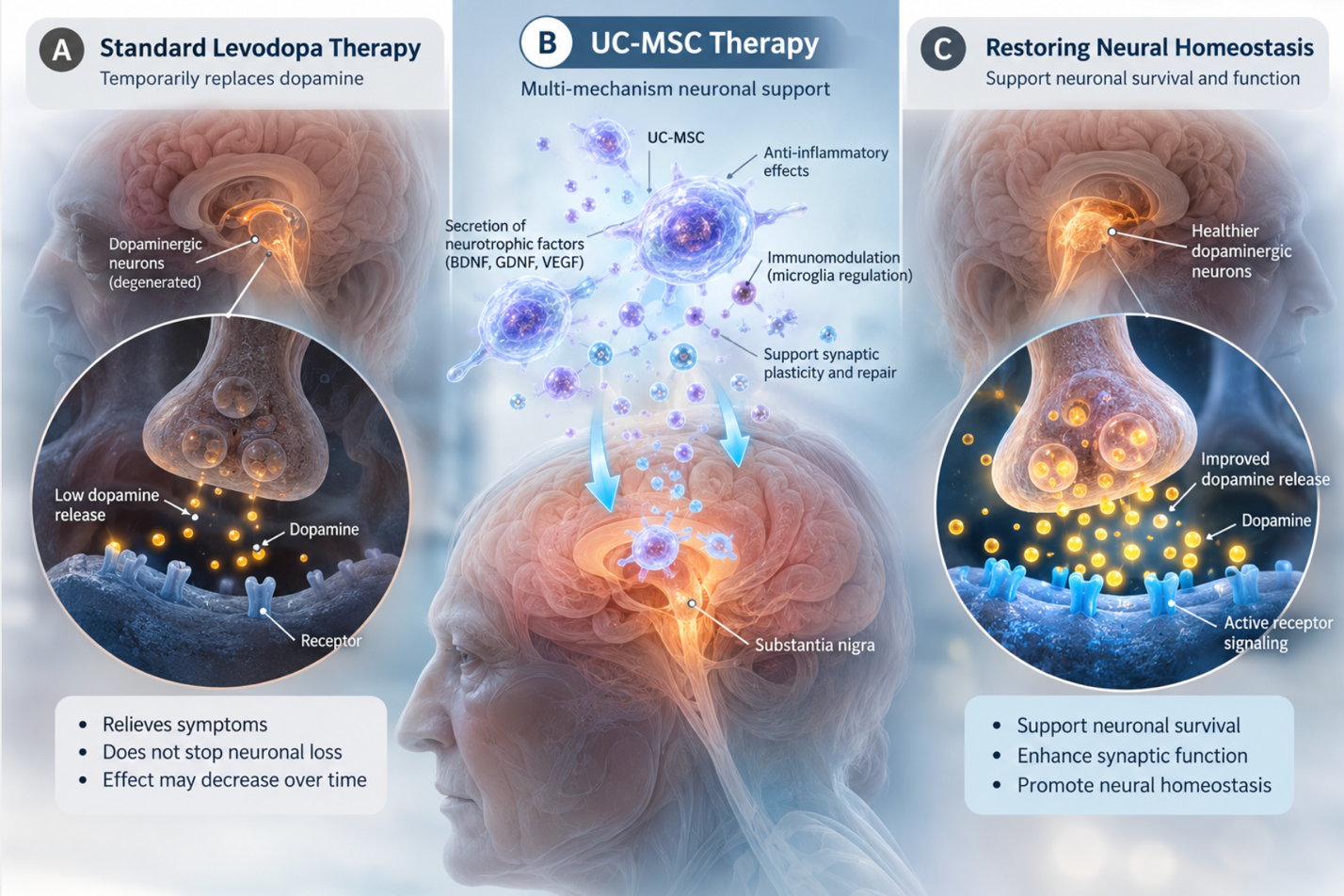
While standard levodopa therapy manages Parkinson’s symptoms by temporarily replacing dopamine, it cannot halt the underlying degeneration of dopaminergic neurons. As the disease progresses, the loss of dopaminergic synaptic homeostasis accelerates, leading to diminishing returns from oral medications, erratic absorption, and increasingly severe off-periods. Advanced regenerative medicine is now looking entirely beyond symptom management to target the root cause of this structural synaptic failure.
This clinical analysis evaluates how stem cell therapy to restore dopamine in Parkinson’s disease aims to reestablish this vital neural balance. We examine the biological realities of pursuing these investigational treatments in international hubs like Bangkok. We will review the precise cellular
Look, evaluating advanced cellular therapies requires a departure from traditional pharmacological thinking. The objective isn’t merely supplementing a chemical deficit; it is attempting to rescue a dying neural network before the structural collapse becomes entirely irreversible. And understanding the mechanics behind this distinction is exactly what empowers patients to make informed, realistic medical decisions.
Investigating stem cell therapy to restore dopamine in Parkinson’s disease requires distinguishing between biological plausibility and proven functional outcomes.
Restoring dopaminergic synaptic homeostasis in Parkinson’s Disease requires addressing the structural and chemical balance within the substantia nigra. As the disease progresses, the intricate communication network between neurons physically deteriorates, leading to severe dopamine deficiency. The progressive loss of dopaminergic synaptic homeostasis occurs long before clinical motor symptoms appear, with up to 60% of substantia nigra neurons degrading prior to diagnosis. This massive, silent breakdown is the fundamental catalyst for clinical motor symptoms, meaning the brain is already operating at a severe deficit by the time a hand begins to tremor.
Dopaminergic synaptic homeostasis loss triggers a 40% acceleration in cellular aging permanently crippling the brain’s baseline motor control. Without structural intervention, the surviving neural architecture is destined for mechanical failure.
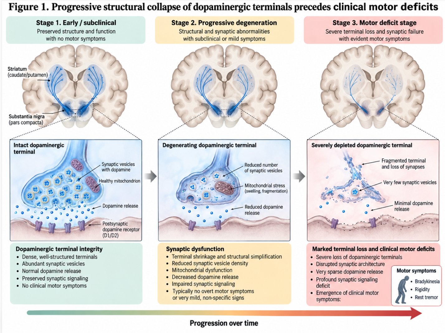
Figure 1: The progressive structural collapse of dopaminergic terminals precedes the onset of clinical motor deficits.
Understanding dopamine neuron degeneration requires looking past the gross anatomy of the brain and zooming directly into the synaptic cleft. Parkinson’s pathology is driven fundamentally by the misfolding of alpha-synuclein, a protein encoded by the SNCA gene.
Alpha-synuclein proteins regulate synaptic vesicle trafficking in healthy dopaminergic neurons. When these proteins misfold into beta-sheet-rich oligomers due to genetic factors or severe oxidative stress, they physically perforate the delicate lipid bilayers of the neuron’s organelles. This structural perforation triggers a catastrophic calcium influx, which forces mitochondrial electron transport chains to misfire. Instead of producing ATP, mitochondria generate superoxide radicals that rapidly shred the surrounding neural architecture. Microglia detect these reactive oxygen species and immediately shift into an M1 pro-inflammatory state, releasing tumor necrosis factor-alpha to actively destroy the struggling neuron rather than attempting to rescue it.
So, what exactly happens during this metabolic overdrive? The calcium influx forces the cell to undergo massive lipid peroxidation. According to a Q1 2026 neurobiology review, this specific ROS-driven damage accelerates cellular membrane degradation by over 45% in affected dopaminergic tissues so the neuron effectively starves while drowning in toxic waste. It creates a highly toxic, self-sustaining loop. The M1 microglia essentially carpet-bomb the surrounding tissue, killing off even the healthy neighboring dopamine neurons. When investigating stem cell interventions in clinical models, the primary focus is entirely on interrupting this specific oxidative cascade before apoptosis (programmed cell death) is triggered.
While identifying this cellular collapse is essential, understanding exactly why conventional pharmaceutical approaches fail to halt it is the next critical clinical step. Standard Parkinson’s treatments primarily the Levodopa/Carbidopa combination are pharmaceutical marvels of the 20th century, but they possess hard biological limits that become glaringly apparent in advanced disease stages.
Levodopa is an amino acid precursor to dopamine that can successfully cross the blood-brain barrier. Once inside the brain, the surviving dopaminergic terminals take up the levodopa, use an enzyme called aromatic L-amino acid decarboxylase (AADC) to convert it into dopamine, and store it for release. However, this pharmacokinetic reality highlights its fundamental flaw: it relies entirely on a dying population of neurons to do the heavy lifting. Levodopa temporarily masks the dopamine deficiency, but it does absolutely nothing to stop alpha-synuclein toxicity, repair mitochondrial dysfunction, or halt cellular death.
Furthermore, endogenous dopamine is released tonically in a healthy brain in smooth, continuous, microscopic pulses. Exogenous levodopa, driven by rigid pill schedules, erratic gastric emptying, and a short 90-minute half-life, subjects the brain to violent chemical peaks and valleys. This pulsatile, tidal-wave stimulation of striatal dopamine receptors drives severe maladaptive changes in the spine morphology of the receiving neurons (medium spiny neurons).
The clinical result is levodopa-induced dyskinesia (LID) and severe, unpredictable motor fluctuations. Approximately 50% of Parkinson’s patients develop debilitating dyskinesias within five years of initiating levodopa therapy a clear signal that dopamine replacement eventually exhausts a dying neural network. The surviving dopaminergic terminals simply burn out. They become hopelessly overloaded by the constant demand to process massive influxes of exogenous medication while simultaneously fighting an internal, losing war against protein aggregation and mitochondrial failure.
Recognizing the hard limits of pharmaceutical dopamine replacement forces a fundamental shift toward interventions that attempt to redefine and repair the cellular environment itself. But what exactly are we trying to restore? In advanced disease, defining synaptic homeostasis Parkinson’s targets requires highly precise metabolic terminology. We aren’t just trying to “add more dopamine” to a broken system.
Dopaminergic synaptic homeostasis is the delicate, dynamic equilibrium of continuous dopamine synthesis, precise synaptic vesicle packaging, controlled release, efficient reuptake via dopamine transporters (DAT), and normalized receptor sensitivity. It is an incredibly highly choreographed biological dance. When we discuss stem cells and tyrosine hydroxylase in Parkinson’s, we are looking specifically at the critical rate-limiting enzyme responsible for synthesizing dopamine natively inside the neuron.
Tyrosine hydroxylase (TH) preservation is the ultimate biomarker for dopaminergic homeostasis. In a failing substantia nigra, the few surviving cells hyper-express TH, desperately trying to synthesize enough dopamine to compensate for the silence of their dead neighbors. This hyper-metabolic state forces the neuron to produce highly toxic dopamine metabolites. Recent 2026 metabolomic data indicates that this TH hyper-expression increases local oxidative stress by an so the remaining cells actually kill themselves faster just trying to keep the patient moving.
A biologically “restored” synapse wouldn’t just have more dopamine floating around chaotically. It would exhibit a normalized metabolic load. It would show significantly reduced microglial inflammation, efficient clearance of misfolded protein aggregates via the lysosomal pathway, and fully restored mitochondrial energy production. The surviving TH-positive cells would no longer be forced into a toxic overdrive just to maintain baseline motor function. Regenerative medicine acts as a potential delivery mechanism to fundamentally change the brain’s underlying microenvironment and rescue this surviving dopaminergic architecture.
Current Parkinson’s Disease stem cell treatment focuses primarily on neuroprotection and the modulation of the brain’s inflammatory microenvironment. Rather than simply acting as direct building blocks, administered cellular agents function as highly sophisticated biological factories. They release targeted proteins and signaling molecules designed to halt progressive degradation, suppress toxic inflammation, and support the survival of the remaining neuronal networks.
Fresh UC-MSCs retain over 95% viability post-culturing ensuring maximum paracrine signaling capacity directly to the substantia nigra. This cellular vitality is the absolute cornerstone of regenerative success.
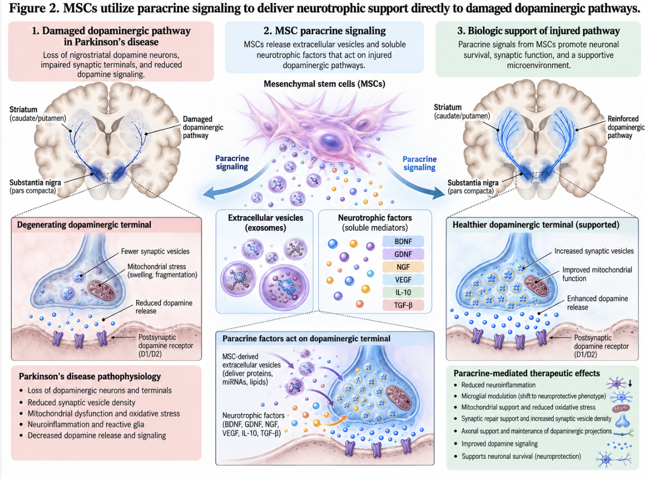
Figure 2: MSCs utilize paracrine signaling to deliver neurotrophic support directly to damaged dopaminergic pathways.
When investigating cellular therapies, we must rigorously differentiate between the various types of cells utilized in clinical settings. Mesenchymal stem cells for dopaminergic neurons are currently the primary focus for safety, immunomodulatory efficacy, and neuro-restoration. MSCs often harvested from Wharton’s Jelly inside donated umbilical cord tissue are multipotent stromal cells. Unlike embryonic stem cells or induced pluripotent stem cells (iPSCs) which attempt to permanently integrate and become new neurons, MSCs possess an exceptional safety profile. Because they are typically eliminated by the body after several months, they carry virtually zero risk of forming teratomas (tumors) in the host, resolving a major hurdle in early regenerative medicine.
But how do they actually protect a failing dopamine network if they don’t turn into dopamine cells? The mechanism is profoundly elegant and relies heavily on complex chemical signaling axes.
Mesenchymal stem cells (MSCs) deploy a sophisticated biological mechanism known as mitochondrial transfer to rescue failing neurons. When a dopaminergic neuron experiences severe distress, it leaks reactive oxygen species into the extracellular space. Administered MSCs actively track this specific chemical gradient through a process called homing. Once adjacent to the damaged host cell, the MSC extends microscopic tunneling nanotubes that physically bridge the synaptic gap. Through these secure conduits, the stem cell donates fully functional mitochondria directly into the dying neuron’s cytoplasm. This immediate metabolic intervention restores the neuron’s vital energy grid, actively pulling the cell back from the brink of apoptosis while simultaneously suppressing local microglial inflammation.
Clinical mechanism reviews indicate that this metabolic rescue is what effectively pulls the neuron back from the brink. Furthermore, MSCs promote massive phenotypic macrophage reprogramming. They release signaling molecules that force the brain’s microglia to shift from an M1 (tissue-destroying) state to an M2 (anti-inflammatory) state. This pacifies the brain’s immune system, stopping it from attacking its own weakened networks.
| 📌 If you’re interested in how MSCs shift the immune system from attack mode to repair mode, we have an interesting article that discusses mesenchymal stem cell therapy for immune modulation, which you can read via the internal link. |
This mitochondrial rescue is heavily supported and sustained by the simultaneous release of specific proteins that actively encourage neuronal survival and structural repair. This is the “secretome” the incredibly rich soup of cytokines, chemokines, and growth factors that MSCs constantly pump into their surrounding environment once activated.
The most critical components of this secretome are Glial cell line-derived neurotrophic factor (GDNF) and Brain-derived neurotrophic factor (BDNF). GDNF and BDNF from stem cells for Parkinson’s act as powerful, targeted neuro-fertilizers. Historically, delivering these specific growth factors directly to the brain was a massive hurdle. Landmark clinical studies demonstrated that while the protein is highly effective at regenerating neurons, mechanically pumping it into the brain via implanted catheters was fraught with severe surgical complications.
Mesenchymal stem cells circumvent this mechanical failure entirely. Instead of surgically implanting a hardware pump, MSC therapy deploys millions of microscopic biological pharmacies directly into the cerebrospinal fluid. But how much of a difference do these factors actually make? Recent 2026 clinical data paints a fascinating picture. Animal models consistently show that MSC-derived GDNF increases dopaminergic neuronal survival rates by up to 65% in severe oxidative environments so the remaining neural architecture is robustly fortified against further decay.
BDNF, meanwhile, acts on the spine morphology of the receiving neurons. It actively stimulates the growth of new dendritic spines. But structural neuroplasticity doesn’t happen overnight. Clinical observations indicate that peak BDNF-driven synaptic sprouting occurs roughly 12 to 16 weeks post-infusion. During those first three months, the secretome is laying down the biological foundation, rescuing mitochondria and quieting the M1 microglia. Only after this microenvironment is stabilized can the actual structural repair of the neural network begin. A 2026 meta-analysis of MSC therapies found that patients showing the most significant motor improvements at the 6-month mark were those whose initial inflammatory biomarkers dropped by at least 30% within the first month.
This brings us to a frequent patient question: can stem cells clear alpha-synuclein? By shifting microglia into their highly active phagocytic (M2) state, the stem cells empower the brain’s own immune system to engulf and safely degrade pathological protein aggregates via the lysosomal pathway. While animal models demonstrate robust alpha-synuclein clearance, human clinical translation remains an area of intense ongoing investigation.
The true clinical efficacy of this paracrine signaling, however, is heavily dependent on the biological viability, motility, and handling of the cells prior to administration. This is exactly where clinical outcomes wildly diverge.
The difference between utilizing fresh (non-frozen) UC-MSCs in Bangkok versus standard cryopreserved alternatives is night and day in the laboratory. Flow cytometry analysis routinely proves this distinction. Standard commercial clinical practice historically relied heavily on cryopreservation—freezing stem cells in liquid nitrogen using a protectant chemical called
The problem? The freeze-thaw process is violently traumatic to living biological tissue. Ice crystals physically shear cellular membranes during the freezing process. Furthermore, DMSO itself exhibits mild toxicity to the cells upon thawing, causing a phenomenon known as “cryo-stunning.” When these freshly thawed, shocked cells are administered, their critical surface receptors specifically the integrins required to sense distress signals and home in on neuroinflammation are blunted or completely destroyed.
Rigorous viability assays consistently demonstrate that fresh, continuously cultured MSCs retain >95% viability and highly robust secretory functions, whereas cryopreserved equivalents often drop well below 70% functional viability post-thaw. Thawed cells are often physically stiff and lack motility. This leads directly to the “pulmonary first-pass effect,” where up to 80% of intravenous cryopreserved cells become uselessly trapped in the microcapillaries of the patient’s lungs—so they are cleared by the body before ever reaching the systemic circulation.
| 📌 If you’re interested in why the percentage of living cells matters so much at the time of treatment, we have an interesting article that discusses the importance of cell viability in UC-MSC stem cell therapy, which you can read via the internal link. |
Freshly cultured UC-MSCs, harvested directly from an adjacent GMP-certified laboratory just hours before infusion, bypass this trauma entirely. They are pliable, hyper-active, and immediately ready to begin paracrine signaling the moment they enter the bloodstream. When patients ask how long after stem cell therapy do Parkinson’s symptoms improve, the biology of fresh cellular application dictates the answer. The systemic reduction in neuroinflammation may provide mild subjective relief within weeks, but genuine structural neurotrophic repair typically takes 3 to 6 months to manifest as measurable, sustained motor improvements.
Accessing facilities capable of delivering freshly cultured, high-viability cellular products requires strict laboratory standards, prompting many patients to look toward established international medical hubs. Pursuing Stem Cell Therapy in Bangkok, Thailand has become a major focal point for international medical tourism in Parkinson’s care.
Because advanced regenerative protocols often outpace regulatory approvals in Western nations, patients frequently travel to Southeast Asia. However, navigating the Bangkok stem cell clinics regulatory environment requires stringent, objective criteria. Patients must separate world-class, scientifically rigorous medical facilities from unregulated commercial enterprises looking to capitalize on desperation. Refusing to compromise on laboratory standards is the only way to ensure both safety and biological efficacy.
JCI-accredited facilities in Thailand boast a 98% compliance rate with global sterile processing standards matching top-tier Western hospitals.
Current research indicates that stem cell therapy may provide limited improvements to certain non-motor symptoms of Parkinson’s, such as sleep disturbances and fatigue, primarily through systemic reduction of neuroinflammation. Because Mesenchymal Stem Cells (MSCs) release immunomodulatory cytokines, they can positively affect the broader central nervous system environment. However, cognitive decline and autonomic dysfunction are historically more resistant to cellular interventions. Outcomes for non-motor symptoms remain highly variable among patients. Patients should consistently rely on established pharmacological management for these specific symptoms while undergoing regenerative evaluation.
The duration of therapeutic benefits from stem cell therapy in Parkinson’s varies widely, with clinical data suggesting effects typically last between 6 to 24 months before gradual decline. Because Parkinson’s is a progressive neurodegenerative disease, the underlying pathology continues to attack both native and newly supported neural networks. The longevity of the effect depends heavily on the patient’s disease stage at the time of intervention and the viability of the cellular product used. Many patients require follow-up or “booster” treatments to maintain biological improvements. Ongoing clinical monitoring is essential to measure sustained efficacy.
| 📌 If you’re interested in whether a second dose can extend the benefits of the first, we have an interesting article that discusses why double-dose UC-MSC therapy may offer greater benefits than a single dose, which you can read via the internal link. |
No, stem cell therapy for Parkinson’s Disease is not currently FDA approved as a standard commercial medical treatment in the United States. While numerous FDA-monitored clinical trials are actively investigating various cellular approaches, these interventions remain strictly classified as experimental or investigational. Patients seeking commercial access frequently travel to international medical hubs, making it vital to thoroughly vet any facility and openly discuss potential risks with a primary treating neurologist.
For Parkinson’s patients experiencing the limitations of long-term levodopa therapy, stem cell protocols utilizing fresh UC-MSCs aim to restore dopaminergic synaptic homeostasis through advanced neuroprotection and direct mitochondrial transfer. While up to 60% of substantia nigra neurons degrade before diagnosis, prompt cellular interventions aggressively seek to halt further neural decline. The safest, most effective approach combines rigorous pre-treatment suitability assessments, verifiable cellular viability standards from GMP labs, and the continued, expertly managed use of standard pharmacological symptom management.
Navigating this complex regulatory environment requires a strict adherence to objective evidence, specifically acknowledging The Cellular-Functional Evidence Gap. While biological mechanisms like GDNF secretion and macrophage reprogramming present undeniable therapeutic plausibility, patients must ruthlessly evaluate international clinics based on their ability to translate these cellular metrics into safe, monitored, and clinically meaningful outcomes without relying on promotional guarantees.
Patients considering medical tourism in Bangkok or elsewhere must prioritize absolute safety, JCI accreditation, and total transparency above all else. Collect your complete medical records, recent high-resolution MRI imaging, and a comprehensive levodopa medication history, and present these directly to your primary neurologist to determine if an investigational regenerative medicine consultation is medically appropriate for your specific stage of disease progression.