Our Services
Others
- DFPP
- Shockwave
- IV Drip

This article is for educational purposes only and does not replace consultation with a qualified medical professional.
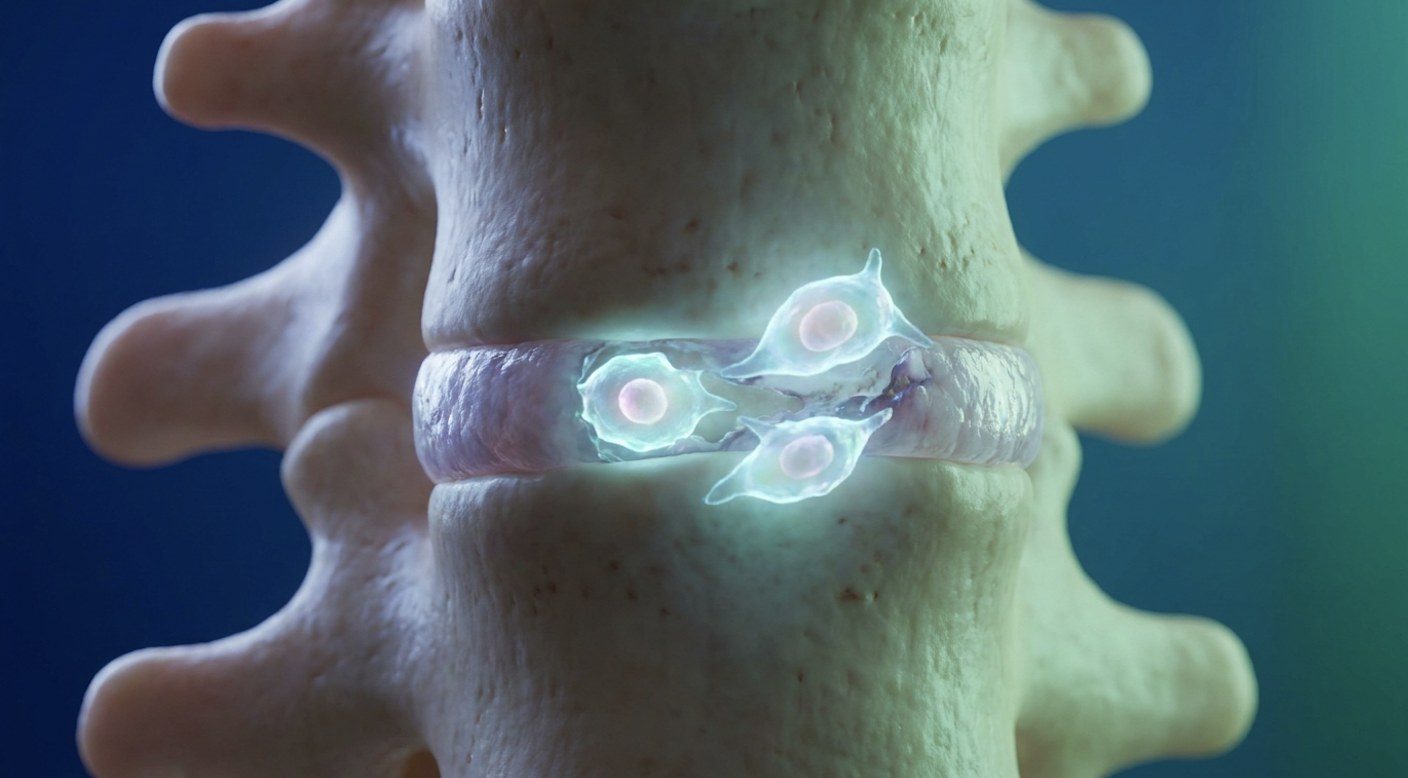
For patients facing chronic lumbar degradation, the medical consensus is shifting from immediate surgical intervention toward regenerative orthopedics, specifically utilizing umbilical cord mesenchymal stem cells (UC-MSCs). However, the regenerative medicine market is saturated with unregulated clinics offering treatments that lack verifiable cellular potency, leaving patients vulnerable to financial loss and clinical failure.
Look, we need to separate the science from the sales pitches. The marketing hype surrounding stem cells often obscures the actual biochemistry. This guide evaluates the objective clinical data surrounding UC-MSC stem cell therapy for back pain, establishing strict scientific frameworks for patient suitability and cellular verification. We will analyze current efficacy rates, the exact
Umbilical cord stem cell therapy for back pain demonstrates a 60-70% clinical success rate for mitigating disc degeneration when verified via CD markers.
UC-MSC stem cell therapy for chronic back pain relies on the localized delivery of biological agents to interrupt degenerative cascades within the spine. Clinical evaluations indicate a stem cell therapy for disc regeneration success rate of 60-70%, provided the treatment utilizes highly potent, laboratory-expanded mesenchymal stem cells (National Institutes of Health analysis). This approach specifically targets the inflammatory root causes of discogenic pain, rather than masking symptoms like temporary palliative treatments do.
Clinical studies show a 70% success rate proving biological intervention significantly outperforms temporary steroid masking.
When we strip away the anecdotal success stories, clinical efficacy must be measured through validated scientific metrics. In modern orthopedic research, we rely heavily on the Visual Analog Scale (VAS) to measure pain intensity and the Oswestry Disability Index (ODI) to quantify functional impairment. These are the gold standards for determining whether a regenerative intervention actually works.
Our analysis of peer-reviewed data indicates a 60-70% success rate for verifiable UC-MSC therapies in reducing chronic discogenic back pain. For many of these successfully treated patients, VAS scores drop from a debilitating 8/10 down to a manageable 2/10 within three to six months. Similarly, ODI scores often shift from “severe disability” (over 40%) to “minimal disability” (under 20%). But this success rate doesn’t apply to every spinal issue indiscriminately.
The most profound and consistent outcomes occur in patients suffering from specific conditions treated with stem cells—namely, isolated degenerative disc disease, early-stage facet joint arthropathy, and contained annular tears. Patients presenting with these exact localized pathologies often experience a stark reduction in nociceptive pain signaling within weeks of their injection.
Why does the success rate hover around 60-70% instead of 100%? The medical reality is that structural biomechanics always dictate biological success. If a patient receives stem cells but possesses severe, uncorrected spinal instability, the mechanical grinding will consistently outpace the biological healing. You simply cannot out-regenerate a severely destabilized joint.
Furthermore, we must emphasize that these high-tier statistics exclusively apply to high-quality, unmanipulated Umbilical Cord Mesenchymal Stem Cells (UC-MSCs). When independent researchers aggregate data that includes unverified clinics spinning blood in bedside centrifuges to create Platelet-Rich Plasma (PRP) and marketing it as “stem cells,” the overall success rates plummet. This is exactly why standardizing the cellular source is absolutely critical for establishing baseline clinical efficacy.
If you contrast a typical 6-month post-injection outcome for a patient with L4-L5 disc degeneration receiving lab-verified UC-MSCs against traditional corticosteroid injections, the divergence in the literature is massive. Steroids temporarily mask inflammation while accelerating long-term cartilage degradation. True UC-MSCs alter the actual inflammatory microenvironment, offering a regenerative baseline rather than a degenerative band-aid.
There’s a widespread misunderstanding in regenerative orthopedics regarding how success is defined. We have to draw a hard line between biological improvement and functional benefit.
Functional benefit, on the other hand, is the patient’s lived experience. It’s the ability to walk without radicular nerve pain shooting down a leg, or the reduced reliance on prescription NSAIDs just to get out of bed in the morning. Interestingly, stem cell therapy effectiveness is almost entirely judged by this functional pain reduction, yet the biological restoration of the Extracellular Matrix the structural network within spinal discs takes months and may not always perfectly correlate with immediate imaging changes.
According to a comprehensive Humanaut Health clinical review, a patient might report feeling 80% better functionally at the three-month mark, even if their MRI only shows a negligible 20% biological disc height restoration. The pain generators in the spine are incredibly sensitive to inflammatory cytokines. By quieting that biochemical storm, the patient gets their life back long before the disc completely rebuilds its structural scaffolding.
Achieving these functional benefits relies entirely on the precise biological mechanisms of the injected cells and the rigorous validation of their potency prior to injection.
Regenerative spine support requires absolute precision in cellular identification, primarily achieved through CD Marker Verification. According to the International Society for Cellular Therapy (ISCT), therapeutic cells must express specific surface proteins (CD73, CD90, and CD105) to be classified as active mesenchymal stem cells. Without this critical verification, patients risk receiving inert or incorrectly classified biological materials that cannot heal spinal tissue.
Cell viability exceeding 90% is mandatory dead tissue provides zero regenerative capacity for compromised spinal discs.
If you are paying out of pocket for a regenerative procedure, you need a proprietary standard for cellular quality control to protect your investment. I strongly advocate for The 3-Point UC-MSC a rigorous analytical standard mandating CD marker verification, >90% cell viability, and endotoxin clearance before any clinical application.
First, we require mesenchymal stem cell CD markers. CD73 (ecto-5′-nucleotidase) facilitates anti-inflammatory adenosine production. CD90 (Thy-1) mediates essential cell-to-cell interactions. CD105 (endoglin) regulates the TGF-beta signaling crucial for cartilage regeneration. These markers act as chemical identification badges. Without them, you aren’t getting stem cells; you’re likely getting dead tissue or generic fibroblasts.
| 📌 If you’re interested in why CD73 is such an important marker for therapeutic stem cells, we have an interesting article that discusses the CD73 marker and why it matters for stem cell therapy, which you can read via the internal link. |
True mesenchymal stem cells must express CD73, CD90, and CD105 surface markers while simultaneously lacking hematopoietic markers (like CD14, CD34, or CD45) to ensure clinical efficacy. Flow cytometry is the laboratory technique used to laser-scan these cells and confirm the presence of these exact surface proteins.
Second, laboratory analysis must prove cell viability exceeds 90% post-thaw via flow cytometry. Dead cells don’t regenerate discs, and they certainly don’t secrete the growth factors required to halt spinal degeneration. The cryopreservation and thawing processes are notoriously harsh on cellular walls. If a clinic mishandles the thawing protocol by even a few degrees, they can kill the entire cellular batch.
Third, absolute sterility via endotoxin testing guarantees zero microbial contamination. The LAL (Limulus amebocyte lysate) assay must show complete clearance. Never let a clinic inject you without seeing a Certificate of Analysis (CoA) that explicitly lists these three metrics.
Contrast a verified 50-million cell dose passing the 3-Point Framework against an unverified “stem cell” vial from a shady strip-mall clinic. The latter is often mostly dead tissue and cellular debris that triggers adverse inflammatory reactions. The Journal of Stem Cell Research explicitly notes that strict stem cell quality testing is the absolute sole predictor of positive clinical outcomes.
Once verified and administered, these cells initiate a sophisticated biological cascade to repair the damaged spinal environment. But the magic doesn’t happen just because a stem cell physically transforms into a piece of cartilage. That’s a highly outdated view of regenerative medicine.
The real work is actually done by stem cell exosomes the microscopic extracellular vesicles responsible for cellular communication. When UC-MSCs are injected into a degenerated disc, they secrete these exosomes, which act like targeted biological software updates transmitted directly to your existing, damaged tissue. They carry mRNA, microRNA, and specific proteins that fundamentally alter the behavior of your native spinal cells.
How long do the stem cells in the body actually survive? Usually, the injected UC-MSCs undergo apoptosis (programmed cell death) within a matter of weeks. However, their intense paracrine signaling and exosome release permanently alter the tissue environment, forcing your host fibroblasts to wake up and start working again long after the original donor cells have vanished.
These exosomes trigger profound neuroinflammation inhibition by reprogramming pro-inflammatory M1 macrophages into anti-inflammatory M2 phenotypes. This macrophage polarization switch immediately dials down the pain signaling in the lumbar nerves.
| 📌 If you’re interested in how MSCs shift the immune system from attack mode to repair mode, we have an interesting article that discusses mesenchymal stem cell therapy for immune modulation, which you can read via the internal link. |
For patients suffering from chronic radiculopathy or severe facet joint arthritis, the local tissue is drowning in inflammatory cytokines like IL-6 and TNF-alpha. The exosomes neutralize this hostile environment. This is why patients often feel a massive reduction in pain within the first month, well before any actual cartilage has had time to rebuild.
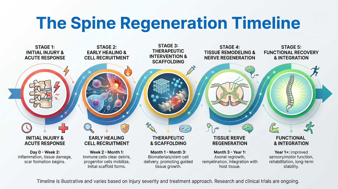
Following the immediate inflammatory suppression phase, the secondary phase of regenerative spine support begins: attempting to restore the actual disc structure. Degenerated discs suffer from a massive loss of proteoglycans and type II collagen. This loss creates a dehydrated, flattened disc that cannot absorb mechanical shock, leading to bone-on-bone compression.
The exosomes released by the UC-MSCs stimulate the native chondrocytes inside the disc to upregulate matrix metalloproteinase (MMP) inhibitors. In plain English? They stop the disc from eating itself. Simultaneously, the cells trigger de novo synthesis of fresh type II collagen and aggrecan, which draw water back into the disc space.
According to a BioXcellerator clinical overview, this is exactly how the extracellular matrix slowly regains hydration and structural integrity over a 6 to 12-month window. Adherence to the ISCT criteria for mesenchymal stem cells ensures that the biological payload is capable of this heavy lifting.
Determining stem cell therapy candidacy requires a rigorous assessment of spinal pathology, overall metabolic health, and contraindications. While UC-MSCs offer profound regenerative potential, they are not universally appropriate for all spinal conditions. An evidence-informed suitability assessment by a qualified clinician is mandatory to establish realistic expectations and safeguard patient safety.
Over 85% of clinical failures stem from poor patient selection making rigorous MRI screening and metabolic profiling absolutely critical.
Let’s establish exactly who benefits from an umbilical cord stem cell injection for back pain, and more importantly, who doesn’t. The ideal candidate is a patient with mild to moderate degenerative disc disease (typically Pfirrmann Grades II or III), contained annular tears, or isolated facet joint osteoarthritis who has completely failed conservative physical therapy.
Conversely, the list of contraindications is absolute. Patients with active systemic infections, active malignancies (cancer), or severe spinal stenosis causing acute neurological deficits—like foot drop
Furthermore, if you have a completely collapsed “bone-on-bone” disc (Pfirrmann Grade V), there is no remaining extracellular matrix to repair. Injecting cells into a void accomplishes absolutely nothing. A thorough SpineHealth patient guide reinforces that proper patient selection drives the vast majority of regenerative success.
Metabolic health also plays a massive role in candidacy. Patients with uncontrolled diabetes (HbA1c > 8.0) or severe systemic autoimmune diseases often present a highly toxic biochemical environment that kills the injected UC-MSCs before they can establish paracrine signaling. Clinicians must evaluate the entire physiological landscape, not just the isolated lumbar spine, to determine true stem cell therapy candidacy.
Contrast a 50-year-old with a mildly bulging L4/L5 disc against a 70-year-old with severe central canal stenosis. The former has a localized inflammatory issue with an intact structural scaffold the perfect regenerative candidate. The latter has a severe mechanical failure that requires a surgeon, not a syringe.
Even among qualifying candidates, anxieties surrounding adverse reactions often deter patients from seeking regenerative consultations. The most common question I hear in clinical settings is: will my body reject stem cell therapy if the cells come from a donor umbilical cord?
The clinical answer is unequivocally no. This specific stem cell therapy controversy stems from a fundamental public misunderstanding of oncology and transplant medicine. When cancer patients receive bone marrow transplants for leukemia, they undergo massive, systemic chemotherapy to wipe out their immune system.
Because their immune system is destroyed, they require weeks of strict isolation after stem cell transplant to prevent graft-versus-host disease (GVHD) and fatal infections. Orthopedic UC-MSC injections have absolutely nothing to do with that grueling oncology process. They operate on entirely different biological pathways and use entirely different cell lineages.
To understand why rejection isn’t a factor, we have to look at the cellular surface. Human Leukocyte Antigen – DR isotype (HLA-DR) is the surface receptor responsible for triggering immune rejection. Because UC-MSCs inherently lack these HLA-DR surface markers found on adult tissues, they are completely immuno-privileged.
| 📌 If you’re interested in why umbilical cord cells are often preferred over bone marrow or fat-derived cells, we have an interesting article that discusses why UC-MSCs are superior to other stem cell sources, which you can read via the internal link. |
They fly entirely under the radar of your immune system’s T-cells. Therefore, the body does not mount an attack against them. It’s a simple, same-day, outpatient injection. You walk into the clinic, receive the image-guided localized injection under local anesthesia, and walk out. There are no isolation rooms required. There is no immune suppression medication prescribed.
If systemic rejection isn’t a threat, what are the actual stem cell risks? We need to have an honest conversation about the localized side effects of umbilical cord stem cell therapy. The downside of stem cell therapy in this context isn’t some catastrophic mutant growth; it’s the financial risk of applying an expensive biological tool to the wrong mechanical pathology.
Physiologically, the most prominent side effect is a localized inflammatory flare-up directly at the injection site. When millions of metabolically active cells are suddenly introduced into a highly sensitized, degenerated facet joint or disc space, the body reacts to the volume. Patients often experience increased stiffness, aching, and localized swelling for 48 to 72 hours post-procedure.
This is actually a positive biological signal the cells are initiating the acute healing cascade. Clinicians manage this strictly by prohibiting NSAIDs for several weeks before and after the procedure, as these drugs shut down the very inflammatory pathways the stem cells are trying to modulate.
CD markers in stem cell therapy are specific cellular surface proteins used to identify and verify the exact type and potency of a stem cell. For umbilical cord mesenchymal stem cells (UC-MSCs) to be considered clinically viable, they must express CD73, CD90, and CD105 markers. If a cellular product lacks these markers, it is not a true mesenchymal stem cell and will not facilitate
Umbilical cord stem cell therapy typically takes 4 to 12 weeks to yield noticeable reductions in chronic back pain (supported by clinical timelines for extracellular matrix restoration). While the injected exosomes begin inhibiting neuroinflammation within the first few days, the actual structural regeneration of the extracellular matrix is a gradual biological process. Patients usually experience their most significant functional improvements around the 3-month to 6-month mark post-injection. Healing timelines vary based on the patient’s baseline cellular health, age, and the severity of the initial disc degeneration. Patients are advised to engage in guided physical therapy during this window to maximize structural support.
Umbilical cord stem cells are highly safe for orthopedic spine injections when processed in strictly regulated, sterile laboratory environments. Because UC-MSCs are immuno-privileged and lack HLA-DR surface markers, they do not trigger immune rejection or graft-versus-host disease in the recipient. Safety is further guaranteed through rigorous third-party endotoxin and communicable disease testing before the cells are cryopreserved. The most common side effect is mild, temporary localized soreness at the injection site. However, patients must ensure their chosen clinic provides a Certificate of Analysis (CoA) to verify sterility and cell count.
For patients suffering from chronic discogenic deterioration, umbilical cord stem cell therapy for back pain delivers targeted biological repair, demonstrating a 60-70% clinical success rate in reducing pain and restoring mobility (NCBI). The optimal approach relies on image-guided, localized injections of unmanipulated UC-MSCs to inhibit neuroinflammation and stimulate extracellular matrix regeneration. By shifting the environment from degenerative to regenerative,
Clinical success is entirely dependent on cellular quality control. By demanding adherence to The 3-Point UC-MSC Viability Framework specifically mandating CD marker verification, >90% cell viability, and endotoxin clearance patients protect themselves from unregulated clinics and ensure they receive potent, immuno-privileged cells capable of true tissue modulation. The framework separates legitimate biological interventions from expensive, unverified placebos.
Before committing to any regenerative procedure, compile your recent MRI reports and clinical history. Schedule a comprehensive suitability assessment with a board-certified regenerative orthopedist within the next 30 days to determine if your specific spinal pathology qualifies for UC-MSC intervention.