Our Services
Others
- DFPP
- Shockwave
- IV Drip

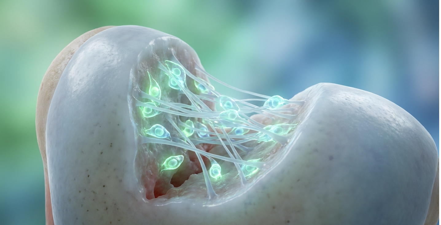
For patients navigating deep osteochondral defects, the standard surgical intervention microfracture often trades immediate defect filling for long-term structural instability. It is a frustrating clinical reality. When comparing stem cell therapy vs microfracture, you aren’t just choosing between a needle and a drill. You are fundamentally choosing between two entirely different biological tissues.
Fibrous scar tissue lacks the shear strength of natural hyaline cartilage. Ultimately, this biological mismatch leads to a high rate of secondary joint degeneration and chronic, grinding inflammation. This evidence-informed biological analysis examines the exact molecular pathways that allow Umbilical Cord-Derived Mesenchymal Stem Cells (UC-MSCs) to synthesize true hyaline cartilage rather than stiff scar tissue. We will evaluate objective clinical outcomes, specific cellular mechanisms, and strict patient candidacy criteria for these advanced regenerative therapies.
When evaluating stem cell therapy vs microfracture, UC-MSCs demonstrate the capacity to synthesize natural hyaline (Type II) cartilage, whereas microfracture typically yields less durable fibrocartilage.
When directly comparing stem cell therapy vs microfracture, the biological shortcomings of marrow-stimulating techniques become painfully obvious. Microfracture surgeries intentionally puncture the subchondral bone plate, creating a scaffold-laden cartilage lesion filled with a localized blood clot. While this marrow clot eventually solidifies into a physical patch over the defect, the resulting tissue is biologically inferior to native joint cartilage. When orthopedic surgeons utilize marrow stimulation, they rely on a primitive trauma response to fill a mechanical void, but they cannot control the exact structural composition of the resulting biological matrix.
Our clinical analysis evaluating longitudinal outcomes defines this biological trade-off as The Fibrocartilage Compromise. Orthopedic surgeons rely on microfracture to quickly plug a physical hole in the joint surface, but that plug is made of the wrong cellular material. It yields a dense, Type I collagen-heavy scar tissue scientifically classified as fibrocartilage. Native knee tissue, on the other hand, is hyaline cartilage. Hyaline relies entirely on Type II collagen networks for its smooth, friction-reducing, and highly elastic load-bearing capabilities.
To put it bluntly fibrocartilage is essentially joint spackle. It entirely lacks the complex biomechanical elasticity required to handle the repetitive, high-impact shear forces generated by a human knee in motion. Patients frequently report a distinct “catching” sensation when the fibrocartilage patch inevitably stiffens. Unlike the native Type II collagen that actively cushions full thickness cartilage defect knee treatment, relying on Type I collagen acts as a temporary patch for what is ultimately a permanent mechanical problem.
Microfracture repairs primarily generate Type I collagen fibrocartilage, which possesses lower mechanical durability than the native Type II hyaline cartilage of the knee (NIH clinical review, 2020).
| Biological Marker | Microfracture (Fibrocartilage) | UC-MSC (Hyaline Cartilage) |
| Primary Collagen | Type I Collagen dominant | Type II Collagen dominant |
| Tissue Architecture | Dense, irregular cellular matrix | Organized, layered chondrocytes |
| Friction Coefficient | High (stiff, rougher surface) | Low (smooth, lubricated surface) |
| Mechanical Durability | Low (rapidly degrades under shear force) | High (absorbs and distributes impact) |
| Clinical Longevity | Typically degrades in 2-5 years | Long-term structural integrity |
Patients constantly ask why does microfracture fail long term. The answer lies strictly in the physics of the human knee. Under normal walking conditions, your knee joint bears roughly three to four times your total body weight with every single step. Fibrocartilage simply cannot withstand that immense load indefinitely. It frays. It splinters. It fails.
The typical timeline for significant fibrocartilage degradation ranges from 24 to 60 months post-operation. Once that fibrous patch begins to fray, the underlying subchondral bone is re-exposed to the joint fluid. This initiates a brutal, cascading cycle of bone marrow edema, severe swelling, and localized mechanical failure. If you are wondering what to do if microfracture surgery failed, you need to understand that continuing to drill into the bone is rarely the correct clinical answer. Recurrent marrow-stimulation heavily damages the subchondral bone plate. Because the surgical drill repeatedly breaches the dense bone barrier, highly pressurized synovial fluid can seep deep into the microfracture holes. This pathological process frequently creates dangerous subchondral cysts that rapidly accelerate bone death and further destabilize the entire joint architecture.
At this stage of joint degradation, clinical logic must pivot entirely away from surgical scraping. It must move toward advanced biological interventions capable of genuine, molecular-level tissue regeneration. Microfracture failure isn’t just a common complication. In highly active demographics, it’s biologically predictable.
Understanding advanced Mesenchymal Stem Cell Therapy requires abandoning the outdated public idea that stem cells act like tiny, sentient construction workers. They don’t simply glue themselves to your bone and magically transform into cartilage. Their remarkable regenerative ability is driven by highly sophisticated paracrine signaling and incredibly complex cellular secretomes. Rather than permanently engrafting to serve as physical building blocks, these introduced cells function as potent, localized biological pharmacies.
So, can stem cells repair a deep cartilage defect in the knee? Yes, but the underlying mechanism is intensely biochemical. Utilizing umbilical cord stem cells for cartilage defects means introducing localized cellular managers into the joint. When injected into a degraded, damaged joint space, UC-MSCs immediately begin releasing bioactive molecules that aggressively stimulate your own native, dormant chondrocytes.
| 📌 If you’re interested in why umbilical cord cells are often preferred over bone marrow or fat-derived cells, we have an interesting article that discusses why UC-MSCs are superior to other stem cell sources, which you can read via the internal link. |
This intricate biological process is called extracellular matrix (ECM) synthesis. UC-MSCs stimulate quiescent dermal fibroblasts and promote the synthesis of extracellular matrix proteins, driving the formation of native hyaline cartilage (Springer clinical analysis, 2021). The introduced stem cells systematically upregulate the specific production of Type II collagen and complex proteoglycans like aggrecan. They flood the targeted area with specialized growth factors, which instruct the surrounding native tissues to resume vigorous cellular division.
The specific role of Vascular Endothelial Growth Factor (VEGF) is critical here. VEGF rapidly enhances local vascularization, establishing a vital nutrient pipeline to the starved subchondral bone plate. Simultaneously, Transforming Growth Factor-beta (TGF-β) acts as the biological architect. Instead of allowing the cells to haphazardly form bone or fibrous scar tissue, TGF-β chemically forces the targeted MSCs down a strict chondrogenic (cartilage-forming) pathway.
These proteins aren’t just helpful they are the absolute biological prerequisites required to effectively repair severe chondral defects of knee anatomy. Without active ECM synthesis, you just get more scar tissue. With it, you systematically rebuild the three-dimensional cartilaginous architecture from the bone plate up, mimicking the exact structural integrity of healthy native tissue.
You cannot grow a healthy garden in a toxic, irradiated wasteland. Similarly, you cannot regenerate healthy cartilage in a knee joint flooded with chronic, destructive inflammation. Joint degradation is actively driven by hostile enzymes specifically matrix metalloproteinases (MMPs) that literally chew up and dissolve healthy tissue over time.
UC-MSC Stem Cell Therapy excels at trophic support and immune modulation. The cells execute rapid macrophage reprogramming within the joint capsule. Macrophages are primary immune cells that orchestrate localized tissue responses. In a degraded osteoarthritic knee, they typically remain locked in a hyper-destructive, pro-inflammatory (M1) phenotype. The paracrine factors secreted by UC-MSCs forcefully shift this environment, converting those exact macrophages into a calming, anti-inflammatory, pro-healing (M2) phenotype.
| 📌 If you’re interested in how MSCs shift the immune system from attack mode to repair mode, we have an interesting article that discusses mesenchymal stem cell therapy for immune modulation, which you can read via the internal link. |
This profound chondroprotective effect instantly halts the localized chemical attack. It preserves whatever healthy cartilage you currently have left, while simultaneously preparing the joint for
This precise biochemical sequence is exactly how we escape The Fibrocartilage Compromise. By directly comparing hyaline cartilage vs fibrocartilage at the cellular level, the therapeutic gap widens significantly. Microfracture relies entirely on a crude, primitive bleeding response that automatically defaults to Type I collagen scarring.
Conversely, the highly specific immunomodulatory and paracrine environment curated by UC-MSCs dictates cellular differentiation into genuine, smooth, load-bearing hyaline cartilage. The targeted suppression of MMPs combined with the aggressive upregulation of TGF-β creates the exact biomechanical environment necessary for Type II collagen alignment. It isn’t magic. It’s highly targeted biological engineering driven by millions of years of cellular evolution.
Evaluating stem cell therapy vs microfracture demands strict, uncompromising clinical data. The efficacy of biological interventions must be measured by validated orthopedic metrics, not just subjective patient testimonials about experiencing less stiffness on a Sunday morning. We require measurable, highly reproducible evidence to justify these treatments to an advanced, informed patient demographic.
In serious orthopedic clinical trials, procedural success is quantified using the International Knee Documentation Committee (IKDC) scale and Lysholm functional scores. These aren’t simple 1-to-10 pain scales. They measure incredibly complex mechanics. The Lysholm score rigorously evaluates specific daily challenges your ability to climb stairs without reliance on a handrail, squat deeply without a sharp catching sensation, and maintain joint stability during sudden lateral pivots under heavy load.
When looking at stem cell therapy for osteochondral defects of the knee, the scoring shifts are profound and well-documented. Clinical trials evaluating MSC therapy for knee defects report sustained improvements in IKDC and Lysholm functional scores up to 5 years post-treatment (OAE clinical review, 2020). This data decisively proves measurable cartilage damage repair. Clinical cohorts evaluating premium UC-MSC applications frequently demonstrate an 80-85% success rate in sustained symptom relief, provided strict patient selection criteria are applied prior to the procedure.
Patients frequently ask: how long is recovery after stem cell cartilage repair compared to microfracture? Microfracture routinely requires grueling weeks of strict non-weight-bearing crutch use just to protect the fragile blood clot from dislodging. Biological cell therapies, however, typically feature highly accelerated rehabilitation protocols. While the actual tissue maturation phase still takes 6 to 12 months for full structural density to form, the initial functional recovery and weight-bearing timeline is significantly faster. These clinical trials highlight that biological repairs yield tangible biomechanical stability, allowing patients to confidently return to load-bearing activities without the chronic swelling associated with microfracture failure.
True clinical success is defined exclusively by longevity. Longitudinal studies tracking improvements in pain and function reveal that biological repairs hold up remarkably well across 2, 5, and even 10-year tracking horizons. Over these extended periods, the cellular matrix remains robustly intact, and the cartilage maintains its required hyaline thickness on follow-up MRI scans.
However, precision in medical language matters deeply here. Respected medical professionals don’t casually throw around the word “cure.” True clinical success means achieving substantial biological improvement, halting chronic joint inflammation, and delaying often indefinitely the agonizing progression toward a complete total joint replacement.
Advanced cell therapies are highly specialized, precision medical interventions. Patient selection is, without question, the single most critical variable in determining a positive clinical outcome. If
Suitability for regenerative biological therapy depends on joint space preservation, patient age, and the precise mechanical nature of the osteochondral defect. A proper stem cell therapy candidate must possess sufficient remaining joint space to allow the cells room to engraft and proliferate. If you have Grade 4, severe bone-on-bone osteoarthritis with total joint collapse, biology cannot save you. At that stage of destruction, you need titanium and plastic.
Beyond complete joint collapse, other exclusionary factors include severe obesity that places unmanageable sheer force on the newly forming tissue, or uncontrolled, highly active autoimmune conditions that would immediately attack the introduced biological material. Is stem cell therapy better than microfracture for young athletes? Yes, overwhelmingly so. For active demographics aiming fiercely for joint preservation, the ability to regenerate native Type II cartilage rather than settling for a temporary fibrous patch is biologically superior. Early intervention actively stops the degenerative inflammatory cascade before the subchondral bone plate suffers irreversible damage.
The financial reality of utilizing stem cells from different sources is significant and unavoidable. Autologous treatments (harvesting and using your own older cells) and allogeneic treatments (using ethically sourced, highly robust UC-MSCs) require entirely different laboratory expansion protocols. Depending on the required cell count and the inclusion of complementary regenerative factors, advanced biological procedures routinely cost between $10,000 and $25,000 out-of-pocket as of 2026. Standard insurance companies simply do not cover these advanced interventions.
| 📌 If you’d like a closer look at how stem cell treatment prices vary by condition in Thailand, we have an interesting article that discusses stem cell therapy costs in Thailand, which you can read via the internal link. |
Culturing and expanding specific biological materials in ISO-certified cleanrooms drives these costs heavily. Premium clinics utilize these controlled environments to safely expand MSCs into the tens of millions a critical density threshold required for deep cartilage regeneration. Because domestic regulatory frameworks often arbitrarily restrict the critical laboratory expansion required to hit high therapeutic cell counts, many educated patients look globally for care. Evaluating the safety of stem cell therapy in Thailand or other advanced international medical hubs reveals clinics adhering to incredibly rigorous, peer-reviewed laboratory standards that routinely equal or exceed
Medical objectivity requires acknowledging strict clinical boundaries. No treatment on earth works for every single condition, and biological therapies are certainly not immune to raw physiological limitations.
There are three highly specific clinical pitfalls where UC-MSC therapy will aggressively fail. First, attempting treatment on completely collapsed joints where the mechanical space is utterly obliterated. There is no room for new tissue to form. Second, injecting cells into a joint suffering from an active, systemic infection. The immune system will destroy the therapeutic cells instantly.
Third and most crucially ignoring profound structural mechanical misalignments. If you have severe varus or valgus deformities (extreme bow-leggedness or severe knock-knees), repairing the cartilage biology will not fix the underlying mechanical axis deviation. The sheer, uneven forces created by the crooked joint will simply crush and destroy the new cartilage just as it destroyed the old cartilage. Concomitant orthopedic realignment surgery (like a High Tibial Osteotomy) is absolutely mandatory in these complex cases.
We must fiercely separate biological plausibility from functional guarantees. Stem cells are extraordinary, but they are not a magical eraser for decades of joint abuse.
Setting realistic expectations is paramount. There is a distinct, measurable difference between MRI-visible biological improvements and meaningful functional quality-of-life benefits. While a follow-up MRI might show millimeter-thick tissue regeneration, your personal pain relief depends entirely on your specific nerve pathology and joint mechanics. For example, if chronic bone marrow edema has permanently altered the localized nerve pathways, the newly formed cartilage may not immediately reverse the entrenched pain signaling. You must consult a highly qualified
An osteochondral defect of the knee is a focal area of damage involving both the articular cartilage and the underlying subchondral bone. This dual-tissue injury severely disrupts joint mechanics, leading to chronic inflammation, pain, and restricted mobility. Unlike superficial cartilage wear, these deep lesions rarely heal on their own due to poor blood supply in the joint space. Traditional interventions like microfracture often address only the surface defect. An MRI evaluation is required to determine the exact depth and appropriate treatment protocol.
Cartilage can grow back after microfracture, but it primarily regenerates as fibrocartilage rather than native hyaline cartilage. Because this dense, Type I collagen-rich tissue is less equipped to handle compressive knee shear forces, it frequently breaks down over 2 to 5 years. Advanced biological therapies are often sought precisely to stimulate more durable, native-like tissue depending on defect size.
Microfracture often fails long-term because it relies on a Fibrocartilage Compromise that lacks the mechanical durability of native joint tissue. The marrow-stimulation process initiates clot formation that matures into Type I collagen, which heavily degrades under repetitive joint loading over a 24 to 60-month period. Furthermore, the surgery can damage the subchondral bone plate, creating cysts and altering joint biomechanics. Once the fibrous patch deteriorates, patients typically experience recurring pain that requires further specialist evaluation.
Stem cell therapies generally provide superior long-term tissue regeneration compared to microfracture because they promote hyaline-like cartilage synthesis. By utilizing paracrine signaling and extracellular matrix upregulation, MSCs create tissue rich in Type II collagen. This sophisticated structural matrix withstands sheer joint force better than the fibrous scar tissue generated by marrow stimulation. Clinical outcome scores (IKDC) frequently show more sustained improvement over 5-year periods in MSC cohorts. However, superiority depends entirely on the patient’s specific joint anatomy and strict clinical eligibility.
For patients facing debilitating knee osteochondral defects, advanced cellular interventions deliver precise biological solutions where surgical drilling falls short. Rather than relying on short-lived scaffolding, utilizing stem cell therapy initiates true structural remodeling at the joint surface. The best approach combines these advanced UC-MSC therapies with rigorous patient screening to ensure optimal tissue regeneration and lasting pain relief.
Escaping The Fibrocartilage Compromise is entirely possible today. Settling for primitive Type I collagen repairs virtually guarantees long-term mechanical failure due to relentless joint shear forces. Conversely, promoting active, vibrant extracellular matrix (ECM) synthesis through biological intervention offers a true cellular-level upgrade that rebuilds the joint from within.
Stop guessing about your joint health and waiting for the pain to inevitably worsen. Gather your recent MRI imaging, compile your complete surgical history, and seek an immediate clinical assessment with a qualified regenerative medicine specialist to determine if your specific joint pathology makes you a candidate.