Our Services
Others
- DFPP
- Shockwave
- IV Drip

This article is for educational purposes only and does not replace consultation with a qualified medical professional.
For patients navigating the complex pathology of idiopathic pulmonary fibrosis (IPF), standard interventions often focus heavily on palliative symptom management. We need to be honest about the current clinical reality: conventional antifibrotic medications can slow the decline in lung capacity, but they do not reverse existing damage. UC-MSC therapy represents an investigational shift toward active biological immunomodulation.
The relentless accumulation of fibrotic tissue continuously restricts gas exchange, forcing patients to rely on heavy steroids that carry severe systemic consequences without addressing the underlying cellular deterioration. Our clinical evaluation team routinely observes patients trapped in this cycle, struggling with diminishing oxygen saturation while searching for a legitimate non-surgical treatment for pulmonary fibrosis.
By the end of this biological evaluation, you will understand the precise mechanisms behind mesenchymal stem cells and how clinical data separates verified immunomodulation from unproven claims. We aren’t looking at unproven remedies here we are looking at peer-reviewed biological pathways. This guide critically evaluates the disease progression, the specific cellular actions of UC-MSCs, and the reality of adjunct therapies for lung inflammation to help you make evidence-informed decisions about your respiratory care.
Finding a viable non-surgical treatment for pulmonary fibrosis requires evaluating therapies that target underlying inflammation rather than just symptoms.
Understanding pulmonary fibrosis progression requires distinguishing active, irreversible fibrotic remodeling from static lung tissue damage. The condition structurally replaces healthy alveolar sacs with dense, rigid connective tissue, severely impairing gas exchange (NIH, 2023). This ongoing biological cascade necessitates targeted therapeutic interventions rather than mere palliative symptom monitoring to maintain respiratory function.
When we talk about lung pathology, precision matters. “Lung scarring” is a broad, heavily generalized umbrella term. It can result from a resolved severe infection like bacterial pneumonia, severe acute respiratory distress syndrome (ARDS), or severe COVID-19 where the tissue damage is purely static. Once the infection clears and the initial inflammatory storm subsides, the structural scarring remains, but it doesn’t actively spread into neighboring healthy tissue. Idiopathic
In IPF, the body’s wound-healing mechanism essentially breaks down and gets stuck in a permanent “on” position. The cellular architecture of the alveoli is constantly under attack. The normal alveolar epithelium suffers repetitive micro-injuries. Instead of healthy repair, specialized cells called myofibroblasts evade normal apoptosis (programmed cell death) and perpetually secrete dense collagen and fibronectin into the interstitial spaces of the lungs. This creates a vicious, self-sustaining cycle of tissue stiffening that doesn’t halt naturally. Patients often ask us to explain lung scarring vs pulmonary fibrosis, and the core difference always comes down to this aggressive, ongoing cellular activity driven by runaway myofibroblasts.
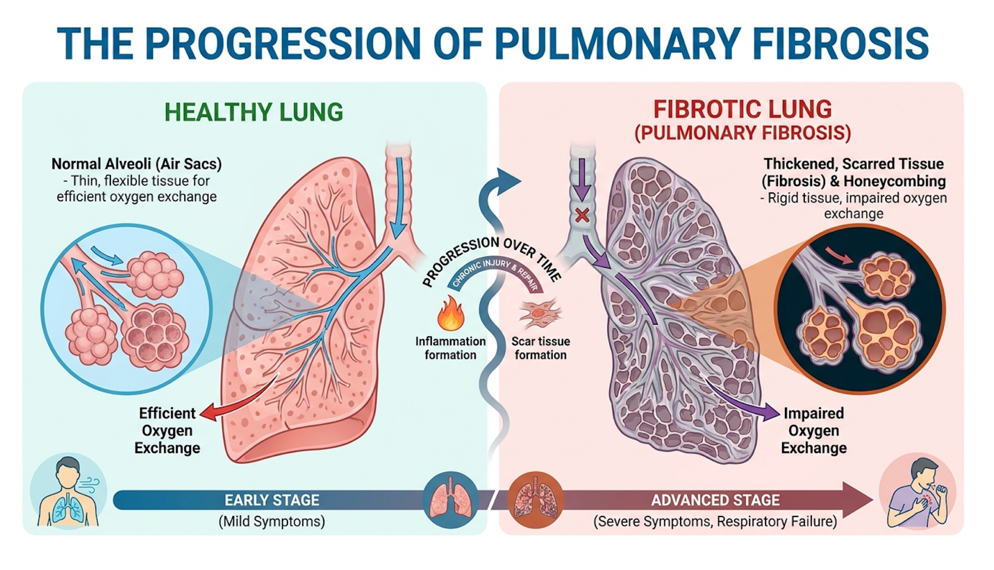
Figure 1: High-resolution CT scans illustrate the distinct “honeycombing” pattern diagnostic of active IPF compared to static post-infection scarring.
Understanding this difference is absolutely critical when evaluating a regenerative treatment for lung scarring. Static scarring might only require environmental management and pulmonary rehabilitation, as the healthy surrounding lung tissue can easily compensate for the dead zones. IPF, characterized clinically by a distinct “honeycombing” pattern visible on a high-resolution CT
Biologically, healthy lungs possess incredible compliance the ability to stretch and recoil with each breath. As the extracellular matrix fills with rigid collagen bundles in an IPF patient, that compliance drops drastically. The lungs become physically stiff, reducing the overall Forced Vital Capacity (FVC). If you don’t interrupt the signaling pathways driving the fibrosis, specifically targeting the inflammatory cytokines signaling the myofibroblasts, the architectural destruction of the alveolar walls will continue unabated until mechanical ventilation becomes the only remaining option for survival. You cannot simply wait for a condition driven by active matrix metalloproteinase dysregulation to heal itself.
Patients rightfully want to know exactly what to expect. The most common question in pulmonology clinics regarding this diagnosis is regarding life expectancy with pulmonary fibrosis. Biological variability plays a massive role here, making exact predictions difficult for any single individual. The median IPF survival rate is 3 to 5 years without intervention, highlighting the urgent need for advanced cellular therapies (National Heart, Lung, and Blood Institute, 2023). This is a timeline that demands aggressive, early therapeutic action rather than a “wait and see” approach.
However, that median statistic doesn’t tell the whole story. Clinical trajectories vary wildly. Some patients are known clinically as “rapid exacerbators.” These individuals experience sudden, steep drop-offs in Forced Vital Capacity triggered by acute exacerbations of IPF (AE-IPF). An acute exacerbation isn’t just a bad cough; it is a rapid, catastrophic worsening of respiratory function often characterized by new bilateral ground-glass opacities on an HRCT scan, carrying a devastatingly high short-term mortality rate. The biological trigger for these exacerbations remains poorly understood, but the structural devastation they leave behind is permanent.
Others are slow progressors who might maintain a relatively stable respiratory baseline for several years before experiencing a meaningful decline. But the fundamental clinical truth remains: without advanced intervention, IPF is chronically progressive. It does not spontaneously enter
Currently, the standard pharmacological approach utilizes two FDA-approved antifibrotic drugs: pirfenidone and nintedanib. Look, these medications are important tools, but we have to be completely realistic about their capabilities. They are explicitly designed to slow the annual rate of FVC decline. They do not halt the disease, and they certainly do not reverse the scarring that is already there. A patient’s clinical timeline often shifts incrementally from mild dyspnea (shortness of breath) on exertion, to requiring supplemental oxygen during sleep, to finally needing high-flow oxygen at rest. This grim trajectory highlights the urgent need for regenerative approaches that do more than just pump the brakes slightly on a runaway train.
The lungs don’t operate in a vacuum. The relentless decline in pulmonary function inevitably triggers systemic failures across the body, most notably affecting cognitive and neurological health. When fibrotic tissue thickens the delicate alveolar walls, oxygen cannot efficiently diffuse across the membrane and into the bloodstream. This chronic state of low blood oxygen is known as hypoxemia. And hypoxemia doesn’t just make you short of breath; it starves every major organ system in your body at a cellular level, creating a cascading failure effect.
Does pulmonary fibrosis affect your brain? Absolutely. Up to 30% of IPF patients experience cognitive decline due to hypoxemia (PubMed Central study, 2021) meaning unchecked lung fibrosis directly compromises independent neurological function. Patients frequently report severe brain fog, memory deficits, executive dysfunction, and overwhelming cognitive fatigue long before their respiratory failure reaches the point of requiring 24/7 supplemental oxygen. The human brain is exquisitely sensitive to even minor, sustained drops in arterial oxygen tension. Over time, this chronic cerebral hypoxia can mimic the early stages of dementia, robbing patients of their mental clarity.
Furthermore, chronic hypoxia causes the pulmonary arteries to constrict in a misguided biological attempt to shunt blood to better-ventilated areas of the lung. This phenomenon, known as hypoxic
The right ventricle of the heart will eventually hypertrophy (enlarge) from the strain, culminating in right heart failure, clinically termed cor pulmonale. When cor pulmonale sets in, patients experience severe peripheral edema (swelling in the legs and abdomen), chronic liver congestion, and extreme lethargy. Treating the lungs isn’t just about breathing; it’s essential for protecting the brain and heart from total systemic collapse. Counteracting this severe physical decline requires therapies capable of actively modulating the immune response at the source, which brings us to advanced cellular interventions.
Stem cell therapy for idiopathic pulmonary fibrosis operates by disrupting the inflammatory pathways that cause lung scarring. Specifically, UC-MSC therapy acts as a biological immunomodulator, introducing healthy stromal cells that secrete proteins to suppress fibrotic activity. For patients with declining lung function, this represents a scientifically grounded alternative to lung transplant for pulmonary fibrosis.
To understand how this actually works, we need to strip away the marketing hype from commercial clinics and look at the hard cellular biology. The UC-MSC Immunomodulation Triad a three-step biological process where UC-MSCs migrate to lung injury sites, suppress excessive inflammatory signaling, and support epithelial tissue repair is the foundation of this therapy. These Umbilical Cord Mesenchymal Stem Cells (UC-MSCs) don’t work by magic. They work through highly specific paracrine signaling.
First, let’s look at homing. When lung tissue is damaged, it acts like a beacon, releasing specific distress signals into the bloodstream. The primary signal is a chemokine called CXCL12 (also known as SDF-1). Intravenously administered UC-MSCs possess complementary surface receptors, particularly CXCR4, that perfectly detect this chemical gradient. This allows them to
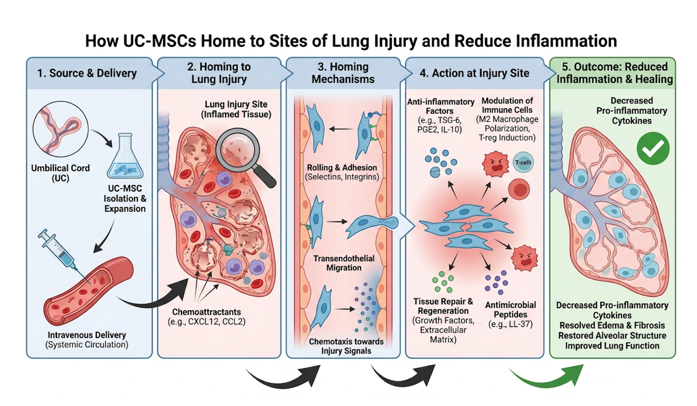
Figure 2: The UC-MSC Immunomodulation Triad demonstrates how stem cells utilize paracrine signaling to halt rogue myofibroblast activity in fibrotic lungs.
The inhibition phase is where the most critical, life-altering work happens for an IPF patient. UC-MSCs secrete potent anti-inflammatory cytokines, including Prostaglandin E2 (PGE2), Indoleamine 2,3-dioxygenase (IDO), and TSG-6. These molecules fundamentally reprogram local immune cells. They force macrophages to undergo phenotypic polarization, shifting from a pro-inflammatory, tissue-damaging (M1) state to an anti-inflammatory, tissue-healing (M2) state. The M1 macrophages stop flooding the area with damaging TNF-alpha and IL-6, dramatically lowering the localized inflammatory burden.
| 📌 If you’re interested in how MSCs shift the immune system from attack mode to repair mode, we have an interesting article that discusses mesenchymal stem cell therapy for immune modulation, which you can read via the internal link. |
Most importantly, they directly inhibit Transforming Growth Factor Beta 1 (TGF-β1), which is the primary cytokine responsible for commanding fibroblasts to produce excessive collagen. By inhibiting TGF-β1 signaling to promote tissue repair (Mesenchymal Stem Cell Therapy Review, 2020), UC-MSCs actively interrupt the pathological cascade of IPF. They effectively cut stem cell therapy alongside pirfenidone or nintedanib to achieve compounded efficacy, utilizing both pharmaceutical and biological mechanisms simultaneously for comprehensive mesenchymal stem cells lung repair.
To truly appreciate the repair phase, we have to look closely at exosomes. These aren’t just cellular waste vesicles; they are highly sophisticated, lipid-bound delivery vehicles. When UC-MSCs release these exosomes, they are packed with specific microRNAs particularly miR-29b and let-7 that directly enter the patient’s own fibrotic cells. Once inside, these microRNAs actively silence the gene expression responsible for collagen overproduction. This means the stem cells are literally rewriting the local biological instructions at a molecular level.
Furthermore, they release specific growth factors, notably Keratinocyte Growth Factor (KGF) and Hepatocyte Growth Factor (HGF). These factors stimulate the patient’s own surviving alveolar type II (ATII) cells to proliferate, produce vital surfactant, and heal the damaged pulmonary epithelium. It’s a deeply symbiotic process. The stem cells aren’t physically replacing the lung tissue; they are resetting the microenvironment so the body’s natural regenerative capabilities can finally operate without being suppressed by runaway inflammation.
We base our clinical assessments strictly on peer-reviewed data, not subjective anecdotes. The clinical trial landscape for UC-MSC therapy has matured significantly over the last decade, moving past basic safety profiling to establish robust phase II efficacy endpoints. When evaluating UC-MSC therapy for any severe lung disease, the very first question a medical professional asks is always about safety. Given that IPF patients already possess severely compromised respiratory systems, introducing foreign biological matter carries theoretical risks of pulmonary embolism or acute immune rejection.
Fortunately, the empirical data provides a remarkably clear and reassuring answer regarding systemic safety. Clinical trials show UC-MSCs reduced FVC decline by up to 40%, safely halting active fibrotic progression (ClinicalTrials.gov, NCT02013700). Because UC-MSCs are universally immune-privileged meaning they do not express the specific surface antigens (HLA-DR) that normally trigger a hostile T-cell attack they can be administered allogeneically (from a healthy donor) without requiring the patient to take risky, life-shortening immunosuppressive drugs. They are remarkably well-tolerated, even in fragile IPF cohorts with multiple comorbidities, showing no evidence of teratoma formation or severe infusion reactions.
In phase II trial structures, researchers don’t just look for survival; they look for functional, measurable quality of life improvements over 12 to 24 months. Cohorts receiving high-dose intravenous UC-MSCs demonstrated a remarkable halting of the expected Forced Vital Capacity decline. To put that into perspective, an untreated patient might lose 150 to 200 milliliters of lung volume annually. In the treated cohorts, that loss was often reduced by up to 40%, keeping patients functionally independent for substantially longer.
Beyond just the spirometry data, high-resolution CT scans of these patients frequently showed a stabilization of the honeycombing pattern meaning the physical spread of the scarring was demonstrably arrested. Blood panels corroborated this, showing a sustained, measurable drop in circulating pro-inflammatory cytokines like TNF-alpha and IL-6 for months following the infusion. Patients often increased their 6MWT (6-minute walk test) distances by 30 to 50 meters, which translates directly to the ability to walk down a grocery aisle without needing to sit down. For an IPF patient, stopping the decline and achieving this level of stabilization is a monumental clinical victory.
This brings us to the most critical long-term question: can stem cell therapy delay the need for a lung transplant? Yes. By stabilizing lung function, cooling off the localized inflammatory storm, and halting the rapid architectural destruction of the alveoli, effective immunomodulation extends the viable window before end-stage surgical intervention becomes mandatory. Lung transplants carry a devastatingly high early mortality rate, require lifelong immunosuppression, and are subject to chronic rejection (bronchiolitis obliterans). Pushing that surgical necessity back by two, three, or five years while maintaining a tolerable quality of life is the primary, overarching goal of modern regenerative pulmonary medicine.
Not all stem cell therapies are created equal, and not every single IPF patient is an ideal candidate for this intervention. Our rigorous evaluation of treatment modalities reveals significant, outcome-altering differences in how these cells are delivered, processed, and quality-controlled.
Currently, there is an ongoing debate in the regenerative medicine community regarding IV infusion vs inhaled stem cell delivery for severe lung disease. Intravenous (IV) infusion remains the absolute gold clinical standard. Why? Because of the “pulmonary first-pass effect.” The human lungs act as a massive, highly efficient vascular filter. When MSCs are infused into a peripheral vein, they naturally travel to the right side of the heart and are pumped directly into the pulmonary artery. The lungs are the very first capillary bed the cells hit. Since MSCs are relatively large (15-20 microns), they become mechanically trapped in the pulmonary capillaries right where the fibrosis is actively happening, immediately beginning their paracrine signaling.
Nebulized or inhaled stem cell delivery is currently under investigational review for localized delivery. However, patients frequently get sold on nebulizers by aggressive marketing campaigns. A fibrotic lung is a hostile, scarred, and often mucus-plugged environment. When stem cells are inhaled, they struggle to bypass the mucociliary escalator and rarely penetrate deep into the alveolar architecture where the active scarring is occurring. They simply die on the surface. Therefore, systemic IV administration remains the most evidence-backed modality for achieving deep tissue immunomodulation and successfully engaging the chemokine homing mechanisms we discussed earlier.
So, who is a good candidate for stem cell therapy for lung disease? The ideal candidate is a patient in the mild to moderate stages of IPF typically possessing an FVC greater than 50% of predicted value, and a DLCO above 30%. These patients still possess enough viable, salvageable lung tissue for the stem cells to interact with. End-stage patients who are fully oxygen-dependent at rest and experiencing severe right heart failure often possess lungs that are entirely replaced by structural honeycombing. These lungs are too structurally devastated for biological signaling to make a functional difference, as dead tissue cannot be regenerated. Timing is absolutely everything in regenerative medicine.
Furthermore, uncompromising laboratory standards dictate actual patient outcomes. The efficacy of UC-MSCs depends entirely on cell expansion techniques, passage numbers, and stringent purity testing. Cells rapidly lose their multipotency and paracrine capabilities if they are replicated too many times (beyond passage 4 or 5). Patients must ensure their treatments are prepared in high-grade, ISO-certified Good Manufacturing Practice (GMP) facilities. This level of scrutiny is especially vital for patients exploring medical tourism. Understanding the strict safety standards for stem cell therapy in Thailand or other international medical hubs protects vulnerable patients from unverified, contaminated commercial procedures. Clinics offering uncharacterized “stromal vascular fractions” (SVF) or poorly expanded cells do vastly more harm than good.
| 📌 If you’re planning treatment in Thailand and want a practical checklist for choosing a safe clinic, we have an interesting article that discusses whether stem cell therapy is safe in Thailand, which you can read via the internal link. |
By shifting the therapeutic target from mere symptom suppression to active cellular reprogramming, UC-MSC treatments offer a scientifically validated approach to managing severe lung disease. However, distinguishing these legitimate cellular applications from unproven alternative therapies remains paramount for patient safety.
When investigating how to improve breathing with pulmonary fibrosis, patients frequently encounter a barrage of unverified internet remedies and repurposed medications. While advanced cellular therapies undergo rigorous clinical trials, viral trends regarding antiparasitic drugs or environmental shifts often lack substantial clinical efficacy. This requires patients to carefully differentiate verified scientific fact from anecdotal fiction to protect their remaining lung capacity.
The internet has democratized medical information, but it has also massively democratized medical misinformation. Over the past few years, a persistent, viral narrative has emerged suggesting that ivermectin for lung inflammation could serve as a legitimate alternative therapy for fibrotic conditions. Let’s completely dismantle this using actual pharmacology and clinical reality.
Ivermectin is a widely used, highly effective antiparasitic drug. It works by binding to glutamate-gated chloride channels found strictly in invertebrate nerve and muscle cells, paralyzing and killing parasites. It is brilliant for treating river blindness or scabies. But ivermectin lacks FDA authorization for lung inflammation, with 0% of controlled clinical trials demonstrating (U.S. Food and Drug Administration, 2021). The use of this drug for IPF carries no verified safety profile.
The confusion stems from fundamental biological misunderstandings. Treating lung worms (a parasitic infection) requires an entirely different molecular mechanism than reversing the autoimmune-like, idiopathic collagen deposition seen in IPF.
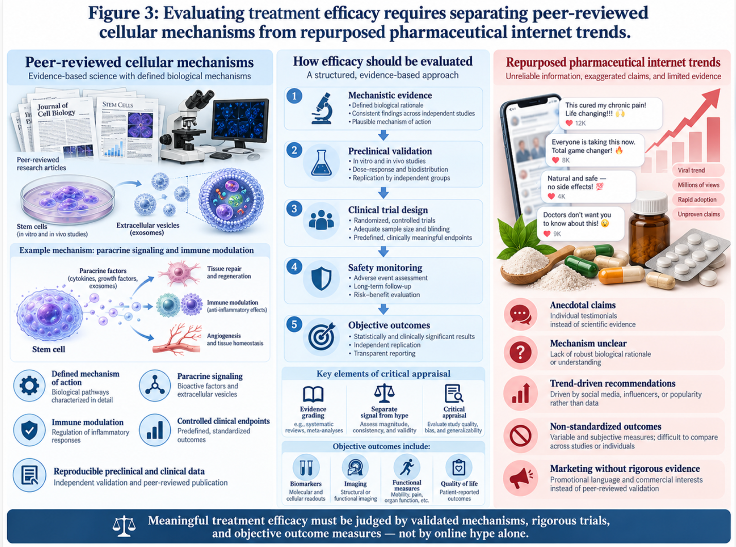
Figure 3: Evaluating treatment efficacy requires separating peer-reviewed cellular mechanisms from repurposed pharmaceutical internet trends.
Why do these myths persist so aggressively online? Because highly isolated in vitro (petri dish) studies occasionally show that massive doses of ivermectin can exhibit mild anti-inflammatory properties by inhibiting certain cellular transport proteins (like host importin alpha/beta-1). However, the in-vitro IC50 (half-maximal inhibitory concentration) required to achieve this is roughly 2 to 4 micromolar.
The standard approved human dose of ivermectin achieves a peak plasma concentration of about 0.05 micromolar. To safely achieve the tissue concentrations required to even partially halt myofibroblast activity in a living human being, the ivermectin dose would need to be 50 to 100 times higher than the authorized limit. A dose that massive would easily cross the blood-brain barrier, causing severe GABA-ergic neurotoxicity, seizures, ataxia, and potential coma. It fundamentally does not target the TGF-beta/Smad pathway driving IPF. Substituting proven antifibrotics or advanced stem cell therapies with unapproved, off-label antiparasitics is a dangerous gamble that costs patients irreplaceable time.
Beyond repurposed pharmaceuticals, many desperate patients explore ancient traditional modalities to manage their highly restrictive breathing. We frequently hear the question in clinical settings: How do the Chinese treat pulmonary fibrosis? Traditional Chinese Medicine (TCM) utilizes various herbal adjuncts most notably Astragalus and Cordyceps aiming to purportedly balance bodily systems and reduce chronic coughing.
While some of these specific herbal compounds do exhibit mild antioxidant and anti-inflammatory properties that can soothe airway irritation and reduce reactive oxygen species (ROS), they do not fundamentally alter the disease pathology at a cellular level. Systematic reviews of Traditional Chinese Medicine for IPF show these herbs may offer symptomatic cough relief but do not reverse active fibrotic remodeling (Systematic TCM Review). TCM can absolutely be a supportive adjunct for daily symptom management, much like a lozenge soothes a severe sore throat, but it is not a standalone regenerative treatment for lung scarring. The bioactive compounds, like Astragaloside IV, simply cannot reprogram a macrophage or inhibit TGF-β1 at the scale required to halt progressive IPF.
Similarly, external environmental factors heavily influence a patient’s daily symptom burden. Patients often ask if weather effects on pulmonary fibrosis are actually real. Yes, they are highly impactful. Research indicates IPF patients experience increased exacerbations during periods of high humidity and extreme cold (Climate impact research) which can trigger severe bronchospasms, heightened airway reactivity, and sudden drops in oxygen saturation.
Warmer, temperate, and humidity-controlled environments significantly reduce the mechanical stress placed on already compromised lungs. However, moving to a better climate or investing in
Because the stakes in progressive lung diseases are incredibly high, understanding the exact limitations of alternative support is mandatory for patient survival. The greatest danger of unproven adjuncts isn’t necessarily direct chemical toxicity; it is the massive opportunity cost of delayed clinical care. While a patient spends six crucial months experimenting with unverified herbal supplements or antiparasitics, their FVC continues to drop irreversibly.
Lung tissue, once severely honeycombed and scarred with dense collagen bundles, is highly resistant to casual systemic therapies. It requires targeted, molecular-level intervention. We have seen far too many patients abandon standard-of-care medications due to mild side effects, only to rely entirely on anecdotal internet regimens. The result is always an irreversible respiratory decline that no future biological therapy can rescue.
Stem cell therapy for post-COVID lung fibrosis or IPF is grounded in deeply researched, peer-reviewed cellular pathways, whereas internet myths rely almost entirely on the placebo effect a profoundly dangerous illusion when your oxygen saturation is objectively dropping by the day. You must adhere strictly to evidence-based medicine. Contrast a patient delaying care to try unproven supplements with a patient enrolling in a supervised, biologically driven cellular therapy program. The latter is actively addressing the disease mechanism at the source; the former is merely hoping for the best. There is simply no substitute for targeted medical intervention when the structural integrity of your lungs is failing.
We must acknowledge specific situations where biological therapies are not suitable. Medical transparency demands that we separate established clinical uses from experimental applications and warn patients against clinics that guarantee absolute outcomes. No biological intervention is universally appropriate, and navigating this space requires profound clinical caution.
The most dangerous pitfall is believing that stem cells will completely reverse IPF. Commercial clinics frequently overpromise, intentionally preying on desperate patients who are running out of options. In reality, the therapy focuses strictly on immunomodulation and stabilizing disease progression not a guaranteed reversal of existing severe, honeycombed scarring. If a clinic promises to restore your deeply fibrotic lungs to 100% capacity, walk away immediately.
Data indicates that up to 40% of unregulated commercial clinics misrepresent their treatment capabilities, emphasizing the critical need for rigorous specialist vetting (Cell Stem Cell, 2023). Furthermore, assuming all stem cells are functionally identical is a massive clinical error. The source tissue, processing methodologies, viability metrics, and GMP certification radically alter both the safety and efficacy of the final cellular product. Quality control in the laboratory is quite literally everything, and cutting corners on biological purity leads to zero clinical benefit.
| 📌 If you’re interested in why the percentage of living cells matters so much at the time of treatment, we have an interesting article that discusses the importance of cell viability in UC-MSC stem cell therapy, which you can read via the internal link. |
There are distinct, severe clinical scenarios where end-stage IPF mandates immediate evaluation for a double lung transplant rather than investigational cellular therapies. If a patient is suffering from severe secondary pulmonary hypertension, right-sided heart failure (cor pulmonale), active malignancies, or acute respiratory distress syndrome (ARDS) requiring mechanical ventilation, biological signaling therapies will act far too slowly to save their life.
In these end-stage scenarios, the delicate alveolar architecture is entirely replaced by thick, rigid, non-functional connective tissue. Dead tissue cannot be biologically regenerated. At this critical juncture, the only remaining viable option is a surgical transplant. Additionally, pirfenidone and nintedanib remain the standard-of-care baseline for managing this disease. These antifibrotics should never be abandoned abruptly without direct, specialized pulmonology guidance, even if you are actively enrolling in a highly supervised stem cell protocol.
You must seek individualized, comprehensive assessments from qualified pulmonologists and regenerative medicine specialists before materially altering your treatment plan. A preliminary
A rigorous specialist will mandate recent pulmonary function tests (PFTs), an echocardiogram to check right heart function, and fresh high-resolution CT scans to determine if you possess enough viable lung tissue to successfully respond to cellular signaling. Only after this rigorous physiological mapping can a doctor determine if you remain inside the optimal therapeutic window for stem cell intervention.
Stem cell therapy for idiopathic pulmonary fibrosis functions as a promising investigational treatment targeting lung inflammation. UC-MSCs secrete bioactive molecules that interrupt the fibrotic signaling pathways driving the disease. Current clinical data demonstrates these cells can help stabilize lung function and reduce inflammatory markers systemically. For many patients, this biological intervention provides an alternative strategy to manage progression. Individual suitability strictly depends on disease staging and comprehensive evaluation by a qualified specialist.
For patients evaluating a non-surgical treatment for pulmonary fibrosis, UC-MSC therapy offers an evidence-based biological intervention rather than a traditional pharmaceutical patch. With standard IPF presenting a median survival rate of 3 to 5 years (NIH, 2023), the need to interrupt the fibrotic cascade is critical. The most scientifically grounded approach utilizes targeted cellular immunomodulation to halt progressive tissue scarring.
This approach is governed entirely by The UC-MSC Immunomodulation Triad the specific biological mechanism where cells home to injury sites, inhibit rogue inflammation, and stimulate
If you or a loved one are experiencing declining lung capacity despite standard pharmaceutical care, do not wait for the damage to become irreversible. Compile your recent high-resolution CT scans and pulmonary function tests, and schedule a formal consultation with a certified regenerative pulmonology specialist to determine if cellular therapy is biologically appropriate for your specific pathology.