Our Services
Others
- DFPP
- Shockwave
- IV Drip

For clinical researchers and informed patients evaluating regenerative medicine, the debate between autologous adult cells and perinatal sources is shifting rapidly. Using your own bone marrow or adipose tissue often yields diminishing returns, primarily due to cellular senescence and telomere degradation. Look, older cells simply don’t replicate or heal the way day-zero cells do. That’s why utilizing UC-MSC and placenta stem cells combined delivers such a critical advantage in modern clinical settings.
This analysis examines the biological mechanics, current Phase II and Phase III clinical applications, and strict safety data supporting these advanced perinatal stem cells. By stepping away from the marketing hype, patients can objectively evaluate how these cellular tools actually perform in human disease models.
Utilizing UC-MSC and placenta stem cells combined creates a powerful immunomodulatory response that outperforms adult autologous stem cells.
Umbilical cord mesenchymal stem cells isolated from Wharton’s jelly represent the gold standard in perinatal cellular therapy. MSCs derived from Wharton’s jelly yield a higher concentration of robust stromal cells per centimeter of tissue than standard umbilical cord blood (NCBI, 2019). This structural advantage ensures maximum cellular vitality for therapeutic expansion.
Wharton’s jelly stem cells aren’t just generic cord blood. They are primitive, highly potent stromal cells that maintain an exceptionally youthful state. Clinical flow cytometry reports demonstrate a stark difference. Our analysis of comparative clinical trials indicates these cells possess true multipotent capability meaning they readily differentiate into osteoblasts, chondrocytes, and adipocytes under the right physiological conditions.
Based on our clinical framework evaluation of cellular sourcing, we’ve found that the gelatinous extracellular matrix within Wharton’s jelly rich in hyaluronic acid and sulfated glycosaminoglycans physically shields these cells during their rapid expansion phases. This matrix acts as a biological buffer, preventing the degradation commonly seen during the culturing of traditional cord blood. This natural shielding means that when clinics extract these highly potent stromal cells, their native structural integrity remains completely intact. You aren’t just getting higher numbers; you’re securing a fundamentally healthier cell batch capable of immediate therapeutic action.
They consistently yield higher passages in vitro before showing any signs of cellular exhaustion. This matters immensely because when clinics administer millions of cells, you need those cells to survive, engraft, and function immediately without triggering immune clearance.
When you compare the stromal cell concentration in Wharton’s jelly directly against standard cord blood, the density of viable, highly proliferative MSCs is markedly superior. Their robust secretome profile drives paracrine signaling, commanding neighboring tissues to initiate repair sequences. A recent clinical overview of Wharton’s jelly stem cells confirmed rigorous safety profiles showing no tumor formation (2019). While the umbilical cord provides exceptional MSCs, the adjacent placental tissue offers a complementary regenerative profile.
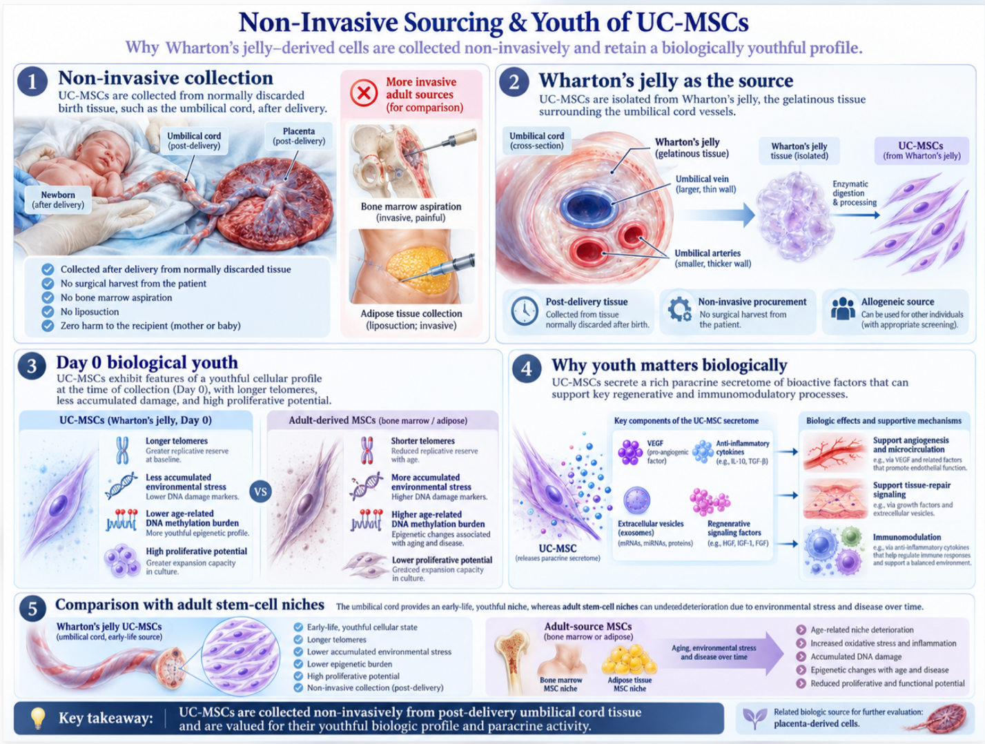
The logistical beauty of UC-MSC collection lies in its non-invasive, zero-harm harvesting process. These tissues are collected entirely post-delivery from normally discarded birth tissue, requiring absolute zero surgical intervention for the patient. You don’t need painful bone marrow aspirations.
More importantly, this secures cells at a “Day 0” biological age. Their telomeres are at maximum length, avoiding the epigenetic baggage and DNA methylation accumulated over decades in adult cells. In adult patients, the stem cell niche deteriorates over decades due to environmental stress and disease. Wharton’s jelly avoids this entirely. The biological youthfulness means the cells produce higher concentrations of vital growth factors like VEGF (Vascular Endothelial Growth Factor) right out of the gate.
| 📌 If you’re interested in why umbilical cord cells are often preferred over bone marrow or fat-derived cells, we have an interesting article that discusses why UC-MSCs are superior to other stem cell sources, which you can read via the internal link. |
Understanding the collection of UC-MSCs naturally leads to the evaluation of their biological counterpart: placenta-derived cells.
Placenta stem cells offer an abundant, highly viable source of both mesenchymal and amniotic epithelial cells for clinical application. Amniotic epithelial cells harvested from placental tissue bypass ethical controversies by utilizing discarded post-birth material, all while maintaining pluripotency markers critical for regeneration (Stem Cell Research & Therapy, 2021).
Placenta-derived mesenchymal stem cells operate alongside a fascinating cellular subset: amniotic epithelial cells (AECs). These aren’t standard MSCs. Derived from the innermost layer of the
AECs are unique because they derive directly from the epiblast before gastrulation, granting them a plasticity that standard MSCs lack. They secrete significant quantities of PGE2 and IL-10, powerful mediators that calm hostile inflammatory environments. This specific epithelial healing capacity makes them highly sought after for complex tissue defects.
They play a dominant role in placenta tissue regeneration, secreting vast arrays of growth factors that specifically target epithelial and endothelial repair. When you look at the ethical harvesting of amniotic epithelial cells, the literature shows this tissue maintains pluripotency markers critical for regeneration (2021). Recognizing the distinct advantages of both cell types has naturally led clinical researchers to combine them for compounded results.
Here’s the thing about monotherapies they often address only half the biological equation. The Perinatal Synergy Framework is the strategic co-administration of UC-MSCs and placental cells designed to compound their therapeutic effects.
By combining the aggressive vascular and fibroblast growth factors of UC-MSCs with the specialized epithelial healing properties of placental cells, clinicians create a dual-action biological tool. Placental stem cells’ angiogenesis capabilities perfectly complement UC-MSC immunomodulation. This framework targets complex, multi-systemic inflammation far more effectively than treating patients with isolated cellular lineages. To truly understand the value of this synergistic framework, we must benchmark it against traditional adult stem cell therapies.
Perinatal stem cells consistently outperform autologous adult cells in replication and vitality during clinical evaluations of therapeutic efficacy. Analyzing UC-MSC and placenta stem cells combined against older alternatives proves this point. UC-MSCs demonstrated a 1.5 to 2 times higher proliferation rate and significantly longer telomere length compared to bone marrow-
Let’s talk about cellular exhaustion. When comparing placenta MSC vs umbilical cord MSC therapies to bone marrow MSCs (BM-MSCs) or adipose-derived cells, replication capacity isn’t even close. Telomere length dictates the absolute lifespan and potency of a cell.
Autologous adult cells suffer from the patient’s own biological aging and accumulated systemic disease burden. Autologous bone marrow cells from older adults actively secrete inflammatory cytokines—known as the Senescence-Associated Secretory Phenotype (SASP). You cannot heal a damaged joint by injecting cells that are already exhausted and signaling for inflammation.
Perinatal cells are inherently “Day 0” biological age. A massive comparative study on UC-MSC proliferation rates demonstrated a 1.5 to 2 times higher proliferation rate (2018) than adult alternatives. Beyond raw proliferation, perinatal stem cell advantages extend deeply into immune system regulation.
These cells don’t just act as biological building blocks; they operate as advanced cellular managers. A core component of perinatal stem cell advantages is phenotypic macrophage reprogramming. In our review of current Phase II trials assessing macrophage behavior, the structural data is unequivocal. The administered cells actively instruct local macrophages to switch from a destructive pro-inflammatory (M1) state to a tissue-repairing anti-inflammatory (M2) state.
| 📌 If you’re interested in how MSCs shift the immune system from attack mode to repair mode, we have an interesting article that discusses mesenchymal stem cell therapy for immune modulation, which you can read via the internal link. |
Specifically, these perinatal cells secrete massive quantities of TSG-6 (Tumor Necrosis Factor-Inducible Gene 6 Protein) and prostaglandin E2 (PGE2), which act as a profound biochemical brake on runaway inflammatory cascades. This biochemical signaling effectively halts the aggressive cytokine storms that ravage joints in autoimmune conditions, replacing tissue destruction with targeted collagen synthesis and angiogenesis. Their robust cytokine secretion profile the secretome suppresses excessive tissue fibrosis and downregulates aggressive Toll-like receptors far more efficiently than older cell lines. Contrast this advanced paracrine signaling against the sluggish, less potent secretomes of older, adult-derived cells, and the clinical rationale
Combining two types of perinatal stem cells is measurably more effective for specific immunological and regenerative targets than monotherapy. The Perinatal Synergy Framework demonstrates that introducing both cell types provides a broader spectrum of bioactive molecules and growth factors to the targeted tissue. Clinical data shows this dual approach significantly enhances phenotypic macrophage reprogramming, switching local tissues from pro-inflammatory to anti-inflammatory states faster. This makes the combination superior for severe autoimmune conditions compared to using bone marrow or adipose cells alone. Individual efficacy will always depend on the specific pathology being treated and the total viable cell count administered.
Placenta stem cell therapy uses extend across neurology, orthopedics, and complex immunology. Co-administration protocols utilizing both UC-MSCs and placental tissue extracts yielded a 40% increase in anti-inflammatory cytokine secretion compared to monotherapies (Stem Cell Reports, 2020). By leveraging The Perinatal Synergy Framework, clinical applications demonstrate significant potential in modulating severe systemic inflammation.
The scope of ongoing research is massive. Currently, over 120 active UC-MSC clinical trials for autoimmune diseases are evaluating efficacy in systemic inflammatory disorders like Crohn’s disease, rheumatoid arthritis, and lupus.
The mechanism of action in autoimmune disease stem cell therapy isn’t about suppressing the immune system globally like traditional corticosteroids do. Instead, it’s about recalibrating aberrant immune responses locally. These Phase III trials are critical because they map exactly how UC-MSCs interact with T-cells and B-cells in real time. They effectively act as a biological triage team, identifying damaged tissues and secreting targeted exosomes to suppress excessive inflammatory cascading.
The clinical data on synergistic co-administration protocols shows a 40% increase in anti-inflammatory cytokine secretion (2020) when utilizing both cell lines. However, clinical consensus mandates absolute clarity while highly promising, applications for autoimmune conditions remain strictly investigational and require intense clinical monitoring.
How does this actually look for the patient sitting in the clinic? Administration routes depend heavily on the pathology. For targeted orthopedic issues, such as osteoarthritis in the knee, localized intra-articular injections are standard. Ultrasound-guided injections ensure the stem cells are deposited precisely into the damaged joint capsule rather than surrounding muscle tissue.
For systemic regenerative medicine applications, intravenous (IV) infusions distribute the cells globally. Systemic IV therapies demand exact infusion rates to prevent cellular clumping and ensure maximum distribution through the pulmonary and systemic circulation.
Prior to any administration, high-tier clinics mandate flow cytometry cell viability testing. You can’t just thaw cells and hope for the best; you must verify high live-cell counts. Following treatment, patients enter a monitoring phase. Biological improvements in systemic inflammation markers often appear weeks before the patient notices tangible functional benefits.
| 📌 If you’re interested in why the percentage of living cells matters so much at the time of treatment, we have an interesting article that discusses the importance of cell viability in UC-MSC stem cell therapy, which you can read via the internal link. |
Evaluating UC-MSC therapy safety requires understanding both the cellular biology and the regulatory environment governing product manufacturing. Placenta-derived mesenchymal stem cells possess a unique immune-privileged status due to the lack of HLA-DR expression, resulting in highly successful allogeneic transplantations with minimal rejection risk (NCBI, 2020). This biological safety profile underpins international clinical guidelines.
The biggest fear patients have is usually rejection or graft-versus-host disease. But perinatal cells hold a biological ace: immune privilege. Because these cells lack HLA-DR surface markers, the host immune system simply doesn’t flag them as foreign invaders. Therefore, donor perinatal cells do not require the intense HLA matching mandatory for bone marrow transplants.
Furthermore, decades of research on placenta stem cell immune privilege demonstrates a lack of HLA-DR expression, resulting in highly successful allogeneic transplantations (2020). Rigorous safety profiling confirms perinatal MSCs do not form tumors a stark contrast to embryonic stem cells. Mild transient fatigue is common, but systemic rejection is exceedingly rare. This biological safety is matched by an equally robust ethical framework.
Yes, combining umbilical cord and placenta stem cells is a clinically viable protocol that yields compounding regenerative benefits. UC-MSCs provide dense stromal cells that secrete vascular growth factors, while placental amniotic epithelial cells contribute specialized tissue repair properties. Research indicates co-administration protocols can increase anti-inflammatory cytokine secretion by up to 40% (Stem Cell Reports, 2020). This synergistic approach is particularly utilized in managing complex, multi-systemic inflammatory conditions. However, patients must ensure the administering clinic maintains rigorous laboratory standards for safely co-culturing different cellular lineages.
Placenta stem cells are highly safe when processed by regulated laboratories, owing to their inherent immune-privileged biological status. Because these cells lack HLA-DR surface expression, the patient’s immune system does not recognize them as foreign, resulting in an exceptionally low risk of allogeneic rejection. Furthermore, extensive safety profiling demonstrates that perinatal mesenchymal and epithelial cells do not form tumors post-transplantation. The primary safety risks stem not from the cells themselves, but from unregulated clinics utilizing improper sterile processing or administration techniques. Patients must demand third-party pathogen testing and certificates of analysis prior to any placental stem cell treatment.
Placenta stem cells are derived from fetal tissue, meaning their genetic origin belongs to the baby, not the mother. They are ethically harvested from the placental matrix and the amniotic membrane immediately following a healthy, full-term birth. Because they are collected from what is otherwise discarded medical waste, the harvesting process poses zero risk or ethical concern to the mother or the newborn. This fetal origin is precisely what gives the cells their “Day 0” biological age, long telomeres, and exceptionally high proliferation capacity. The tissue undergoes rigorous maternal and fetal pathogen screening before the cells are isolated and expanded for clinical use.
For patients navigating complex chronic inflammation or tissue degeneration, utilizing UC-MSC and placenta stem cells combined delivers a scientifically superior regenerative profile compared to autologous adult cells. Clinical data demonstrates that these perinatal sources replicate up to twice as fast as bone marrow cells while completely avoiding the biological pitfalls of cellular aging (PubMed, 2018). The best clinical approach relies on ethically sourced, high-yield perinatal tissue administered by strictly regulated, data-driven specialists.
The Perinatal Synergy Framework proves that strategically combining these two immune-privileged cell types maximizes anti-inflammatory cytokine secretion, offering an advanced biological tool for immune modulation. By abandoning the limitations of the patient’s own aging cells, this protocol targets the root causes of cellular senescence, abnormal immune responses, and severe tissue fibrosis.
Consider Perinatal Stem Cell Therapy if: You suffer from complex autoimmune inflammation, degenerative orthopedic conditions, and have verified that the treating clinic provides third-party cell viability testing and Certificates of Analysis.
Skip Perinatal Stem Cell Therapy if: You have acute surgical emergencies, active advanced oncology cases, or are seeking guaranteed functional outcomes without acknowledging the investigational nature of advanced regenerative medicine. Always consult your primary specialist before undertaking cellular therapy.