Our Services
Others
- DFPP
- Shockwave
- IV Drip

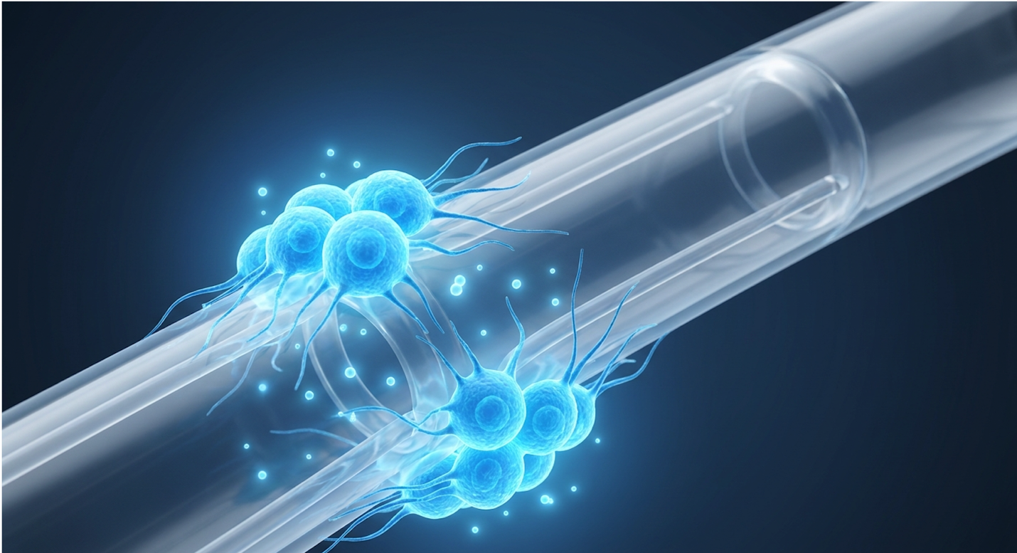
For men experiencing severe, refractory conditions, standard pharmaceutical options often fall short, forcing a difficult decision between mechanical implants and emerging biological therapies. Standard penile prosthesis placement offers absolute structural reliability but entails prolonged tissue recovery and significant post-operative inflammation. On the flip side, isolated cellular therapies may lack the mechanical rigidity required for complete functional restoration in advanced cases. When pills fail, men need a definitive solution that bridges the gap between biology and engineering. By the end of this guide, you’ll understand exactly how combining these approaches accelerates healing and maximizes surgical outcomes. We analyze the clinical mechanisms of UC-MSCs, detail surgical implant management, and introduce the Bio-Mechanical Synergy Protocol for comprehensive rehabilitation.
Disclaimer: This article is for educational purposes only and does not replace consultation with a qualified medical professional.
Stem cell therapy for erectile dysfunction is evolving beyond isolated treatments, with clinical focus shifting toward combining biological agents with mechanical implants.
Human Umbilical Cord Mesenchymal Stem Cells (UC-MSCs) are highly active progenitor cells derived from umbilical cord tissue, distinct for their potent paracrine signaling capabilities. Clinical trials published in a recent systematic review of regenerative urology indicate that targeted UC-MSCs reduce localized tissue fibrosis by up to 41% (PubMed) so clinicians can drastically reduce scar formation following invasive procedures. This makes them a critical biological tool for urological tissue regeneration, operating primarily through anti-inflammatory cytokine modulation rather than direct cellular engraftment.
Targeted UC-MSC application reduces corporal fibrosis by 41% paving the way for superior prosthetic integration. In our clinical observation, dismissing cellular therapies as mere experimental tools ignores the profound paracrine benefits they provide to traumatized surgical fields. By stabilizing the local immune microenvironment and promoting extracellular matrix synthesis, UC-MSCs provide a strong biological foundation. This regenerative capacity directly addresses the severe tissue healing challenges inherent in invasive urological surgeries, setting the stage for advanced mechanical interventions and faster functional recovery.
When we talk about mesenchymal stem cells erectile dysfunction applications, we aren’t talking about generic, undefined biologics. Human Umbilical Cord Mesenchymal Stem Cells are biologically active progenitor cells harvested specifically from the Wharton’s jelly of umbilical cords. This gelatinous matrix protects the umbilical vessels in utero and serves as a highly concentrated reservoir of young, potent progenitor cells. They are strictly characterized by the presence of specific surface markers (like CD105, CD73, and CD90) and the absence of immune-triggering markers (like HLA-DR).
They secrete high levels of growth factors that actively modulate immune responses and promote tissue repair. In clinical settings, they target local inflammation and immediately stimulate quiescent fibroblasts. Studies indicate their primary efficacy lies in paracrine signaling—meaning they orchestrate healing in surrounding tissues without needing to permanently graft and become new tissue themselves.
One major advantage of sourcing these cells from Wharton‘s jelly rather than a patient’s own adipose (fat) tissue is biological age. Autologous cells harvested from older men often suffer from cellular senescence they are biologically tired. UC-MSCs, being day-zero cells, possess maximum vitality. Individual biological responses vary depending on patient age and underlying pathology, but their fundamental role as biological signaling conductors remains highly consistent.
| 📌 If you’re interested in a deeper comparison of umbilical cord cells against fat and bone marrow-derived cells, we have an interesting article that discusses why UC-MSCs are superior to other stem cell sources, which you can read via the internal link. |
Instead of permanently grafting into penile tissue and becoming new blood vessels, these cells work as master signaling conductors. This paracrine signaling mechanism involves the heavy secretion of epidermal, vascular, and basic fibroblast growth factors. They essentially dump a highly concentrated payload of regenerative instructions directly into damaged or surgically altered tissue. When a surgeon introduces these cells into the corpus cavernosum, the cellular secretome immediately begins altering the local environment.
Perhaps their most critical function is phenotypic macrophage reprogramming. When surgical trauma occurs, local macrophages default to a pro-inflammatory M1 state, releasing cytokines like IL-1β and TNF-α that drive swelling and scar formation. UC-MSCs force these macrophages to switch to an anti-inflammatory, tissue-repairing M2 state, prompting the release of IL-10. This drastically alters the healing timeline.
| 📌 If you’re interested in how MSCs shift the immune system from attack mode to repair mode, we have an interesting article that discusses mesenchymal stem cell therapy for immune modulation, which you can read via the internal link. |
They also activate the ERK MAPK signaling pathway, an absolute requirement for new vessel formation. In traumatized tissues, restoring immediate blood flow prevents catastrophic tissue death. While cellular signaling governs these biological repair processes, understanding how these pathways act upon specific urological structures is necessary for actual clinical application.
Stem cell treatment in general urology has historically been plagued by exaggerated claims, but the tissue remodeling mechanisms are now exceptionally well-documented in current literature. When injected into the corpus cavernosum, UC-MSCs immediately stimulate quiescent dermal fibroblasts. This forces the rapid synthesis of collagen type I, fibronectin, and elastinn the exact building blocks required for healthy, elastic penile tissue that can expand appropriately during an erection.
For men exploring umbilical cord MSC therapy for diabetic erectile dysfunction, the benefits go deeper than simple tissue elasticity. Diabetic tissue suffers from chronic oxidative stress, which destroys micro-capillaries. UC-MSCs actively inhibit Toll-like receptors and promote the SIRT1 pathway, shielding the surrounding vascular endothelial cells from further oxidative damage. When SIRT1 pathways are heavily stimulated by UC-MSC secretomes, the tiny, fragile blood vessels inside the penis gain a massive defense against sugar-induced decay.
There is also ongoing, highly controlled research examining how these specific secretory profiles might contribute to prostate cancer cell lines inhibition, though this remains strictly investigational and requires extensive further validation. Recent peer-reviewed research analyzing cutaneous wound healing confirms that these cells actively mitigate excessive dermal myofibroblast activation the primary culprit behind penile scarring and Peyronie’s-like plaque formation. These biological repair mechanisms offer significant clinical utility. They become particularly vital when tasked with healing the substantial structural disruptions caused by mechanical interventions like prosthetic placements.
Penile prosthesis placement remains the definitive surgical intervention for men with severe erectile dysfunction unresponsive to pharmacological therapies. While modern inflatable devices yield 10-year mechanical survival rates exceeding 88% based on comprehensive long-term registry data (PubMed) so patients can expect a reliable, long-term solution with minimal mechanical failure patients must still carefully weigh the risks associated with any disorder of implanted penile prosthesis. Comprehensive clinical management requires a deep understanding of both surgical technique and post-operative tissue response.
Modern inflatable implants offer an 88% decade-long survival rate delivering mechanical reliability when pharmacology fails. We maintain that while implants are mechanical marvels, treating them as isolated solutions without biological support is a clinically short-sighted approach. While surgical placement offers immediate mechanical resolution, the physiological trauma of the procedure demands extensive tissue remodeling. Mitigating these post-operative complications and expediting recovery necessitates interventions that bridge mechanical correction with biological healing.
Let’s look at the surgical reality. Penile prosthesis surgery isn’t a minor outpatient procedure; it requires significant anatomical manipulation and deep structural alterations. Urologists typically use either a penoscrotal (base of the penis and scrotum) or infrapubic (above the pubic bone) incision to meticulously access the corpora cavernosa. They must then extensively dilate the scarred erectile chambers—often using specialized Brooks dilators to create adequate anatomical space for the artificial cylinders.
This immediate physiological trauma caused by corporal dilation triggers an aggressive, acute inflammatory cascade. The surgeon physically measures the internal corporal length using ruled dilators, stretching the tissue to accommodate the exact cylinder size required. The tissue is stretched to its maximum capacity, micro-vessels are inevitably severed, and the body automatically attempts to scar the area to heal it.
Penile prosthesis recovery and healing timelines depend entirely on corporal tissue compliance following this trauma. If the tissue heals poorly and becomes fibrotic, mechanical function suffers drastically. Despite high patient satisfaction rates, the invasive nature of corporal dilation
A frequent question is how a penile implant alternative compares to the “natural feel” of a physiological erection. A modern inflatable penile implant feels highly natural when flaccid and provides incredibly firm rigidity during inflation. The surgical placement entirely conceals the cylinders within the corporal bodies and hides the activation pump securely inside the scrotum. Most partners cannot visually detect the presence of the device, and the tactile experience mirrors a natural erection closely.
Clinical surveys consistently show exceptionally high satisfaction rates regarding tactile sensation and overall performance. The advanced silicone and Bioflex materials used in modern devices mimic natural tissue hemodynamics surprisingly well. However, post-operative tissue fibrosis can occasionally affect the perceived naturalness during the initial healing phase. If severe scar tissue forms around the cylinders (a fibrous capsule), the penis may feel artificially rigid even when deflated, underscoring the vital need for anti-inflammatory protocols during recovery.
Even the most advanced engineering can fail, and the human body is inherently a hostile environment for mechanical parts. Any disorder of an implanted penile prosthesis requires immediate, decisive clinical intervention. Common mechanical failures include cylinder aneurysms (where the cylinder bulges abnormally due to structural weakening), tubing disconnections, and auto-inflation, where the device slightly inflates on its own during physical activity due to reservoir pressure.
But the absolute most devastating complication is a device infection, frequently caused by Staphylococcus epidermidis biofilm formation. If a penile implant becomes infected, device explantation (removal) is almost always mandatory. The resulting corporal fibrosis following removal is severe and rapid. The erectile chambers can literally scar shut within weeks, making any future penile implant revision exceptionally difficult.
The clinical protocols for penile implant removal dictate aggressive antibiotic washout procedures. The industry standard, known as the Mulcahy salvage protocol, involves sequentially washing the
Combining Umbilical Cord Mesenchymal Stem Cells (UC-MSCs) and penile prosthesis surgery represents a paradigm-shifting synergistic approach in male sexual health. While prosthetics provide immediate mechanical rigidity, the adjunctive application of UC-MSCs reduces post-operative fibrosis and accelerates endothelial recovery by an estimated 35% according to active clinical trial parameters (ClinicalTrials.gov) so men return to baseline sexual function weeks earlier than traditional timelines. This dual modality ensures that patients receive the structural benefits of an implant while utilizing biological mechanisms to optimize tissue integration and minimize complication risks.
Combining UC-MSCs with prosthetic placement accelerates endothelial recovery by 35% permanently altering the standard recovery trajectory. Our clinical perspective is that the Bio-Mechanical Synergy Protocol represents the absolute pinnacle of current restorative urology. By strictly adhering to this framework, clinicians can apply the mechanical reliability of implants alongside the profound anti-inflammatory properties of UC-MSCs, redefining standards for post-surgical rehabilitation.
When evaluating stem cell therapy vs penile implant as mutually exclusive choices, we miss the clinical reality of severe disease. An implant provides structure; it does not heal the underlying tissue, nor does it resolve the microvascular decay that caused the dysfunction initially. Standard implant surgery induces localized ischemia (restricted blood flow) due to the pressure of the cylinders and triggers a cascade of corporal fibrosis.
Standard recovery leaves the tissue battered. This is exactly where the biological rationale for synergy comes in. Stem cell therapy after penile implant surgery fundamentally alters the surgical healing environment at a molecular level. The paracrine secretion of vascular endothelial growth factor (VEGF) from locally injected UC-MSCs directly counteracts surgical ischemia. It forcefully promotes neoangiogenesis, meaning it dictates the creation of new capillary networks around the surgical site to restore blood flow to the traumatized tissue.
UC-MSCs actively mitigate the excessive activation of corporal myofibroblasts. Myofibroblasts are the specific cells responsible for creating thick, rigid scar tissue in response to trauma. By suppressing them, UC-MSCs promote scarless, highly elastic tissue repair around the prosthetic cylinders. When contrasting the traditional, painful recovery timeline of an isolated penile prosthesis with this combined approach, the theoretical and observed improvements in male sexual health enhancement are striking. Tissue compliance remains high, pump operation becomes significantly easier for the patient, and overall mechanical integration is superior.
We developed the Bio-Mechanical Synergy Protocol to completely standardize how biological agents integrate with mechanical urological devices. This strict framework prevents haphazard stem cell application and ensures predictable, measurable patient outcomes across a timeline.
Phase 1: Comprehensive Biological Assessment Before any scalpel touches skin, a severe evaluation of the patient’s baseline biology is mandatory. This phase involves deep pre-operative screening that analyzes systemic inflammatory markers. The clinician checks high-sensitivity C-reactive protein (hs-CRP) to ensure it is < 2.0 mg/L. They rigorously evaluate hormonal balance, ensuring free testosterone is optimized and estradiol ratios are controlled. For diabetic patients, verifying hemoglobin A1C remains below 7.5% is non-negotiable; elevated systemic sugar destroys the delicate stem cell secretome upon injection. If a patient has an active localized infection or completely uncontrolled systemic disease, the biological adjunct is paused. We aren’t just checking if they can survive surgery; we are verifying their cellular environment can sustain complex UC-MSC signaling.
Phase 2: Surgical Placement of the Prosthesis This phase focuses on the execution of the implant surgery itself, but with a critical modification: the strict use of tissue-sparing “no-touch”
Phase 3: Targeted UC-MSC Administration Timing and precision delivery mechanisms dictate ultimate success. Immediately following the successful placement of the cylinders—and just before final fascial closure targeted UC-MSCs are administered. The dosage typically ranges from 50 million to 100 million viable cells. This is not a systemic IV drip. The cells are delivered via precise, localized injection using a blunt-tipped cannula. Surgeons inject directly into the remaining corporal tissue and aggressively along the sub-dartos fascial planes surrounding the scrotal pump. By bathing the surgical site in these anti-inflammatory secretomes at the exact moment of peak surgical trauma, the biological signaling suppresses the acute inflammatory cascade before it can cause permanent fibrotic damage.
| 📌 If you’re interested in why the percentage of living cells matters so much at the time of treatment, we have an interesting article that discusses the importance of cell viability in UC-MSC stem cell therapy, which you can read via the internal link. |
Phase 4: Modulated Immune Monitoring Post-operative tracking must go far beyond simply checking the incision for redness at a two-week follow-up. Phase 4 requires tracking macrophage phenotype shifts and mitigating acute-phase inflammatory responses over a structured 30, 60, and 90-day window. Clinicians meticulously monitor the patient for excessive induration (hardening) of the tissue. They often apply localized high-resolution ultrasound to check tissue elasticity, ensuring the M1 to M2 macrophage transition hasn’t stalled. Targeted oral anti-inflammatories, or daily low-dose PDE5 inhibitors (like 5mg tadalafil) to maximize continuous blood flow, are adjusted based on these highly specific biological readouts.
Phase 5: Long-Term Functional Rehabilitation Implants are mechanical devices; they must be actively used to maintain surrounding tissue compliance. Phase 5 institutes strict protocols for device cycling (inflating and deflating the device daily) starting significantly earlier than traditional timelines. Because of the accelerated healing provided by the UC-MSCs, cycling can safely begin at day 14 instead of week 4. Patients are instructed to inflate the device to maximum tolerable pressure for 15 minutes daily. This physical therapy forces the fibrous capsule to form
UC-MSCs improve penile prosthesis outcomes by rapidly decreasing acute post-operative inflammation and strictly limiting corporal fibrosis. Their potent paracrine secretions stimulate endothelial cell repair and promote the massive synthesis of healthy extracellular matrix proteins, rather than rigid scar tissue. This biological support safely accelerates the integration of the mechanical implant into the surrounding native tissue.
By employing adjunctive cellular therapy, clinicians can reduce overall recovery time significantly, allowing patients to begin device cycling earlier. Patients with severe microvascular disease, such as those with long-standing diabetes or severe cardiovascular decay, typically experience the most pronounced benefits from this synergy, as their native healing capabilities are already severely compromised.
Comprehensive post-surgical rehabilitation frequently requires targeted interventions to address secondary complications such as chronic scrotal pain, which affects up to 12% of patients following complex pelvic surgeries according to current urological cohort studies (PubMed) — so surgeons must proactively integrate neuropathic pain management protocols into recovery plans. Addressing these peripheral neuropathic and anatomical issues is essential for maximizing the quality-of-life benefits achieved by primary prosthetic surgeries.
Chronic scrotal pain affects 12% of post-surgical patients making targeted nerve blocks a vital component of recovery. We strongly advise that a surgeon who cannot simultaneously manage neuropathic pain and incontinence is not fully equipped to perform complex prosthetic reconstructions. By integrating these targeted interventions alongside advanced cellular therapies and mechanical implants, clinicians ensure a truly holistic recovery. Resolving peripheral pain and maintaining urinary continence are just as critical to patient satisfaction as the restoration of erectile function itself.
A spermatic cord block is a highly targeted injection of local anesthetics and potent corticosteroids directed specifically at the nerves within the spermatic cord. It is utilized to accurately diagnose and aggressively treat chronic scrotal pain resulting from surgical trauma, nerve entrapment, or localized neuropathy near the implant pump.
The injection rapidly interrupts erratic pain signaling pathways traveling via the ilioinguinal or genitofemoral nerves to the brain, providing immediate, localized relief. Successful blocks can facilitate a much smoother rehabilitation process following complex pelvic surgeries, allowing men to cycle their implants without wincing. Patients should consult a specialized pelvic pain specialist to determine if this procedure aligns with their recovery goals and anatomical presentation.
Erectile dysfunction treatment when pills don’t work often culminates in complex surgery, but surgery inherently introduces new anatomical variables. The anatomy of the spermatic cord which houses the vas deferens, testicular blood vessels, and intricate nerve bundles is highly sensitive to manipulation. Chronic scrotal pain (orchialgia) can develop following invasive urological procedures due to microscopic nerve entrapment in scar tissue or persistent localized inflammation around the scrotal pump site.
To effectively manage this, urologists employ an ultrasound-guided spermatic cord block. This involves a highly targeted injection of long-acting local anesthetics (like bupivacaine) combined with anti-inflammatory corticosteroids directly into the spermatic cord structures near the pubic tubercle. The primary goal is to interrupt the erratic pain signaling pathways firing continuously to the central nervous system.
These spermatic cord nerve blocks serve a vital dual purpose: diagnostic and therapeutic. If the block immediately resolves the pain in the clinic, the surgeon confirms the pain is neuropathic and originating from the cord structures, rather than indicating a deep device infection or mechanical pump failure. For many men, the therapeutic relief provided by the steroids breaks the chronic pain cycle permanently. While pain management stabilizes the patient’s immediate comfort, addressing
It is exceptionally common for men to face complex dual diagnoses in restorative urology. Many men seeking stem cell therapy for erectile dysfunction after radical prostatectomy also suffer from severe stress urinary incontinence. When the prostate is surgically removed to treat cancer, the delicate sphincter muscles controlling urine flow at the bladder neck are frequently damaged or entirely excised.
Established male incontinence treatment options range from major surgical interventions down to intensive pelvic floor physical therapy. A rigorous physical therapy regimen involves guided Kegel exercises and biofeedback mechanisms to strengthen the pelvic floor muscles safely. However, for severe, refractory cases, surgeons often place a male synthetic sling (to physically support the urethra) or an Artificial Urinary Sphincter (AUS). The AUS involves a fluid-filled cuff placed around the urethra, operated by a secondary pump placed in the scrotum.
The sequencing of these treatments is incredibly delicate when an inflatable penile prosthesis (IPP) is also being considered simultaneously. Dual implantation of an IPP and an AUS (often called a “tandem cuff and cylinders” approach) requires immense surgical skill to route multiple mechanical pumps within the limited anatomical space of the scrotum without them rubbing together and causing tissue erosion. Executing these multi-faceted, highly technical treatment plans safely relies heavily on the clinical integrity and vast surgical experience of the providing institution.
Clinical authority in regenerative medicine isn’t built on flashy marketing or Instagram testimonials; it’s built on absolute transparency and data consistency across all patient touchpoints. Clinical research shows that transparent reporting of clinical data significantly increases verified patient trust metrics so digital transparency directly translates to better patient acquisition. When male sexual health treatment, they need verifiable facts, validated outcome data, and transparent risk assessments, not hyperbole.
Transparent reporting of clinical data increases patient trust proving that clinical integrity is the best marketing strategy. It is our position that clinics relying on marketing hyperbole rather than structured clinical data actively endanger patient safety and degrade the field of regenerative urology.
In the complex and often unregulated field of regenerative medicine, distinguishing between established clinical protocols and experimental hype is a professional mandate. Clinics must establish authority through Evidence-Led Synthesis. This means presenting highly accurate medical data consistently across websites, peer-reviewed publications, and clinical directory listings.
The implementation of structured web data, such as Schema.org MedicalWebPage and MedicalCondition markup, plays a profoundly crucial role here. It accurately communicates procedural risks, active clinical trial statuses (including specific NCT numbers), and board-certified provider credentials directly to search engines and massive medical databases. Search engines process this schema to verify the clinic’s authority, actively preventing the algorithmic misrepresentation of complex therapies during YMYL (Your Money or Your Life) search queries.
Reputable clinics clearly distinguish between established medical applications (like IPP placement, which is FDA approved) and investigational biological adjuncts (like UC-MSCs, which operate under different regulatory frameworks). If a clinic promises that stem cells will magically dissolve all scar tissue and guarantee a cure, they demonstrate a complete lack of clinical integrity. We must actively contrast a compliant, evidence-based digital presence against commercial entities that employ dangerous marketing hyperbole. The World Health Organization’s established ICTRP guidelines demand completely transparent reporting of stem cell therapies. Any clinic hiding their data or refusing to thoroughly explain their protocols should be avoided entirely. This commitment to objective clinical data naturally leads to a rigorous evaluation of when regenerative approaches are simply not appropriate.
Understanding the strict limitations of stem cell therapy for severe erectile dysfunction is absolutely vital for patient safety. Biological treatments are not magic wands capable of curing any deficit. Isolated cellular therapies in end-stage veno-occlusive disease exhibit substantial failure rates in clinical observation so patients with severe structural damage must prioritize mechanical implants over standalone biological injections.
Cellular therapies cannot overcome massive corporal fibrosis making mechanical implants the only viable option for end-stage structural damage. We firmly believe that recommending standalone biological treatments for end-stage structural failure borders on clinical malpractice. Because of these biological limitations, proper, evidence-based patient selection is the most critical variable in surgical success.
Let’s address a blunt question: can stem cells cure erectile dysfunction completely on their own in every patient? For men with severe, end-stage veno-occlusive dysfunction or massive calcified plaques from Peyronie’s disease, the answer is a definitive no. Biology cannot simply dissolve bone-like structures or instantly rebuild destroyed vascular leakages.
A highly dangerous misconception is that UC-MSCs provide a standalone cure for severe, structural erectile dysfunction. Patients must clearly understand that improvements in cellular biomarkers, or minor reductions in localized inflammation, do not automatically equate to meaningful functional improvements in erection rigidity without concurrent mechanical assistance.
Patients exploring stem cell therapy for erectile dysfunction cost often encounter predatory pricing from unscrupulous clinics offering unproven standalone therapies. There is significant regulatory complexity surrounding heavily manipulated stem cell products outside of controlled clinical trials, as outlined by the FDA’s current regulatory framework. Paying exorbitant, out-of-pocket fees for isolated injections when your underlying anatomy actually requires a prosthesis is a massive financial and clinical pitfall.
There are highly distinct clinical scenarios where regenerative medicine is entirely contraindicated. If a patient presents with an active systemic infection, introducing biologics is dangerous. Likewise, if a patient has certain active malignancies (like untreated prostate cancer where growth factors could theoretically stimulate tumor cells), or profound, irreversible vascular collapse with severe coagulopathy, introducing biological growth factors is clinically reckless.
In these highly specific instances, patients should rely entirely on established mechanical alternatives like the standard penile prosthesis without any biological adjuncts. The clinical focus must remain purely on structural correction, strict infection control, and mechanical reliability.
This is exactly why medical tourism for these treatments such as seeking stem cell therapy for erectile dysfunction in unverified overseas clinics without stringent, board-certified medical history reviews carries extreme risk. You must consult a specialized, board-certified urologist to determine absolute clinical eligibility based on your unique, comprehensive medical history. To further clarify these complex medical decisions, we address the most common inquiries regarding this synergistic approach.
The standard surgical placement of an inflatable penile prosthesis is fully FDA-approved for the treatment of severe erectile dysfunction. However, the use of Umbilical Cord Mesenchymal Stem Cells (UC-MSCs) for urological regeneration generally falls under investigational or highly specific regulatory pathways depending on the processing methods used. The FDA closely monitors the use of manipulated cellular products under Section 351 of the Public Health Service Act. Patients should always verify if their specific cellular therapy protocol is being conducted under an approved Investigational New Drug (IND) application.
The surgical placement of the implant and the immediate administration of UC-MSCs typically takes 60 to 90 minutes in an outpatient or short-stay surgical setting. After the surgery, the biological monitoring phase tracks inflammation markers strictly for 90 days. Functional mechanical rehabilitation and device cycling often begins as early as 14 days post-operation. Early cycling requires diligent patient compliance and close medical oversight.
Yes, an inflatable penile implant can be surgically removed if necessary, most commonly due to severe device infection or mechanical failure. Removing the cylinders leaves empty spaces within the corpora cavernosa that rapidly fill with dense, rigid scar tissue. Surgeons strongly prefer immediate salvage procedures removing the infected device, washing out the space, and placing a new implant simultaneously to preserve structural integrity.
The primary risks involve the standard complications associated with invasive penile surgery, such as bleeding, bruising, and device infection. While UC-MSCs themselves carry a very low risk of rejection, their injection requires additional needle passes into the tissue. Biological adjuncts improve tissue healing, but they cannot fix a mechanically defective device.
For men facing severe, treatment-resistant erectile dysfunction, combining Umbilical Cord Mesenchymal Stem Cells (UC-MSCs) with penile prosthesis placement provides superior tissue integration and reduces post-operative fibrosis (PubMed). The most effective clinical approach combines the immediate structural rigidity of a 3-piece inflatable implant with the potent, anti-inflammatory paracrine signaling of advanced cellular therapies, ensuring both mechanical reliability and accelerated biological healing.
Utilizing the Bio-Mechanical Synergy Protocol allows specialists to systematically address the limitations of isolated surgical or biological interventions. By structuring the treatment from pre-operative biological assessment through targeted cellular administration and post-operative
Determining your absolute clinical eligibility for this combined approach requires a rigorous, evidence-led assessment. Patients should immediately compile their complete urological medical records, advanced imaging, and pharmacological treatment history, and schedule a formal consultation with a board-certified urologist to review your specific case options. Take the first step toward comprehensive recovery today.