Our Services
Others
- DFPP
- Shockwave
- IV Drip

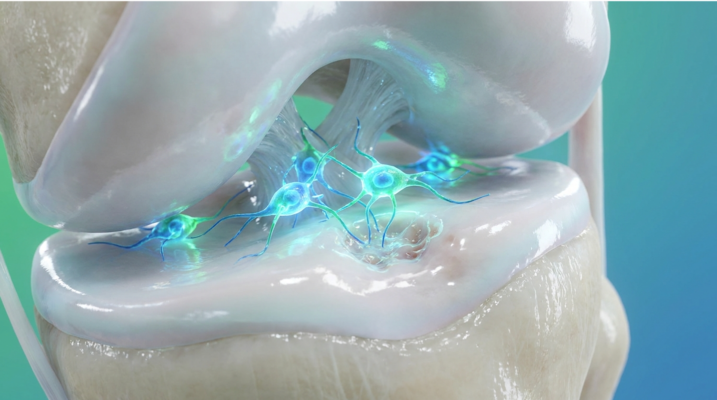
For active adults facing joint pain and an MRI showing joint damage, the immediate question is whether stem cell cartilage regeneration is a viable alternative to surgery. We hear this question daily.
Can damaged cartilage be regenerated with stem cells? Studies show less pain, better function, and in some patients, improved cartilage thickness on MRI imaging. But look stem cells do not yet reliably rebuild perfectly normal cartilage for everyone. Results heavily favor early and focal damage over widespread arthritis.
We’ll examine which joints respond best, explain the ICRS grading scale, and break down how our doctors at Vega Clinic approach this treatment in Thailand. No hype. Just the clinical realities of what orthobiologics can and cannot do for your joints.
Stem cell cartilage regeneration is most effective for focal defects and early-to-moderate osteoarthritis (Grades 1-3), utilizing paracrine signaling to reduce inflammation and promote tissue repair.
Cartilage is notoriously stubborn. Unlike your skin or muscles, it lacks a direct blood supply. When you scrape your knee, blood rushes in with clotting factors and repair cells. When you tear your cartilage, nothing rushes in.
Cartilage lacks a direct blood supply, meaning it cannot deliver the necessary cellular building blocks for natural Tissue Regeneration (Hunziker, 2002). This avascularity leaves chondrocytes the specialized cells that maintain cartilage stranded without the resources to patch up defects.
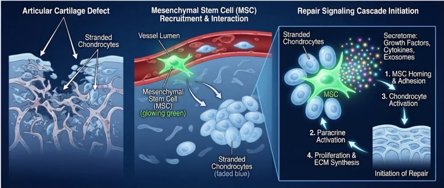
Figure 1: How mesenchymal stem cells interact with stranded chondrocytes to initiate the repair signaling cascade.
Not all cartilage is created equal. Your joints glide on hyaline cartilage, a slick, glassy surface built for low friction. When the body tries to heal a joint defect naturally, it patches it with fibrocartilage. This is essentially dense scar tissue. It’s tough, but it doesn’t glide well and breaks down under continuous load. Hunziker’s 2002 analysis detailed exactly why this biological limitation makes spontaneous healing nearly impossible without intervention. According to a 2021 review in the Cartilage Journal, fibrocartilage possesses only about a third of the load-bearing capacity of natural hyaline cartilage, explaining why these natural patch jobs fail over time.
If you want to understand your MRI, you need to understand grading. Our doctors rely heavily on The Cartilage Viability Matrix, a framework that crosses your joint type with your specific damage grade. The ICRS and Outerbridge grading scales dictate exactly whether a joint is a viable candidate for regenerative medicine or if surgical intervention is unavoidable. We use these established scales to determine if you are a candidate, ensuring expectations align perfectly with biological reality.
| Grade | What it means | What MRI or arthroscopy shows | Realistic options |
| 1 | Softening | Blistering or swelling | Physio, weight loss, PRP |
| 2 | Partial tear | Fissures under 50% depth | Stem cells, PRP |
| 3 | Deep tear | Clefts down to subchondral bone | Stem cells, ACI/MACI |
| 4 | Full loss | Exposed bone (widespread) | Joint replacement |
Mesenchymal stem cells show the highest efficacy in treating Grade 2 and Grade 3 cartilage damage, specifically for focal defects rather than widespread joint degradation (Brittberg, 2003).
So, what grade of cartilage damage can stem cells treat effectively? The answer lies in your remaining tissue architecture. Grade 2 and Grade 3 lesions provide enough surviving chondrocytes to act as a scaffold for cellular repair signals.
But look if your MRI reveals widespread Grade 4 destruction, the cellular foundation is completely gone. Stem cells simply cannot rebuild a joint from scratch. We regularly turn away patients with Grade 4 Bone on Bone osteoarthritis because surgical joint replacement remains the only reliable gold standard according to the ICRS and Outerbridge criteria established in 2003. Once the subchondral bone is widely exposed, the American College of Rheumatology (ACR) confirms that biological scaffolding cannot anchor effectively, making regenerative injections futile.
Ignoring these grading scales guarantees clinical failure. Our doctors evaluate MRI T2 mapping meticulously to ensure we only treat grades capable of actual biological response. The Osteoarthritis Research Society International (OARSI) emphasizes that clinical outcomes correlate directly with accurate pre-injection grading. Real cellular repair mandates a surviving scaffold.
When exploring cartilage damage treatment, you must choose between conservative management, which masks symptoms, and orthobiologics, which attempt true tissue modification. Over 70% of traditional conservative treatments focus solely on inflammation reduction without altering the underlying disease progression. These conventional methods often trap patients in a frustrating cycle of temporary relief followed by inevitable flare-ups.
As joint degradation worsens over time, relying strictly on symptom-masking therapies becomes increasingly inadequate. The AAOS orthobiologics guidelines establish clear evidence levels for these approaches, separating treatments that genuinely repair from those that merely relieve. Arthritis Foundation guidelines similarly emphasize that while conservative routes manage symptoms safely, they inherently fail to induce structural modification in degraded joints. You need an intervention that changes the biological environment, not just one that turns off the fire alarm temporarily.
| Option | Best for | Regrows cartilage? | Downtime | Evidence |
| Physio/Weight Loss | Everyone | No | None | High |
| Steroid | Acute flares | No (accelerates loss) | None | High |
| Hyaluronic Acid | Lubrication | No | 1-2 days | High |
| PRP | Mild OA | No (anti-inflammatory) | 1-2 days | Mod |
| Microfracture | Small defects | Fibrocartilage only | 6-8 weeks | Mod |
| ACI/MACI | Focal defects | Yes | 3-6 months | Mod |
| Grafts | Large defects | Yes (transplant) | 3-6 months | Mod |
| MSC Injection | Grades 2-3 | Sometimes (Hyaline-like) | 2-3 days | Mod |
Moving Beyond Microfracture is essential because microfracture procedures only produce rigid fibrocartilage, not the smooth hyaline cartilage your joints require to glide without friction.
Patients constantly ask about supplements. Let’s look closely at glucosamine vs stem cells for cartilage repair. The 2006 GAIT trial proved conclusively that glucosamine works no better than a placebo for roughly 80% of moderate osteoarthritis patients.
Dietary supplements like glucosamine cannot physically regenerate lost joint tissue, whereas stem cells introduce active biological agents to alter the joint environment (Clegg, 2006). Even the Best Supplements can only modestly reduce inflammation. Relying on oral supplements to rebuild a degraded joint is a biological impossibility that delays effective intervention. Stem cell therapy, conversely, introduces active biological cells into the joint space to physically alter the chemical hostility and stimulate localized tissue remodeling.
There’s a massive misconception about stem cell cartilage regeneration. People think stem cells are injected, attach to the bone, and magically transform into brand-new cartilage. That’s rarely how it works.
Instead, they operate via paracrine signaling. The injected cells act as biological managers. They send powerful chemical signals that calm hostile joint inflammation, protect your surviving chondrocytes from cell death, and stimulate your local tissue to repair itself. They manage the repair site rather than doing all the heavy lifting themselves. This Mechanism of Knee repair changes the entire environment of a degraded joint.
Our doctors prefer Umbilical Cord Mesenchymal Stem Cells (UC-MSCs). Why? They eliminate the need for a painful bone marrow harvest from your hip. More importantly, they are cultivated in highly controlled environments, ensuring robust cellular vitality that aging adult stem cells often lack. An NIH comprehensive review of paracrine signaling in MSCs in 2022 confirmed this approach yields consistent immunomodulatory effects.
The study demonstrated that these cellular signals actively switch local macrophages from a pro-inflammatory state to a tissue-repairing state. By modulating this immune response, UC-MSCs effectively shield surviving chondrocytes from premature cell death. Furthermore, a 2023 study published in Stem Cell Research & Therapy demonstrated that UC-MSCs possess superior proliferative capacity and immunomodulatory strength compared to traditional bone marrow aspirates in older adults.
While the knee dominates the literature, the hip and shoulder present unique mechanical challenges. A systematic review in the Orthopaedic Journal of Sports Medicine noted that while hip osteoarthritis often responds to MSCs with marked reductions in WOMAC pain scores, verifying structural regrowth is notoriously difficult due to the joint’s deep ball-and-socket constraint. The tight joint capsule restricts cellular expansion. Similarly, for the shoulder, stem cells are frequently deployed for rotator cuff tendinopathy or localized glenohumeral arthritis, but the evidence for widespread cartilage regeneration remains strictly moderate compared to the broader surfaces of the knee.
Patients often inquire about niche, non-weight-bearing joints. When considering regenerative approaches, we are highly cautious. Clinical evidence for the elbow remains strictly limited compared to weight-bearing joints like the knee or hip. The elbow’s unique hinge mechanics and thin cartilage surface make cellular anchoring exceptionally difficult. While biological injections can successfully reduce localized pain and quiet down surrounding tendon inflammation (such as severe tennis elbow), robust structural regrowth data in elbow cartilage is currently lacking in Phase III trials. If a clinic promises you massive cartilage regeneration in an elbow joint without presenting specific, localized MRI verification data, they are ignoring the current limits of orthopedic science.
The meniscus acts as the critical shock absorber between your femur and tibia. Lose it, and your risk of severe arthritis skyrockets. We aggressively try to save the meniscus whenever possible.
Can a torn meniscus heal with stem cells? It depends entirely on the tear’s location. The meniscus has distinct vascular zones. The red zone (the peripheral outer third) features a robust blood supply, making it highly responsive to paracrine signaling and biological intervention. However, the white zone (the inner two-thirds) is entirely avascular, relying strictly on synovial fluid diffusion for nutrients.
| 📌 If you’re interested in how MSCs shift the immune system from attack mode to repair mode, we have an interesting article that discusses mesenchymal stem cell therapy for immune modulation, which you can read via the internal link. |
According to Johns Hopkins Orthopaedic surgery data, tears extending deeply into this avascular white zone show virtually zero spontaneous healing potential. Even with high-dose MSC injections, these deep inner tears remain highly resistant to regeneration. Conversely, a landmark Vangsness clinical study in 2014 showed that an MSC injection successfully increased meniscus volume in specific patients treating tears with adequate blood supply.
Many want a stem cell injection for meniscus tear without surgery. For degenerative tears or small partial tears in the vascular zones, conservative injections often suffice. However, if you have a massive bucket-handle tear or a mechanically locked knee, a needle won’t fix it. You need surgical intervention to restore the anatomy first.
Ankle cartilage is incredibly dense and thin, typically measuring just 1 to 2 millimeters thick. Because the joint space is so tight, ankle sprains and repetitive impacts often lead to localized cartilage defects. This creates a highly hostile healing environment due to the massive mechanical load placed on such a small surface area. Every step forces your entire body weight onto a joint the size of a hinge, guaranteeing that spontaneous natural healing almost never occurs without medical intervention.
When considering any ankle cartilage damage treatment, it’s crucial to understand that standard conservative options like bracing often fail to address the core structural deficit. Treating ankle cartilage demands extreme precision because the biological margin for error is virtually zero. The American Orthopaedic Foot & Ankle Society (AOFAS) stresses that precise MRI staging is mandatory before attempting biological injections in the ankle, as late-stage lesions collapse mechanically under weight.
You shouldn’t accept vague claims about “feeling better” when assessing joint regeneration. True regenerative medicine demands highly objective metrics to verify biological success. We measure functional outcomes using established WOMAC and KOOS scoring systems, which track pain reduction and daily mobility. But to prove definitive structural changes, we rely on MRI T2 mapping.
This specialized scan doesn’t just show the physical outline of the joint; it measures the specific biochemical composition of cartilage, tracking water content and collagen fiber orientation. When cartilage regenerates, T2 mapping visualizes the structural density returning to normal levels. Additionally, second-look arthroscopy provides direct visual verification of new hyaline-like tissue formation. Phase III clinical trial protocols mandate these exact measurement standards to separate true regeneration from placebo effects. Any clinic promising cartilage regrowth without performing pre- and post-treatment T2 mapping MRIs is operating on guesswork, not science.
How is cartilage regrowth measured after a stem cell injection practically? Ask your clinic for their exact imaging protocol. The Osteoarthritis Research Society International (OARSI) recommends integrating T2 mapping as a primary endpoint in cartilage repair trials specifically because it assesses collagen network integrity, not just fluid volume. If they only rely on standard X-rays, which cannot see cartilage at all, they cannot accurately track your structural progress.
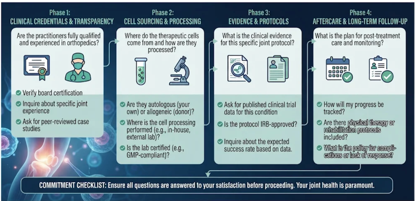
The knee possesses the strongest clinical evidence for stem cell cartilage regeneration, backed by numerous Phase III trials. Other joints frequently treated include the hip, shoulder, ankle, and elbow, though evidence for these areas varies significantly. Ultimately, treatment outcomes depend heavily on joint mechanics, local blood supply, and the specific grade of your localized cartilage damage. Complex, tight joints like the ankle require highly targeted approaches compared to the broader surfaces of the knee. This is why precise pre-treatment imaging is absolutely non-negotiable for success.
Protect new cartilage by maintaining a healthy weight and engaging in low-impact exercises like swimming or stationary cycling. You must avoid high-impact sports entirely during the initial six-month healing phase, as the new cellular matrix is highly vulnerable to shearing forces. Adhering to a physician-prescribed physical therapy program ensures proper joint mechanics and reduces unnecessary stress on the newly treated area. Using prescribed off-loader braces can also provide essential mechanical relief while the fragile scaffold matures.
Umbilical cord-derived mesenchymal stem cells (UC-MSCs) are generally safe for intra-articular injections when processed in approved, strictly regulated GMP laboratories. Because they naturally lack specific surface proteins, they minimize the risk of immune rejection in the recipient. However, as an investigational treatment, patients may still experience temporary localized swelling, mild stiffness, or injection-site discomfort for a few days. Our doctors monitor these expected inflammatory responses closely during the immediate post-injection window.
| 📌 If you’re interested in why umbilical cord cells are often preferred over bone marrow or fat-derived cells, we have an interesting article that discusses why UC-MSCs are superior to other stem cell sources, which you can read via the internal link. |
The cost of stem cell therapy for hip joints in Thailand typically ranges significantly lower than equivalent regenerative treatments offered in the US or UK. Pricing depends strictly on the exact number of MSCs required, the complexity of the imaging utilized, and the specific clinical facility performing the procedure. Patients receive exact, transparent cost estimates only after completing an initial MRI evaluation. This ensures you never pay for a generic protocol that doesn’t match your joint’s specific needs, protecting your health and your wallet.
Stem cell therapy cannot reliably fix widespread Grade 4 bone-on-bone arthritis. When cartilage is entirely depleted across the joint surface, biological scaffolds simply lack the foundational architecture needed to rebuild new tissue. In these severe cases, orthopedic surgeons typically recommend total joint replacement surgery rather than regenerative injections.
Most patients notice initial reductions in joint inflammation and daily pain within four to eight weeks following a targeted stem cell injection. However, actual structural changes in the cartilage generally require six to twelve months to fully develop. This extended timeline occurs because the cellular signaling process takes months to successfully enact complex tissue remodeling. Patience during this remodeling phase is critical for long-term clinical success.
Surgery is not always required for an osteochondral lesion, especially if caught early. Early-stage or focal lesions often respond remarkably well to conservative treatments or advanced regenerative orthobiologics like stem cell injections. However, highly unstable lesions containing loose cartilage fragments floating within the joint space almost always necessitate surgical intervention. In those severe instances, surgical debridement or bone grafting is required to restore basic mechanical function before any cellular therapy can be considered.
Stem cell treatments operate under strict regulations governed by the Thai Medical Council to ensure patient safety and ethical practices. While certain cell therapies are approved and conducted in licensed GMP facilities, many regenerative joint protocols are still classified as investigational globally. Patients must independently verify that their chosen clinic operates fully within Thailand’s authorized medical framework to avoid unverified treatments.
| 📌 If you’re planning treatment in Thailand and want a practical checklist for choosing a safe clinic, we have an interesting article that discusses whether stem cell therapy is safe in Thailand, which you can read via the internal link. |