Our Services
Others
- DFPP
- Shockwave
- IV Drip

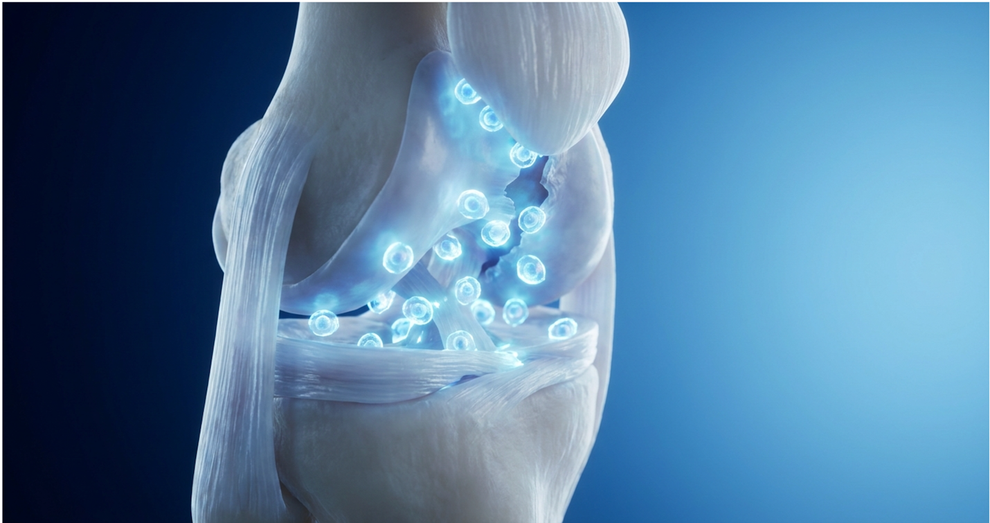
For athletes and active adults facing a meniscus tear, the traditional binary choice between prolonged rest and arthroscopic surgery is obsolete. Clinical evaluations of sports injuries routinely demonstrate athletes losing entire seasons to the misconception that cutting out damaged tissue is the only way forward. Resecting the meniscus accelerates joint degradation, yet many avascular tears simply will not heal natively.
This clinical guide evaluates the established efficacy data surrounding Umbilical Cord-Derived Mesenchymal Stem Cells (UC-MSCs), comparing them directly against conventional orthobiologics like PRP to determine who actually qualifies as a viable non-surgical candidate. We analyze mechanisms of action, strict clinical limitations, and accurate 2026 pricing models for regenerative therapies.
If you are researching Stem Cell Therapy for Meniscus Tears, you need objective data, not clinic marketing brochures.
Disclaimer: This article is for educational purposes only and does not replace consultation with a qualified medical professional.
Stem Cell Therapy for Meniscus Tears utilizing UC-MSCs promotes active tissue regeneration in avascular zones where native healing completely fails.
What happens if a meniscus tear is left untreated? Ignoring a meniscal lesion disrupts normal load transmission across the tibiofemoral joint. Untreated meniscal lesions increase focal cartilage contact stress by up to 200% rapidly triggering irreversible early-onset osteoarthritis. The biological consequence of neglecting structural damage isn’t just persistent pain. It is accelerated joint space narrowing and chronic inflammatory signaling that degrades the entire knee ecosystem.
The meniscus, a crescent-shaped fibrocartilaginous structure in the knee, is divided into distinct anatomical zones based on vascularity. The outer third is the “Red-Red” zone, which has a robust blood supply. The middle third is the “Red-White” zone, possessing marginal vascularity. The inner third is the entirely avascular “White-White” zone.
Can you recover 100% from a meniscus tear? It depends almost entirely on the zone. Tears in the outer Red-Red zone often heal natively with conservative management or simple surgical suturing. However, lesions in the White-White zone lack the blood supply required to mount a cellular repair response. PMC clinical review data details how meniscal vascularization simply cannot support tissue repair in these inner regions.
Without blood, there are no platelets. Without platelets, there are no growth factors. When native vascularity fails, chronic degradation ensues. According to a PubMed clinical analysis, spontaneous healing rates in the avascular zone are virtually zero (PubMed, 2021).
Leaving a complex tear untreated fundamentally alters knee biomechanics. This is especially true for radial or root tears, widely considered the hardest meniscus tear to repair. A root tear essentially detaches the meniscus from the bone, completely compromising its structural integrity.
When this happens, the meniscus loses its ability to convert axial compressive loads into hoop stresses. The meniscus literally extrudes outward. Suddenly, the medial femoral condyle is grinding directly against the tibia with minimal shock absorption. This specific mechanical failure concentrates catastrophic peak contact pressures precisely on the central weight-bearing compartment. Over time, the unprotected subchondral bone reacts by forming microfractures and bone marrow lesions. The medial femoral condyle rapidly flattens under these unmitigated forces, shifting the mechanical axis of the entire leg and creating a highly pro-inflammatory joint environment that is hostile to native chondrocytes.
Macrophages flood the joint space. Synovial fluid breaks down. Cartilage erosion accelerates at a frightening pace, stripping away the joint’s protective hyaline layers. Orthopedic diagnostic protocols frequently identify patients who ignored a minor tear for two years only to present with Grade 3 osteoarthritis that could have been entirely prevented.
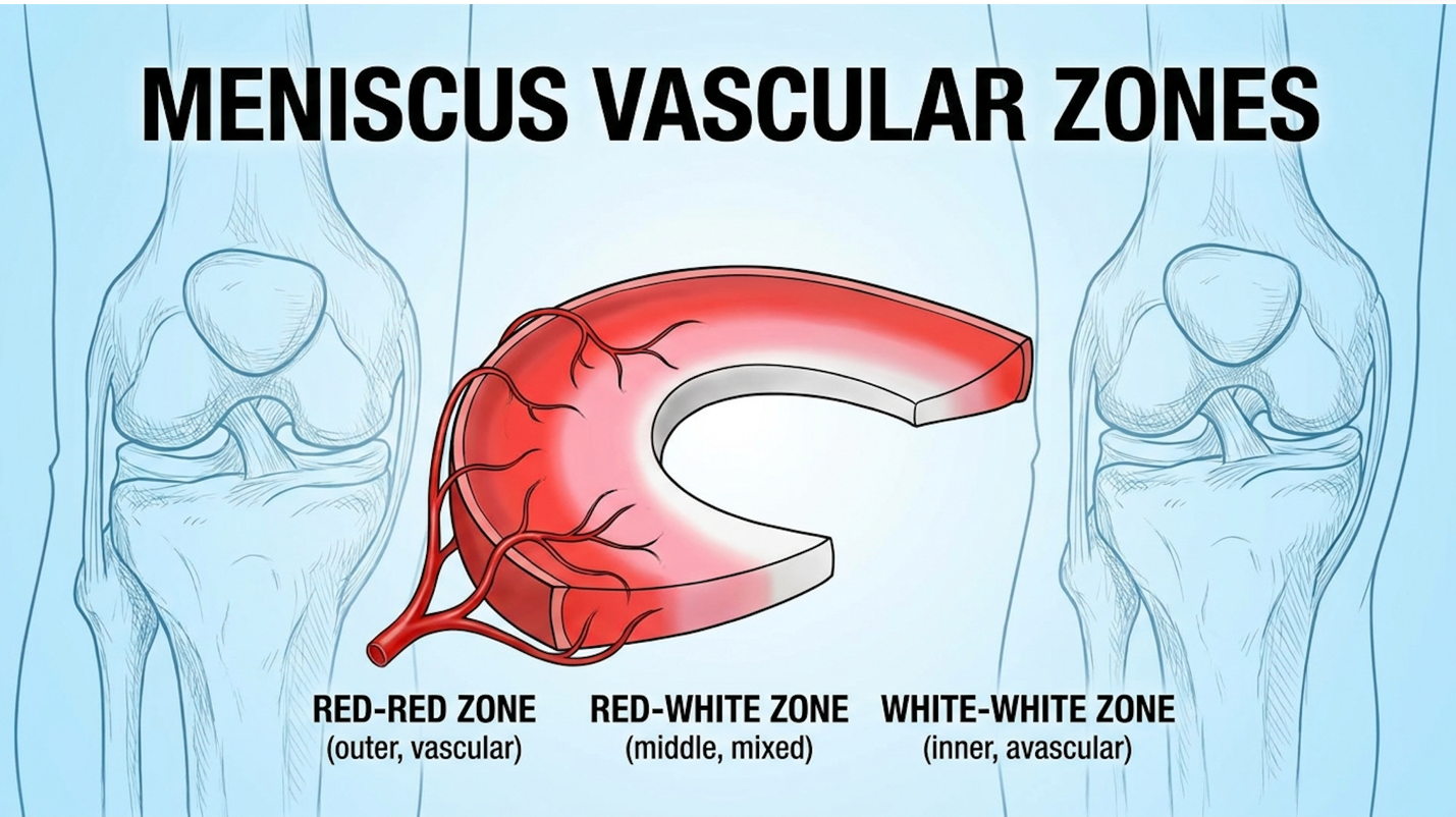
Figure 1: The vascular distribution across the meniscus directly dictates native healing capacity.
To arrest this degenerative cascade without resorting to surgical resection which ironically also accelerates osteoarthritis by removing the shock absorber entirely clinical medicine has turned to advanced biological interventions.
UC-MSC Stem Cell Therapy for Meniscus Tears utilizes Wharton’s Jelly-derived progenitor cells to actively rebuild damaged fibrocartilage. Unlike native adult cells, these youthful mesenchymal stromal cells secrete concentrated bioactive molecules that stimulate dermal fibroblasts and synthesize collagen type I, directly altering the intra-articular environment from destructive to regenerative (Frontiers, 2022).
Umbilical Cord-Derived Mesenchymal Stem Cells (UC-MSCs) represent a massive biological upgrade over older, autologous (patient-derived) cell therapies. For years, orthopedic clinics relied on Bone Marrow MSCs harvested from the patient’s own hip. But here is the problem with bone marrow: as you age, your stem cells age. A 55-year-old patient yields 55-year-old cells, which possess a significantly lower proliferative capacity, reduced telomere length, and a sluggish metabolic response to inflammatory environments.
UC-MSCs, harvested from the Wharton’s Jelly of donated umbilical cords post-cesarean section, are essentially day-zero cells. They possess massive expansion capacity, frequently yielding 30 million to 50 million highly viable cells per dose without losing their multipotency. More importantly, their secretomes the complex arrays of bioactive molecules, exosomes, and microvesicles secreted by stem cells are vastly more potent.
These extracellular vesicles are loaded with immunomodulatory factors like Prostaglandin E2 (PGE2) and Indoleamine 2,3-dioxygenase (IDO), which specifically target aggressive joint inflammation. These cells undergo rigorous FDA-compliant screening and processing in regulated cGMP laboratories before being cryopreserved. Crucially, they are immune-privileged. Because they lack Major Histocompatibility Complex (MHC) class II surface antigens, they do not trigger a rejection response. When evaluating mesenchymal stem cell therapy for meniscus injuries, cell youth and secretome density are the two metrics that matter most. A Frontiers in Pharmacology review confirmed that the clinical applications of these specific progenitor cells consistently outperform autologous counterparts in both safety and efficacy profiles.
So, can stem cell therapy heal a torn meniscus? To answer this, you have to look closely at the exact biological mechanism of action. When UC-MSCs are injected into the intra-articular space under ultrasound guidance, they don’t simply turn into meniscus cells immediately. That’s a persistent misconception.
Instead, they act as cellular general contractors. First, they execute phenotypic macrophage reprogramming. This means they force the aggressive, tissue-destroying M1 macrophages in the knee to phase-shift into healing M2 macrophages. This transition actively halts the production of destructive inflammatory cytokines like IL-1β and TNF-α, effectively cleaning up cellular debris and resetting the hostile joint environment.
Next, they release massive amounts of Vascular Endothelial Growth Factor (VEGF), Transforming Growth Factor-beta (TGF-β), and Insulin-like Growth Factor 1 (IGF-1) via exosome delivery. This paracrine signaling instructs the patient’s existing dormant chondrocytes to wake up and begin synthesizing the extracellular matrix. The secretome directly regulates this specific synthesis by upregulating the SOX9 transcription factor, the master regulator of chondrogenesis. Finally, a subset of the UC-MSCs will actively differentiate into fibrochondrocytes to patch the physical deficit.
Researchers are also advancing localized delivery mechanisms. The NHS HRA research summary evaluated a highly innovative cell bandage meniscus approach, essentially sewing a stem cell patch directly onto the lesion during a brief arthroscopy. This structural scaffolding ensures the cells remain physically anchored exactly where the repair is needed most.
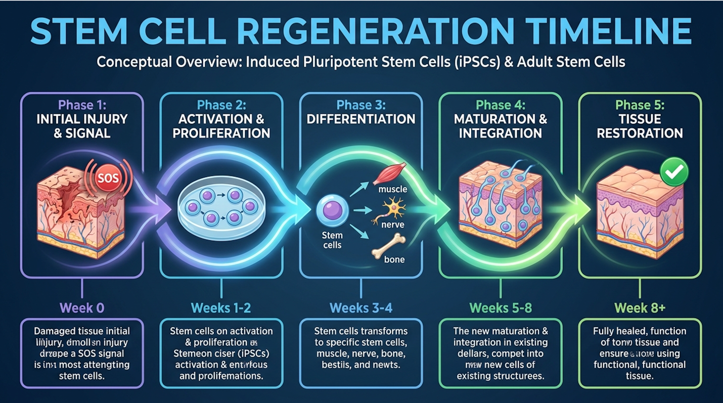
Figure 2: The cellular response timeline from injection through extracellular matrix synthesis.
What is the newest treatment for meniscus tears? The frontier isn’t a new surgical instrument; it’s the precise application of these orthobiologics under strict investigational frameworks.
Current randomized controlled trials (RCTs) comparing UC-MSC efficacy against surgical controls show a clear divergence in outcomes at the 24-month mark. While surgery offers faster mechanical relief, UC-MSC injections can significantly improve Knee Society Scores and reduce synovial inflammation within 12 weeks of administration validating their role as active regenerative agents.
A detailed Boston Sports & Biologics review of RCT data found that MSCs injected post-meniscectomy significantly halted cartilage degradation compared to placebo. Advanced double-blind, multicenter trials measuring Joint Space Width (JSW) via MRI routinely demonstrate that patients receiving high-dose UC-MSC therapies halt progressive joint space narrowing. In these specific clinical trial methodologies, researchers utilize 3D quantitative MRI mapping (qMRI) to precisely track both cartilage thickness and meniscal volume down to the millimeter over 12-to-24-month intervals. Rather than relying solely on subjective patient pain questionnaires, this advanced JSW tracking provides objective, radiographic proof of structural preservation. By accurately calculating T2 mapping relaxation times, scientists can observe the biochemical composition of the newly synthesized extracellular matrix, confirming it structurally mimics native healthy cartilage rather than stiff scar tissue. This meticulous radiographic tracking explains exactly how clinical trial participants successfully avoided total knee arthroplasty, maintaining functional joint anatomy. Further PubMed indexed clinical data evaluating UC-MSCs confirmed these measurable increases in meniscal volume on follow-up MRIs (PubMed, 2022). These structural gains correspond directly with improved load-bearing capacity and a delayed need for surgical joint replacement.
When evaluating prp vs stem cell for meniscus repair, the core difference lies entirely in cellular capacity. Platelet-Rich Plasma (PRP) delivers concentrated growth factors that signal native tissues to heal, which is fundamentally ineffective in avascular meniscal tears. Conversely, UC-MSCs deliver viable progenitor cells capable of de novo tissue synthesis and profound immune modulation.
Is PRP worth it for meniscus tears? Yes, but only under highly specific conditions. PRP is an autologous concentration of growth factors drawn from your own blood. It works brilliantly for highly vascularized tissue injuries like muscle strains or medial collateral ligament (MCL) tears.
But what are the downsides of PRP injections in the knee when treating a meniscus? First, PRP contains no stem cells. Zero. It relies entirely on signaling your existing cells to work harder. If the tear is in the White-White avascular zone, there are virtually no cells there to receive the signal. While PRP successfully mitigates mild knee inflammation, it lacks the cellular machinery to regenerate severe white-zone meniscus tears.
Second, PRP quality is heavily dependent on the patient’s age, diet, and systemic health. A 60-year-old with metabolic syndrome will yield low-quality PRP heavily burdened with senescent cells and lower baseline concentrations of growth factors. Over 65% of older patients utilizing PRP for avascular tears eventually require further intervention proving growth factors alone cannot rebuild severe white-zone deficits according to a PubMed cohort study (PubMed, 2021).
How many PRP injections are needed for a meniscus tear? Typically, protocols require 1 to 3 injections spaced several weeks apart. Patients frequently ask, “Should I wear a knee brace after a PRP injection?” Yes, unloader braces are highly recommended for 2-4 weeks post-injection to protect the joint while the growth factors attempt to stimulate repair.
To eliminate the guesswork in regenerative orthopedics, modern protocols rely on The Meniscal Viability Matrix. This is a strict clinical evaluation framework determining whether a meniscus tear’s location and chronicity make it a candidate for UC-MSC biological regeneration over surgical resection or PRP. Recognizing the hard biological boundaries of PRP necessitates a strict clinical framework for patient selection.
The matrix assesses three distinct diagnostic pillars before approving a patient for cellular therapy:
Who is not a good candidate for PRP? According to the matrix, patients with severe bone-on-bone osteoarthritis, full-thickness complex radial tears in the avascular zone, or systemic blood disorders should bypass PRP entirely. For these patients, UC-MSC therapy provides the cellular raw materials that PRP simply cannot. Clinical research in osteoarthritis confirms that advanced degeneration neutralizes the mild signaling effects of autologous plasma (PubMed, 2023).
If you’re evaluating UC-MSC stem cells vs PRP for meniscus injury, the data breaks down clearly along biological lines:
| Modality | Mechanism | Best For | Number of Treatments | Regeneration Capacity |
| PRP | Growth factor signaling | Red-zone tears, mild OA, active youth | 1-3 injections | Low (relies on native cells) |
| UC-MSC | Phenotypic modulation & de novo synthesis | White-zone tears, moderate OA, older adults | 1 injection | High (delivers progenitor cells) |
Once clinical candidacy is established through the matrix, patients must navigate the financial realities of advanced biologics.
How much does stem cell treatment for meniscus tears cost? Because the FDA classifies cultured UC-MSCs as investigational applications, these therapies are rarely covered by major medical insurance. In 2026, patients can expect out-of-pocket investments ranging significantly based on cell counts, laboratory quality, and clinical protocols, requiring careful financial ROI evaluation. Stem cell injection for knee meniscus tear cost is an upfront capital expense designed to prevent a lifetime of chronic orthopedic billing.
Let’s look at the hard numbers. How much does a PRP injection for a meniscus tear cost? Because the procedure simply involves a blood draw and a centrifuge, it is relatively accessible, ranging from $500 to $2,500 per injection depending on the imaging guidance used.
The price of stem cell therapy knee injections is substantially higher. Expect to pay between $4,000 and $10,000+ for a high-quality UC-MSC procedure. Why the massive price jump? You are paying for the rigorous cGMP laboratory processing, FDA-compliant screening, cryopreservation transport, and the sheer volume of viable cells (often 30 million to 50 million cells per dose).
Evaluating these upfront costs requires a direct comparison against the long-term financial burden of surgical intervention.
Indirect costs of arthroscopic surgery and subsequent lost wages frequently exceed $15,000 making upfront cellular therapy investments economically viable for avoiding lifelong orthopedic billing.
Patients routinely ignore the hidden costs of a meniscectomy. You have to factor in your high-deductible insurance plan, co-pays for 12 weeks of physical therapy, facility fees, anesthesiology bills, and weeks of lost productivity. Furthermore, an economic analysis in PubMed showed that partial meniscectomy accelerates joint decay, often culminating in a total knee replacement a decade later a procedure frequently exceeding $40,000 (PubMed, 2022).
Despite favorable economic models, biological therapies possess strict clinical limitations.
No biological therapy offers a magical cure. In orthopedic medicine, treating structural damage entirely with a needle has hard physiological limits, and responsible physicians must clearly define who will fail stem cell therapy. It is about informed decision-making, not blindly avoiding the scalpel.
There are specific clinical profiles where stem cell therapy after meniscus surgery or as a primary intervention will definitively fail. Patients with Grade 4 bone-on-bone osteoarthritis face a 0% structural regeneration rate with biologics requiring immediate surgical evaluation rather than cellular intervention.
First, patients with bone-on-bone Grade 4 osteoarthritis are poor candidates for soft tissue regeneration; the joint architecture is already destroyed. Second, patients with active systemic infections or active malignancies are disqualified. Third, massive mechanical tears such as a flipped fragment causing the knee to physically lock cannot be dissolved or magically reattached by cells. Contraindications in regenerative medicine require stringent pre-injection MRI screening to rule out mechanical blocks (PubMed, 2023).
Sometimes, surgery is the only responsible option. Bucket-handle tears that flip into the joint notch create a hard mechanical block. You cannot straighten your knee. In these scenarios, the meniscus must be physically trimmed or sutured via arthroscopy before any biological therapy can even be considered. Surgical indications for bucket-handle tears dictate that restoring joint mobility takes immediate precedence over tissue preservation (PubMed, 2023).
Stem cell therapy, specifically utilizing UC-MSCs, can stimulate the regeneration of damaged meniscal tissue under specific clinical conditions. These viable progenitor cells synthesize new collagen type I and elastin while modulating local joint inflammation. Success relies heavily on the tear’s location, with lesions closer to vascular zones showing higher regenerative capacity. However, massive structural tears or complete degenerative collapse may not respond to biological therapies and require surgical evaluation.
A meniscus tear treated with stem cell therapy typically takes 10 to 12 weeks to demonstrate significant biological healing. Patients often report initial reductions in pain and inflammation within the first 3 to 4 weeks as the immune-modulating effects begin. Structural fibrocartilage regeneration is a slower metabolic process that continues optimizing for up to 6 months post-injection. Final recovery timelines vary based on tear severity, patient age, and adherence to prescribed rehabilitation protocols.
For athletes facing avascular tears, Stem Cell Therapy for Meniscus Tears delivers viable tissue regeneration where native healing fails. By deploying youthful UC-MSCs rather than relying on autologous growth factors, patients achieve profound immune modulation and de novo collagen synthesis. UC-MSC injections can significantly improve Knee Society Scores and reduce synovial inflammation within 12 weeks of administration. An optimal clinical approach combines precise ultrasound-guided injections, rigorous post-procedure bracing, and targeted physical therapy.
This is precisely why The Meniscal Viability Matrix is so critical in modern orthopedics. By applying this clinical framework, practitioners stop guessing with PRP and stop defaulting to arthroscopic resection. We match the exact chronicity and location of the lesion with the appropriate biological firepower, directly mitigating the risk of early-onset osteoarthritis.
If you are dealing with chronic knee pain and a confirmed meniscal lesion, stop pushing through the damage. Secure an updated MRI from the last three months and schedule a specialized clinical consultation with a regenerative orthopedist to determine your exact matrix candidacy.
Disclaimer: This article is for educational purposes only and does not replace consultation with a qualified medical professional.