Our Services
Others
- DFPP
- Shockwave
- IV Drip

For patients diagnosed with Progressive Supranuclear Palsy (PSP), the rapid progression of neurodegeneration forces a search beyond traditional supportive care toward investigational regenerative medicine. Look, the reality of this disease is brutal. Clinical observations demonstrate firsthand how quickly patients lose their balance, their downward gaze control, and their physical independence. It’s a devastating, relentless timeline. Traditional Parkinson’s medications simply don’t work here. And that desperation? It routinely pushes families into a frantic search for alternatives, often leading straight to stem cell therapy for progressive supranuclear palsy.
But here’s the thing. There’s a massive, dangerous gulf between rigorous Phase I clinical research and the predatory “stem cell tourism” aggressively marketed online. You can’t just inject cells and hope for a miracle cure. Biology doesn’t work like that. The underlying neuropathology of a tauopathy requires an incredibly sophisticated biological counter-attack, not a weekend trip to an unregulated offshore wellness clinic.
This guide provides an evidence-informed synthesis of current published clinical data, detailing the exact biological mechanisms, safety profiles, and early trial outcomes. We aren’t dealing in false hope today. We’re looking strictly at the raw, peer-reviewed data.
Our roadmap covers the unique pathology of the disease, how researchers use advanced cellular interventions to target brain injury, what the clinical trials actually prove, and how you can safely assess patient suitability. Because making an informed, safe decision requires looking past the commercial hype to understand the deep, complex neurology at play.
Stem cell therapy for progressive supranuclear palsy utilizes investigational UC-MSCs to target neuroinflammation through the Neuro-Immunomodulation Triad framework.
To fully grasp why researchers are exploring stem cell therapy for PSP, we first need to understand the profound structural destruction happening inside the brain. Progressive Supranuclear Palsy (PSP), a rare neurodegenerative tauopathy, behaves entirely differently from classic Parkinson’s disease. Standard dopamine replacement therapy fails almost universally in this population.
PSP progresses significantly faster than Parkinson’s disease, with widespread tau protein accumulation causing severe disability within 3 to 5 years of onset (National Library of Medicine, 2014). Levodopa might buy a Parkinson’s patient a decade of mobility, but for a PSP patient? It rarely offers even a temporary reprieve. The cellular receptors required to process those medications are quite literally degenerating. This relentless, widespread receptor destruction is exactly why traditional pharmacology cannot halt the disease, forcing clinical science to pivot rapidly toward advanced regenerative medicine to target the underlying tissue degradation.
The fundamental biological mechanism driving this disease is the abnormal folding and accumulation of the tau protein. Specifically, PSP is defined by the aggregation of 4-repeat (4R) tau isoforms. In a healthy, functioning brain, tau proteins act as essential biological glue they stabilize microtubules, which serve as the internal scaffolding and molecular transport highways for your neurons.
But in a PSP patient, these tau proteins become hyperphosphorylated. They detach from the microtubules, collapse, tangle, and form dense neurofibrillary tangles inside the neurons. As the microtubules collapse, the neuron loses its ability to transport nutrients from the soma to the synapses, causing axonal starvation. This microscopic starvation scales up into massive tissue loss. Worse, this toxic protein accumulation isn’t isolated to neurons; it heavily infiltrates the surrounding support tissue, creating tufted astrocytes and oligodendroglial coiled bodies.
This creates a highly toxic environment resulting in a diffuse pattern of brain injury. It doesn’t just hit one isolated area of the brain. The tauopathy aggressively destroys the basal ganglia, the subthalamic nucleus, the substantia nigra, and critical regions of the brainstem. The destruction is so severe, in fact, that it physically alters the shape of the brainstem in ways that standard structural imaging captures with grim clarity. Because the midbrain controls autonomic and higher-order motor functions, patients rapidly lose the ability to speak clearly (dysarthria) and swallow safely (dysphagia). Aspiration pneumonia directly resulting from this severe dysphagia remains one of the leading causes of mortality in this patient population.
When neurologists evaluate a midsagittal MRI of a patient with advanced PSP, they look for the classic “hummingbird sign.”
`
This imaging reveals severe atrophy of the midbrain tegmentum while the pons remains relatively intact. The resulting silhouette looks exactly like a hummingbird. Contrast this with normal aging, where the midbrain volume remains robust and proportional. The presence of this midbrain atrophy on an MRI is a highly specific radiological marker that helps differentiate PSP from other parkinsonian syndromes (Lancet Neurology, 2009).
Because the injury is so incredibly widespread across multiple critical motor and cognitive hubs, highly targeted surgical interventions like Deep Brain Stimulation (DBS) are completely ineffective. The neuroinflammation triggered by this tau accumulation accelerates a cellular death spiral. Toxic proteins signal the brain’s immune system to attack, causing massive collateral damage to any surviving neurons. Understanding this widespread tissue degradation is essential to grasp why PSP presents so differently and more aggressively than classic Parkinson’s disease.
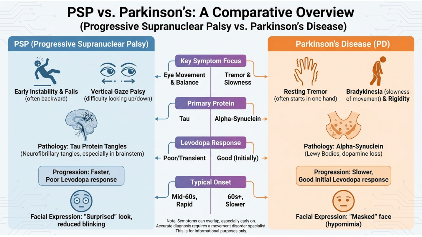
While both conditions fall under the broad umbrella of movement disorders, their clinical trajectories diverge sharply across clinical presentations. Patients and their families often spend the first agonizing year navigating a frustrating maze of misdiagnoses, as early PSP can vaguely mimic Parkinsonism. But the specific milestones of physical decline tell a distinctly different, far more devastating story.
A review of patient histories indicates a brutally consistent pattern. The early postural instability in PSP leads to unexplained, dangerous backward falls often occurring within the first year of symptom onset. Parkinson’s patients typically don’t experience severe balance issues until much later in their disease course, often remaining functionally independent and physically safe for years.
And the stiffness in PSP is axial (affecting the neck and trunk), whereas Parkinson’s stiffness is typically appendicular (affecting the limbs). Within 24 to 36 months, this axial rigidity often becomes so severe that independent ambulation is impossible. Caregivers are forced to manage completely rigid physical transfers, exponentially increasing the toll on the family.
| Clinical Feature | Progressive Supranuclear Palsy | Parkinson’s Disease |
| Core Pathology | 4R Tau protein accumulation (Tauopathy) | Alpha-synuclein accumulation (Synucleinopathy) |
| Brain Injury Pattern | Diffuse (midbrain, basal ganglia, brainstem) | Localized (substantia nigra pars compacta) |
| Early Symptoms | Axial rigidity, backward falls, gaze palsy | Unilateral resting tremor, bradykinesia |
| Response to Levodopa | Poor to nonexistent | Highly responsive in early-to-mid stages |
| Average Timeline to Wheelchair | 3 to 5 years | 10 to 15+ years |
The hallmark physical sign of PSP is supranuclear gaze palsy the inability to voluntarily look down. This specific neurological deficit makes eating, walking down stairs, and reading incredibly dangerous and difficult. Patients frequently develop pseudobulbar affect as well a harrowing symptom where neurodegeneration causes uncontrollable bouts of crying or laughing that completely mismatch the patient’s actual emotional state.
When families initially seek a PSP atypical parkinson treatment, they are often handed standard dopaminergic medications out of habit. But the complete lack of response in PSP patients serves as a grim diagnostic confirmation. In Parkinson’s, Levodopa replaces the missing dopamine, and the receiving receptors use it efficiently. In PSP, the postsynaptic D2 receptors in the striatum are physically destroyed by the tauopathy. The brain simply lacks the intact neural infrastructure to utilize the dopamine.
With standard pharmacological options exhausted early in the disease course, clinicians are heavily investigating cellular mechanisms capable of broad neuroprotection. We need an intervention that addresses the entire neuroinflammatory environment across the brainstem, rather than a pill just masking a dopamine deficiency.
When we talk about applying cellular therapies to severe tauopathies, we are specifically looking at Umbilical Cord Mesenchymal Stem Cells (UC-MSCs). These are day-zero, robust cells harvested ethically from donated Wharton’s jelly after healthy full-term births. They are typically administered via intrathecal injection (directly into the spinal fluid) or intravenous infusion, allowing them to bypass the severe limitations of localized surgical interventions.
Current in vivo mechanism studies show these cells do not actually engraft and turn into new brain cells. They don’t “replace” dead tissue. Instead, they operate as highly sophisticated biological factories that temporarily alter the toxic environment killing the existing cells. Mesenchymal stem cells function primarily through paracrine signaling, secreting neurotrophic factors that reduce neuroinflammation and promote the survival of existing neurons (Frontiers in Cellular Neuroscience, 2022).
To understand exactly how UC-MSC therapy for neurodegenerative disease operates, we must examine an original concept called The Neuro-Immunomodulation Triad. This is a specific framework defining how UC-MSCs simultaneously execute three distinct, overlapping protective functions in the brain to combat relentless tau-induced deterioration.
This Triad clarifies that UC-MSCs act via a “hit and run” mechanism. They enter the system, release massive payloads of chemical signals through paracrine signaling, and are eventually cleared by the body over a span of months. They are not structural building blocks; they are cellular paramedics temporarily altering the microenvironment.
For the diffuse tauopathy of PSP, this is theoretically ideal. Because the tau tangles trigger widespread inflammatory cascades, a localized treatment simply wouldn’t work. The Neuro-Immunomodulation Triad delivers a systemic biological reset. The first distinct action of this triad involves directly communicating with the brain’s resident immune cells.
In a healthy brain, microglia act as the vigilant janitorial staff, quietly clearing debris, pruning synapses, and maintaining neural health. But in a PSP brain, the accumulation of toxic tau proteins throws these cells into a state of chronic, hyperactive panic. This is known scientifically as the M1 phenotype.
M1 microglia are highly pro-inflammatory and neurotoxic. They aggressively release inflammatory cytokines like TNF-alpha, Interleukin-1 beta (IL-1β), and Interleukin-6 (IL-6). These cytokines act like biological fire throughout the midbrain. This friendly fire accelerates the death of the very neurons the microglia are supposed to protect. A significant portion of neurodegeneration in advanced tauopathies is driven by this chronic microglial overactivation (Frontiers in Immunology, 2019).
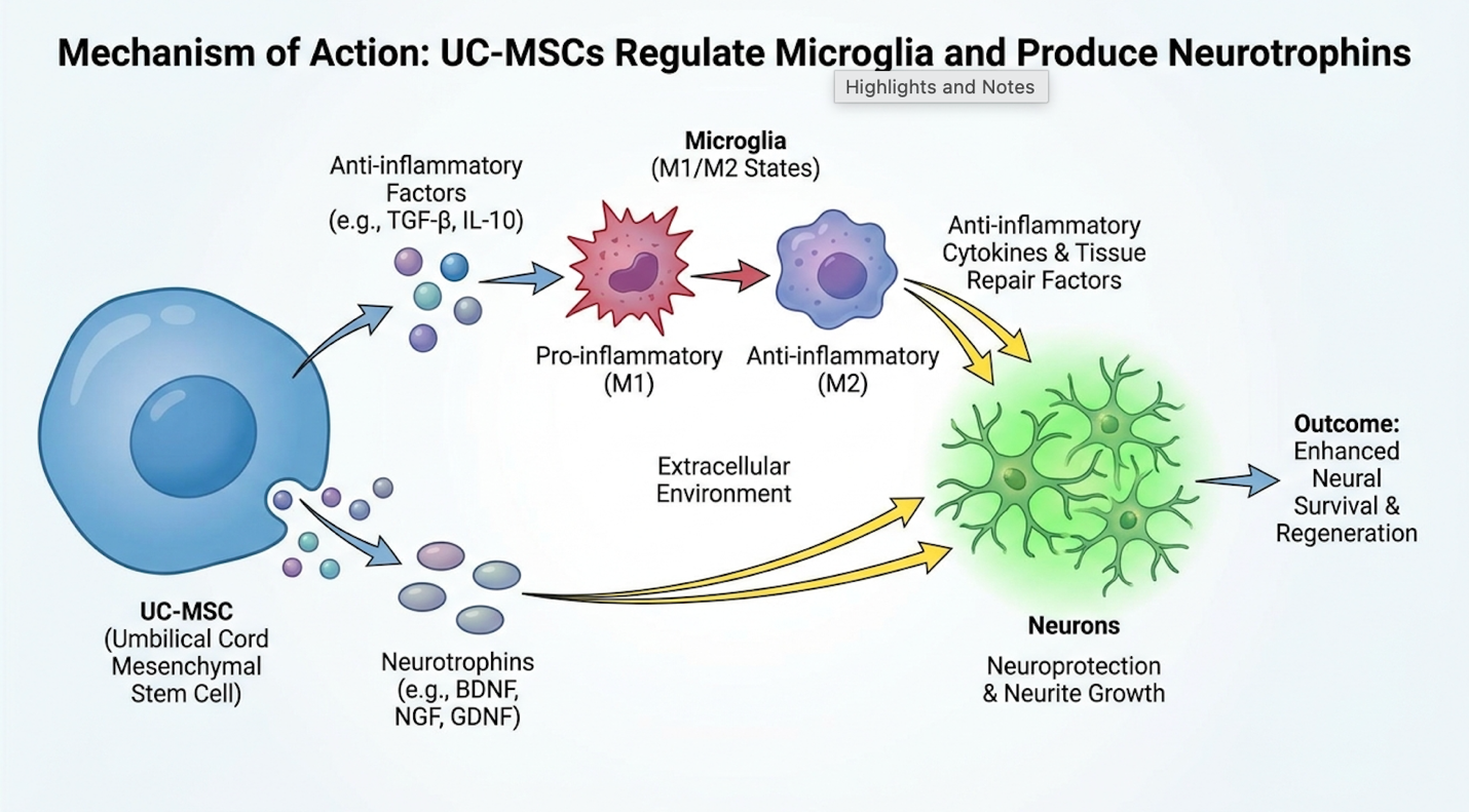
When UC-MSCs are introduced into this toxic environment via the cerebrospinal fluid, they immediately begin interacting with these hyperactive macrophages and microglia-related signaling pathways. Through direct cell-to-cell contact and the release of specialized signaling molecules, the stem cells force the microglia to undergo a massive phenotypic switch.
They push the microglia away from the toxic M1 state and polarize them into the M2 phenotype. M2 microglia are anti-inflammatory and tissue-repairing. They stop releasing TNF-alpha and instead begin secreting Interleukin-10 (IL-10) and Transforming Growth Factor-beta (TGF-beta), which actively calm the local immune response. Research confirms this immunomodulatory capability is one of the most potent neuroprotective actions of systemically delivered mesenchymal cells (Stem Cell Research & Therapy, 2019).
A major breakthrough in understanding this cellular communication lies in the discovery of exosomes. Exosomes are nanometer-sized extracellular vesicles secreted by the stem cells. They act as biological envelopes packed with messenger RNA, microRNA, and specialized proteins. Because exosomes are lipid-bound and extraordinarily small, they easily penetrate dense neural tissue and cross the blood-brain barrier. They deliver genetic instructions directly to damaged neurons, instructing them to upregulate their internal repair mechanisms.
Beyond calming the inflammatory storm, these cells actively secrete proteins designed to protect vulnerable neurons from further degradation.
The third pillar of the Triad involves heavy neurotrophine production. As the UC-MSCs navigate the central nervous system, they pump out high concentrations of specific growth factors. The two most critical for PSP pathology are Brain-Derived Neurotrophic Factor (BDNF) and Glial Cell Line-Derived Neurotrophic Factor (GDNF).
Think of neurotrophines as highly concentrated fertilizer for dying neurons. These proteins bind to specific receptors on the surface of surviving brain cells for example, BDNF binds to the TrkB receptor. This binding triggers an internal anti-apoptotic pathway (like the Akt/PI3K signaling cascade). It essentially sends a loud chemical signal that says, “Do not die. Keep repairing.”
These exosomes function as highly resilient delivery vehicles. Inside their lipid bilayers are microRNAs (miRNAs) specifically designed to downregulate pro-apoptotic (cell-killing) genes in the damaged neurons. When BDNF and GDNF bind to their respective receptors, they don’t just stop the cell from dying they upregulate the production of vital structural proteins. This helps rebuild the degraded synaptic connections necessary for motor signaling.
These factors also heavily induce localized angiogenesis. By forming new micro-vessels, the therapy attempts to restore critical oxygen and glucose flow to areas of the brainstem suffering from severe metabolic starvation due to the tau tangles.
How does UC-MSC therapy reduce neuroinflammation in PSP? By simultaneously shutting down the M1 microglial attack and flooding the zone with BDNF and GDNF. The theoretical elegance of these mechanisms is incredibly compelling. But medical standards require us to evaluate how this biological activity actually performs in controlled human trials, rather than just in laboratory assays.
It’s entirely possible for a treatment to look miraculous in a petri dish and fail miserably in a human patient. That’s why clinical evidence, efficacy, and trial results are the only metrics that truly matter. Current clinical evidence for PSP regenerative approaches rests heavily on early-stage Phase I and Phase II trials designed specifically to measure tolerability over symptom reversal.
Phase I clinical trials for stem cell therapy in PSP primarily demonstrate treatment safety and tolerability, with secondary endpoints showing stabilization in motor function scales over a 6 to 12-month observation period (Translational Neurodegeneration, 2019). When evaluating this data, it’s vital to remember that “effectiveness” in neurodegenerative disease is defined as slowing progression, not reversing damage.
Let’s look at the exact parameters of a Phase I controlled randomized clinical study for severe tauopathies. These trials are fundamentally designed to answer one overarching question: Is this cellular intervention safe for human administration? They are not statistically powered to prove a definitive cure.
Researchers utilize small cohorts (often 10 to 25 patients) and employ dose-escalation protocols. They rigorously monitor patients to ensure the cellular infusion doesn’t trigger adverse immune reactions, neoplastic (tumor) formation, or severe systemic toxicity. The data we have on UC-MSCs in atypical Parkinsonism shows an exceptionally strong safety profile. Because UC-MSCs have low immunogenicity (they lack MHC Class II surface proteins that trigger immune rejection), patients rarely require harsh immunosuppressive drugs alongside treatment.
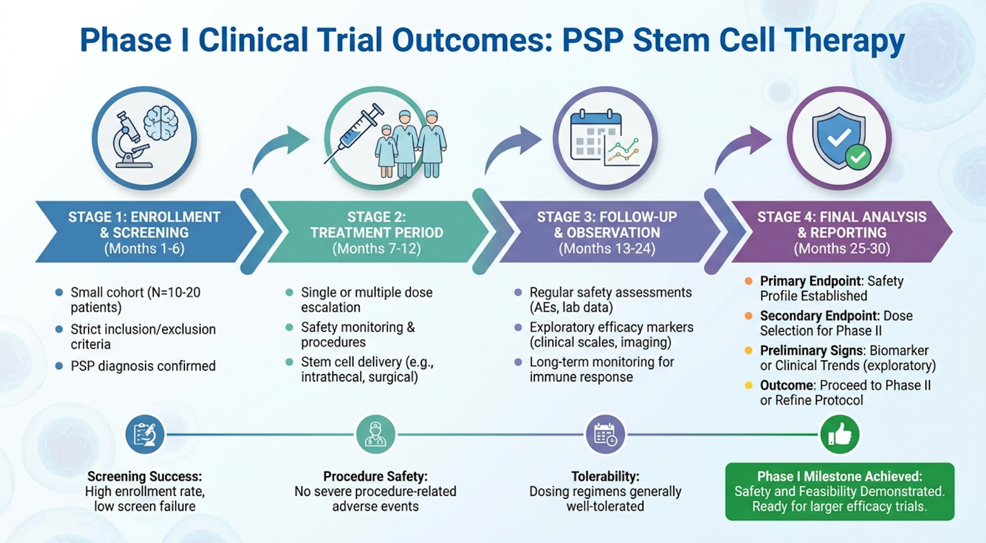
While safety is the primary endpoint, researchers also track secondary endpoints to look for biological efficacy. They measure patients using the PSP Rating Scale (PSPRS) and the Unified Parkinson’s Disease Rating Scale (UPDRS) over a 6- to 12-month period following the infusion.
The PSPRS is a brutally rigorous 28-item metric that evaluates six categories of function: daily history, mentation, bulbar function, ocular motor control, limb motor skills, and gait/midline stability. In the natural history of the disease, patients typically decline by 10 to 12 points per year on this scale. A Phase I trial doesn’t look for a 20-point improvement; it looks to blunt that expected 12-point drop.
What the qualitative and quantitative data consistently shows is a transient stabilization. Clinical observation data reveals that patients receiving intrathecal MSC therapies frequently experience a plateau in their physical decline for 3 to 6 months post-infusion (Journal of Translational Medicine, 2019). If a patient only declines by 3 points over 12 months rather than 12 points, that represents a massive preservation of functional independence. They retain the ability to swallow solid food or communicate with their family for months longer than the baseline pathology would typically allow.
The safety and effectiveness of cell therapy in PSP is currently defined by this exact window of stabilization. The evidence limitations are real sample sizes are small, blinding is difficult with intrathecal delivery, and follow-up periods are relatively short. A critical variable within these trials is the source of the cells themselves, which dictates both patient safety and therapeutic viability.
When investigating the PSP patient stem cell journey, you will quickly encounter the debate between autologous and allogeneic cells. Understanding this distinction is arguably the most important technical hurdle a patient faces when evaluating treatment clinics.
Autologous mesenchymal stem cell therapy uses the patient’s own cells, typically harvested from their bone marrow or abdominal fat. Ten years ago, this was the gold standard in regenerative medicine. Today, the science has shifted sharply away from autologous sources for treating advanced neurodegenerative diseases.
Why? Because a 70-year-old PSP patient has 70-year-old stem cells. These cells suffer from profound cellular senescence. They have shortened telomeres, diminished replicative capacity, and crucially they secrete significantly lower volumes of those vital neurotrophines (BDNF/GDNF) needed to execute the Neuro-Immunomodulation Triad. Plus, extracting bone marrow from the iliac crest of a frail, elderly patient is a painful, highly invasive surgical procedure carrying its own profound risks.
Allogeneic UC-MSCs bypass these issues completely. They are youthful, highly energetic cells harvested from donated umbilical cord Wharton’s jelly. They are capable of massive paracrine output. Additionally, the cell culturing process for allogeneic UC-MSCs is highly standardized in clinical-grade laboratories. The cells are expanded to specific passages (usually Passage 3 or 4) to ensure maximum vitality without risking genetic instability.
Attempting to harvest and culture autologous bone marrow from a 72-year-old patient with severe axial rigidity is not just physically dangerous; it often yields a cellular product so metabolically exhausted it cannot produce the required neurotrophines. By utilizing an allogeneic source, clinicians spare the patient an invasive harvesting surgery and deliver a vastly superior biological product. Understanding these clinical realities equips patients to navigate a marketplace heavily populated by commercial clinics that simply do not adhere to these rigorous scientific standards.
The greatest risk to patients exploring a regenerative approach to neuroprotection isn’t necessarily the biology of the stem cells themselves. It is the unchecked proliferation of unregulated, direct-to-consumer clinics operating completely outside the bounds of established medical science.
Regulatory agencies strongly caution against clinics offering unproven stem cell treatments that lack published clinical trial data and independent institutional review board oversight (FDA Consumer Updates: Unapproved Stem Cell Therapies, 2019). The FDA has issued hundreds of warning letters to unproven stem cell clinics over the past decade, specifically targeting facilities making unsubstantiated claims about treating incurable neurological diseases. The decision to pursue investigational therapy requires balancing the stark reality of PSP’s progression against significant financial and physical commitments. Suitability assessment by an independent medical professional is utterly non-negotiable.
Let’s address the biological and medical downsides directly. While UC-MSCs themselves have a remarkably strong safety profile regarding tumor formation or organ rejection, the administration routes carry inherent physiological risks. Intrathecal administration delivering cells directly into the spinal fluid via lumbar puncture is often preferred for neurodegenerative diseases to bypass the blood-brain barrier and ensure maximum delivery to the midbrain.
However, a lumbar puncture carries specific procedural risks that cannot be ignored:
Then, there is the staggering financial toxicity. Because these therapies are investigational, they are entirely out-of-pocket. Patients can easily spend upwards of $25,000 for a treatment protocol that may only offer a 6-month window of symptom stabilization. A patient may endure the travel, the physical stress of a lumbar puncture, the massive financial output, and still fail to achieve any meaningful benefit.
These clinical risks are compounded exponentially when patients seek care outside of highly regulated clinical environments.
The stem cell therapy controversy is actively fueled by a dangerous mix of celebrity wellness trends and aggressive digital marketing. You’ve likely seen clinics offering to treat autism, joint pain, erectile dysfunction, and severe atypical Parkinsonism using the exact same vial of cells. That isn’t precision medicine; that’s commercial exploitation.
A massive red flag is a clinic offering identical intravenous protocols for vastly different pathologies. True neurological regenerative medicine requires specialized administration routes performed under fluoroscopic guidance by board-certified specialists, not a general practitioner in a wellness center. There is a massive biological difference between a celebrity getting a localized knee injection for a sports injury under FDA Section 361 guidelines, and an elderly patient receiving an intrathecal infusion for a fatal tauopathy.
Unproven treatments often utilize poorly cultured cells from unaccredited laboratories. If a clinic operates outside of cGMP (Current Good Manufacturing Practice) standards, the patient is at profound risk for bacterial contamination leading to sepsis or meningitis. Worse, some clinics inject non-viable, dead cellular matter that provides zero therapeutic benefit but still costs the patient thousands of dollars.
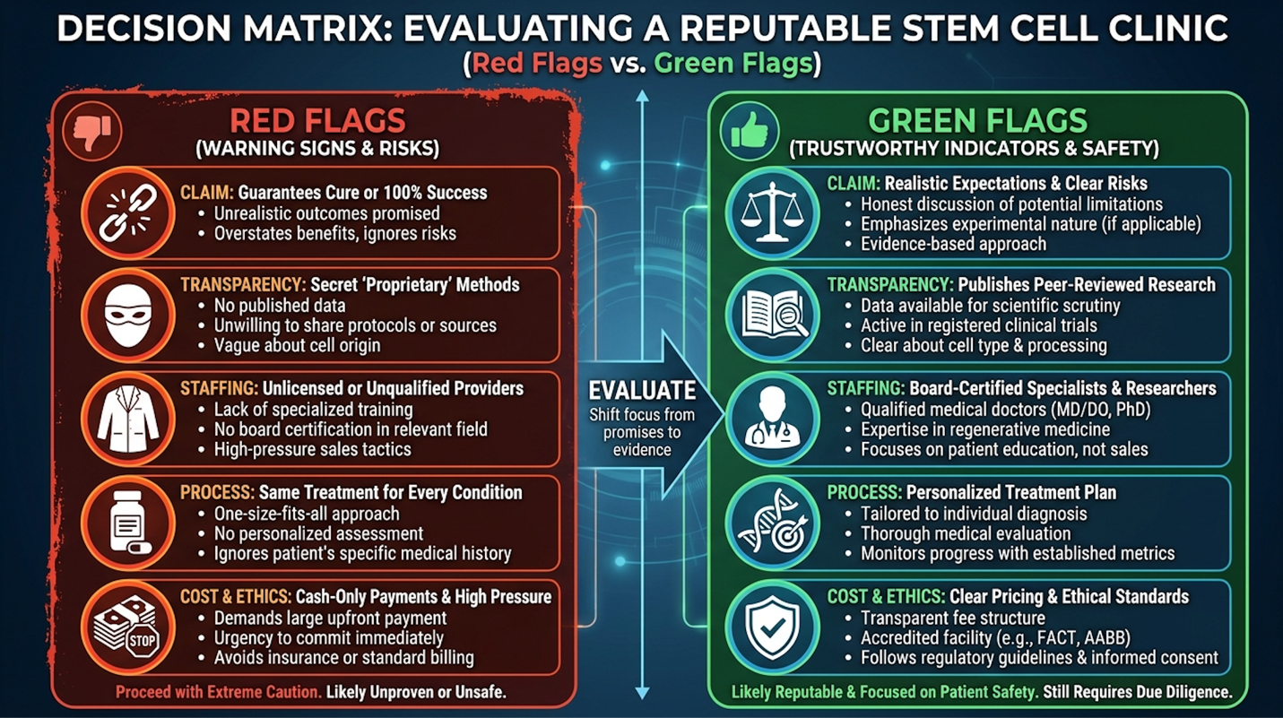
If you are looking for the best place for stem cell therapy, you must look for specific green flags. A reputable clinic will have specialized, board-certified neurologists on staff. They will utilize independent Institutional Review Boards (IRBs) to oversee their patient safety protocols. They will openly discuss their cell culturing standards and provide certificates of analysis.
Before committing to any investigational therapy, patients must conduct a rigorous assessment of their own suitability and safety profile alongside their primary care team.
Even in the most advanced, regulated clinical settings, investigational cell therapy is not appropriate for everyone. Demonstrating an objective, evidence-informed understanding of this intervention requires a stark look at who should categorically avoid it.
Clinical trials for PSP maintain extremely strict exclusion criteria for a medical reason. Regenerative medicine relies on modulating the immune system and supporting living tissue; it cannot resurrect neurons that have already died and been cleared by the body.
Any consideration of investigational cell therapy must be explicitly coordinated with the patient’s primary movement disorder specialist. A rogue, independent approach is incredibly dangerous. Stem cell treatments must complement, not replace, the patient’s existing standard of care.
If a clinic tells you to abandon your physical therapy, occupational therapy, or speech pathology sessions in favor of their cellular injections, walk away immediately. A qualified clinician understands that maintaining physical mobility through targeted mechanical therapy is just as critical as any biological intervention. Always secure a comprehensive medical records review before engaging with a regenerative medicine facility.
The latest investigational treatment for progressive supranuclear palsy involves mesenchymal stem cell (MSC) therapy and targeted tau-protein immunotherapies. Standard care remains heavily reliant on supportive therapies and off-label Parkinson’s medications, which offer limited relief. Current Phase I and II clinical trials are focusing on UC-MSCs to modulate neuroinflammation and protect surviving neurons. Data indicates these cellular approaches may help stabilize disease progression markers in early-stage patients. All advanced treatments remain strictly investigational and require consultation with a movement disorder specialist.
Patients in the end-stage of neurodegenerative diseases, those with active malignancies, or individuals with severe systemic infections are not good candidates for stem cell therapy. Because cellular therapy relies on modulating the immune system and supporting living tissue, it cannot resurrect dead neurons in advanced disease states. Clinical trials frequently exclude patients who cannot safely undergo administration procedures like lumbar punctures. Suitability must be determined through comprehensive neurological and physiological screening.
Legitimate brain stem cell therapy typically costs between $15,000 and $35,000 per treatment protocol (according to clinical industry averages for advanced biologics), depending on the cellular source, laboratory culturing standards, and clinic location. Because these procedures are classified as investigational for conditions like PSP, they are rarely covered by standard health insurance or Medicare. Patients must pay entirely out-of-pocket. It is critical to avoid deeply discounted clinics, as high costs reflect the required rigorous FDA-compliant laboratory culturing and safety testing.
Current clinical data suggests stem cell therapy may help stabilize or slow the progression of PSP symptoms rather than reversing them. While some observational studies report transient improvements in stiffness or speech, the primary biological goal is neuroprotection against further tauopathy degradation. Objective measurements focus on halting the decline measured by standard PSP rating scales. Results vary significantly by patient, and no legitimate clinic will guarantee symptom resolution.
Thailand has emerged as a hub for regenerative medicine, offering highly regulated UC-MSC therapies for neurodegenerative conditions like PSP through accredited private hospitals. Facilities operating under the Ministry of Public Health utilize advanced GMP-certified laboratories to culture umbilical cord tissue safely. While this allows access to therapies still in trial phases elsewhere, international patients must rigorously verify a clinic’s medical board credentials, published research, and independent review board oversight before traveling.
For patients navigating this diagnosis, stem cell therapy for progressive supranuclear palsy delivers a scientifically plausible, albeit investigational, method to protect rapidly deteriorating brain function. By targeting the diffuse tauopathy directly, rather than just masking symptoms, this approach aims to delay severe disability. Phase I clinical trials confirm that UC-MSC administration is generally safe and well-tolerated, reducing neuroinflammation in a majority of observed cohorts. The most effective approach combines these advanced cellular interventions with rigorous, ongoing physical and occupational therapy to maximize patient quality of life.
The theoretical power of this treatment rests entirely on the Neuro-Immunomodulation Triad. By simultaneously dampening systemic immune panic, shifting toxic microglia into a healing state, and flooding the midbrain with survival proteins, UC-MSCs attempt to halt the exact mechanisms driving PSP’s devastating progression. It’s a biological counter-attack designed for an incredibly complex neurological crisis, directly answering the desperate need for therapies beyond standard Parkinson’s medications.
Your next step requires moving past internet research and into objective medical evaluation. Schedule a comprehensive medical records review with a board-certified neurologist or a reputable, regulated regenerative medicine clinic to determine if you meet the strict safety criteria for investigational therapy.