Our Services
Others
- DFPP
- Shockwave
- IV Drip


“This literature review summarizes recent biomechanical and tribological research on how the non-Newtonian shear-thinning behavior of synovial fluid and the biphasic permeability of articular cartilage work together to minimize friction and support mechanical loads, and how the disruption of this fluid-tissue interaction contributes to the progression of osteoarthritis.”
Diarthrodial joints are subjected to extreme physiological loads, but at the same time exhibit extremely low friction and wear over a lifetime. Such biological performance is based on a coupled mechanical system of non-Newtonian synovial fluid and porous, permeable articular cartilage. Healthy synovial fluid demonstrates well-defined shear-thinning pseudoplasticity, with high zero-shear viscosity at rest, but lower viscosity at high articulation speeds. Articular cartilage functions as a biphasic poroelastic material in which interstitial fluid pressurization absorbs the majority of externally applied compressive loads, thereby protecting the solid extracellular matrix from damaging mechanical stresses. This review of the literature synthesizes the computational, rheological and tribological investigations of the cooperative role of the rheology of synovial fluid and permeability of cartilage in normal and osteoarthritic states. Fluid shear-thinning homogenizes shear stresses across the articular gap under physiological articulation, and strain-dependent reductions in cartilage permeability trap interstitial fluid to preserve load support. In osteoarthritis, molecular degradation of hyaluronic acid and lubricin compromises the non-Newtonian fluid dynamics, shifting the lubricant toward a Newtonian-like behavior with reduced cushioning capability. At the same time, enzymatic degradation of the collagen-proteoglycan network increases tissue hydraulic permeability, leading to rapid fluid exudation and loss of interstitial fluid pressurization. This leads to an increase of the contact gap permeability such that the protective boosted lubrication films cannot be formed and the mechanical loads are transferred to the solid matrix. This cascade switches the tribological regime from fluid-supported articulation to solid-solid boundary contact which predisposes the cartilage to middle-zone fatigue failure and surface delamination. The elucidation of such coupled biofluid-tissue mechanisms provides crucial insights into the design of biomimetic tribosupplements and therapeutic approaches for retarding osteoarthritic degeneration.
For instance, the knee and hip are diarthrodial joints, which are complex biomechanical systems capable of supporting contact pressures over ten megapascals with friction coefficients on the order of 0.001 to 0.02(Jaiswal & Shah, 2025). This extremely low friction surpasses engineered mechanical bearings and is maintained over millions of motion cycles per year(Liao, Miramini, Liu, & Zhang, 2020). Two interacting entities, synovial fluid, a complex biological lubricant filling the joint cavity, and articular cartilage, an avascular, hydrated soft tissue lining the articulating bone surfaces, are entirely responsible for the maintenance of joint longevity through their specialized properties(Gerard A. Ateshian, Zhang, & Hung, 2026).
Traditionally, the theory of joint lubrication has been developed in the framework of classical engineering paradigms, e.g., pure hydrodynamic fluid-film lubrication or rigid boundary film models. However, both experimental and theoretical studies have shown that single classical mechanisms cannot describe the dynamic behavior of biological joints during physiological gait cycles(Rajankunte Mahadeshwara et al., 2024). Articular cartilage is a soft, highly compliant, porous tissue with asperities on the surface larger than the typical thickness of a lubricant film. Therefore, full-film hydrodynamic separation cannot be maintained under heavy contact loads. Rather, natural joint articulation is a result of coupled fluid-tissue interaction between the non-Newtonian flow behavior of synovial fluid and the biphasic poroelastic mechanics of articular cartilage(Eschweiler et al., 2021).
In a healthy state, synovial fluid is a non-Newtonian pseudoplastic fluid that exhibits shear-thinning behavior, meaning that the viscosity decreases dynamically with increasing shear rates. Articular cartilage can also be thought of as a porous medium, containing a fluid phase (interstitial water and electrolytes) trapped within an elastic solid extracellular matrix (type II collagen fibrils and aggregating proteoglycans)(Mow, Kuei, Lai, & Armstrong, 1980). Under joint loading conditions, the flow of fluid through the porous matrix leads to interstitial fluid pressurization that supports over 90% of the applied contact load and greatly reduces the effective friction coefficient across the surface(DeMoya et al., 2024).
Osteoarthritis (OA) is a debilitating multifactorial joint disease associated with progressive mechanical degradation of cartilage and pathological changes in synovial fluid composition. Historically, OA has been considered a structural ‘wear-and-tear’ phenomenon. More recently, the biomechanical literature has identified OA as a breakdown of the synergistic fluid-tissue machinery(Jaiswal & Shah, 2025). Pathological degradation of the macromolecules of the synovial fluid together with enzymatic destruction of the cartilage extracellular matrix changes the fluid flow in the intra-articular contact gap and fluid percolation through the tissue. This review collates the current literature on the non-Newtonian rheology of synovial fluid, poroelastic permeability of articular cartilage and their mechanistic interaction during normal load bearing and osteoarthritic degradation(Jaiswal & Shah, 2024).
Synovial fluid is an ultrafiltrate of blood plasma enriched with macromolecules secreted by synoviocytes and superficial chondrocytes. The main determinant of its unique physical properties is hyaluronic acid (HA), which is a high-molecular-weight linear glycosaminoglycan consisting of repeating disaccharide units of D-glucuronic acid and N-acetylglucosamine(Wu et al., 2017). In healthy human joints, the concentration of HA is 1.5-4.0 mg/mL and its molecular mass is 2-10 Megadaltons. Due to its polyanionic character and extended wormlike chain conformation, HA forms extensive intermolecular entanglements and transient physical networks in aqueous solution(Mathieu, Conrozier, Vignon, Rozand, & Rinaudo, 2009; Rajankunte Mahadeshwara et al., 2024).
These HA entanglements co-operate with important synovial proteins such as proteoglycan-4 (PRG4, or lubricin) and surface-active phospholipids (SAPL) and plasma proteins including globulins and albumin. At low shear or at rest, these macromolecular complexes are highly entangled giving rise to a large internal flow resistance and a high zero shear viscosity (μ0 ~ 10 – 100 Pa • s)(Jaiswal & Shah, 2025). As the shear rate and mechanical motion increase, the long-chain HA polymers unravel, disentangle and align parallel to the direction of flow. This disruption of the microstructure results in a rapid, non-linear decrease in the apparent viscosity and is called the classic shear-thinning (pseudoplastic) non-Newtonian profile(Jaiswal & Shah, 2024; Rajankunte Mahadeshwara et al., 2024).
This is directly related to the structural state of these macromolecular networks, which determines the rheological response of the fluid in different mechanical regimes, as in Figure 1. The increased viscosity provides a cushioning effect of the fluid which resists squeeze-film expulsion at low articulation speeds when the joint surfaces are pressed together by static body weight. Conversely, rapid joint sliding causes a dynamic reduction in viscosity, which reduces fluid friction and viscous drag and allows fluid to flow through tight contact gaps without imposing high fluid shear stresses on the cartilage surface(Jaiswal & Shah, 2025).
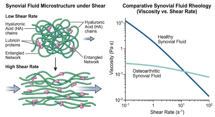
Figure 1. Non-Newtonian Dynamics and Rheology of Synovial Fluid. Schematic of shear-induced alignment of HA and lubricin networks from an entangled state (left). The right panel shows the rheological plot with the characteristic shear thinning viscosity of healthy synovial fluid compared to the reduced viscosity of osteoarthritic fluid at different shear rates.
Generalized Newtonian constitutive equations are used to quantify shear-thinning behavior in computational simulations of biofluid flow(Jaiswal & Shah, 2025). The simplest model is the Power-Law (Ostwald-de Waele) model:
where represents dynamic viscosity, is the shear rate, m is the fluid consistency index, and n is the flow behavior index. For pseudoplastic fluids, n < 1, the smaller the value of n the greater the degree of shear thinning. The literature matching the experimental rheological data confirms that the healthy synovial fluid shows the flow behavior indices of n ≈ 0.25 – 0.35 over the physiological shear regimes (0.05 s-1 s-1)(Liao et al., 2020).
For the plateau region at very low and high shear rates, sophisticated models like Carreau and Cross models are being used in the computational fluid dynamics (CFD) formulations(Wu et al., 2017):
where is zero-shear viscosity, is infinite-shear viscosity ( 0.001 – 0.01 Pa•s), and is a characteristic relaxation time constant. Numerical experiments based on these constitutive models show that healthy non-Newtonian synovial fluid provides a stable and uniform shear stress distribution along the cartilage interface, regardless of sudden changes in sliding velocity(Jaiswal & Shah, 2025).
In osteoarthritis joints, synovial fluid composition is altered by inflammatory cytokines (IL-1β and TNF-α), reactive oxygen species and increased enzymatic activity (hyaluronidases). HA molecules are depolymerized and enzymatically cleaved, reducing the average molecular weight from > 6 Megadaltons down to 0.5 – 2 Megadaltons. During this time, synovial effusion lowers the concentration of macromolecules and reduces the concentration of HA to less than 1.0 mg/mL and lubricin availability(DeMoya et al., 2024; Jaiswal & Shah, 2024; Wu et al., 2017).
This molecular breakdown has profound consequences on the non-Newtonian fluid dynamics. Rheological measurements demonstrate the loss of shear-thinning ability of osteoarthritic synovial fluid. The zero-shear viscosity decreases by up to two to three orders of magnitude ), and the flow behavior index n moves toward unity , showing a shift toward Newtonian-like behavior. Without a high zero-shear viscosity, the fluid can no longer resist rapid expulsion under static squeeze loads, resulting in premature contact gap closure and increased mechanical contact between opposing cartilage surfaces(Liao et al., 2020).
Articular cartilage is a structured biphasic material consisting of an incompressible fluid phase (water and dissolved electrolytes, which makes up 68% to 85% of the total wet weight) that can pass through an elastic solid extracellular matrix (ECM)(Mech & Rizvi, 2025). The solid matrix is mainly a dense network of type II collagen fibrils (10% to 20% wet weight) that give the matrix tensile stiffness, and large aggregating proteoglycan complexes (aggrecans, 5% to 10% wet weight) with negatively charged glycosaminoglycan (GAG) side chains that produce high fixed charge density and osmotic swelling pressure(Mech & Rizvi, 2025).
Cartilage is a viscoelastic material, and its mechanical response is inherently time-dependent, controlled by the interaction of the solid matrix with the fluid flow. The biphasic theory, initially developed by Mow et al., 1980 (Mow et al., 1980), assumes the tissue to be a mixture of solid and fluid phases, in which the total applied contact stress is balanced by the stress in the deformed solid matrix and the interstitial fluid pressure (p):
When an external compressive load is applied to the tissue surface, as shown in Figure 2, interstitial fluid cannot escape instantaneously through the dense, narrow pore space of the collagen-proteoglycan matrix. This flow resistance generates an immediate high hydrostatic pressure in the interstitial fluid, which is called Interstitial Fluid Pressurization (IFP)(Gerard A. Ateshian et al., 2026; DeMoya et al., 2024).
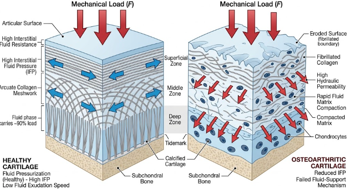
Figure 2. Biomechanics of Articular Cartilage: Healthy vs. Osteoarthritic States. Diagram comparing the structural and biomechanical responses of healthy and osteoarthritic cartilage under compressive loading. The healthy tissue exhibits effective fluid pressurization and intact zonal architecture, whereas the osteoarthritic tissue demonstrates surface fibrillation, rapid fluid loss, and matrix compaction.
Experimental measurements of pressure with micro-electromechanical sensors show that IFP carries over 90% to 95% of the total applied contact loads during the early stages of compressive loading and dynamic articulation(Hui, McCarty, Masuda, Firestein, & Sah, 2012). Biphasic friction formulations govern the effective friction coefficient over the articular surface, which is directly modulated by the fractional fluid load support :
where is the equilibrium solid-solid boundary friction coefficient after full dissipation of fluid pressurization, and α is a boundary lubricant fraction. When is close to 1.0, the interstitial fluid carries almost all of the contact load, leading to almost no solid matrix contact and very low friction (Jaiswal & Shah, 2025).
The dissipation of IFP is dictated by fluid exudation, which is regulated by the hydraulic permeability (k) of the cartilage matrix. Hydraulic permeability is the resistance of the tissue to fluid percolation due to a hydrostatic pressure gradient , and is described by Darcy’s Law(Gerard A. Ateshian et al., 2026):
In healthy cartilage the intrinsic hydraulic permeability is very low, between Moreover cartilage permeability is deformation-dependent and nonlinear. With increasing compressive strain upon loading, the collagen-proteoglycan matrix is compacted and the internal pore radius and fixed charge spacing is reduced(Mech & Rizvi, 2025). This strain-dependent compaction exponentially reduces hydraulic permeability:
where is initial zero-strain permeability and M is a dimensionless coefficient of compaction. This compaction mechanism limits fluid exudation under heavy compression, traps pressurized fluid within the tissue, and sustains fluid load support during prolonged loading phases(G. A. Ateshian, 2009).
Matrix metalloproteinases (MMPs) and aggrecanases degrade the superficial collagen architecture and cleave aggrecan molecules in osteoarthritic cartilage. Depletion of fixed negative charges and loss of integrity of superficial zone leads to reduction in osmotic swelling pressure and loosening of structural meshwork(G. D. Jay & K. A. Waller, 2014).
Thus, intrinsic hydraulic permeability is greatly augmented in the osteoarthritic tissue. Experimental studies report increases in hydraulic permeability of 2-fold to over 6-fold in damaged human and animal cartilage with values exceeding (Liao et al., 2020). When mechanical load is imposed, the hyper-permeable matrix allows interstitial fluid to be exuded out of the tissue rapidly. Therefore, IFP decays fast resulting in fast collapse of fluid load support . Without fluid load support, compressive and shear forces are directly transmitted to the solid extracellular matrix, resulting in increased matrix strain, increased micro-scale friction and increased structural wear(DeMoya et al., 2024; Rajankunte Mahadeshwara et al., 2024).
The combination of non-Newtonian flow of the synovial fluid in the narrow intra-articular contact gap and the fluid percolation through porous cartilage is used to understand the joint articulation. This coupled domain is represented using computational fluid dynamics (CFD) frameworks that solve the Navier-Stokes equations in the fluid film gap and the Brinkman-extended Darcy equation in the porous cartilage(Jaiswal & Shah, 2025):
where u and v are the fluid velocities in the gap and the porous matrix, respectively, is the non-Newtonian dynamic viscosity, K is the intrinsic permeability tensor of the cartilage and is the effective porous medium viscosity.
The overall gap permeability as in Figure 3, is a function of the micro-scale geometry of the contact gap (described by surface roughness asperities and gap height h) and the lubricant viscosity. The gap permeability is the ease with which the trapped synovial fluid flows laterally out of the contact zone, under applied fluid pressure gradients, (Liao et al., 2020).
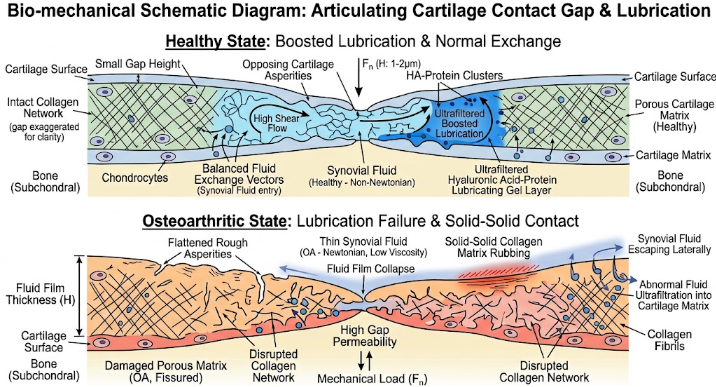
Figure 3. (A) Lubrication of Normal and Osteoarthritic Joints. Comparison of a protective fluid film in healthy cartilage to lubricant failure and solid contact friction in osteoarthritis.
In healthy joints under dynamic articulation, a wide gap permeability variation . is seen for non-Newtonian synovial fluid. Fast sliding or intermittent loading, the steep shear-thinning slope of healthy fluid maintains a high gap permeability, which permits fluid entrainment from the leading edge and encourages the formation of an elastohydrodynamic film(DeMoya et al., 2024; Liao et al., 2020).
Under heavy sustained compressive contact (e.g. prolonged standing) the gap height h decreases and fluid pressure gradients are diminished. As the non-Newtonian shear rate is lowered the viscosity of the synovial fluid approaches its high zero-shear plateau As a consequence, the gap permeability of healthy fluid drops sharply over several orders of magnitude to a critical point where the gap permeability equals the permeability of cartilage tissue (Dowson & Jin, 1986).
This balance is the point at which “boosted lubrication” is achieved. At this stage the trapped fluid is equally opposed by the lateral flow through the gap and the vertical flow into the cartilage matrix. High-molecular-weight HA macromolecules (>2 MDa) are unable to cross the tight pore network of healthy cartilage (pore size 2-6 nm). Water and small electrolytes are preferentially filtered into the tissue matrix (ultrafiltration)(Liao et al., 2020). This leaves an ultra-concentrated, very viscous gel layer of HA and lubricin complexes trapped directly on the articular surface. The thicker gel layer provides a stiff boundary lubricant and prevents direct contact of matrix asperities during final squeeze out(Mech & Rizvi, 2025).
In osteoarthritic joints, this synergistic fluid-tissue mechanism breaks down through a destructive feed-forward cascade:
| Biomechanical Parameter | Healthy Joint State | Osteoarthritic Joint State |
| Rheology of synovial fluid | Pronounced shear-thinning non-Newtonian High zero-shear viscosity(μ0 = 10 – 100 Pa⋅s) | Newtonian-like profile flattened Low zero-shear viscosity(μ0≤ 0.1 Pa⋅s) |
| Hyaluronic Acid (HA) State | High molecular weight (2-10 MDa)High concentration(1.5 – 4.0mg/mL) | Fragmented molecular weight (0.5 – 2 MDa)Diluted concentration (< 1.0 mg/mL) |
| Cartilage Hydraulic Permeability (k) | Extremely low 10-15 m4⋅s Strain-dependent compaction | Surges 2- to 6-fold(> 5.0× 10⁻¹⁴ m4/N⋅s)Matrix degradation & rapid fluid exudation |
| Interstitial Fluid Load Support (Wp/W) | Sustained > 90 – 95 %under dynamic load | Rapid pressure decay (Wp/W to 0) Early solid matrix overload |
| Contact Gap Mechanics | Augmented lubrication threshold (Kr≈ Kc)Forms HA-protein boundary gel layer | High gap permeabilityLateral escape of fluid prevents formation of protective gel |
| Primary Mechanical Wear Mode | Ultra-low friction μ≤ 0.005High fatigue resistance | Surface delamination and middle-zone collagen fatigue failure |
A key idea from current biomechanical literature is the distinction between the coefficient of friction and structural wear. Much of the classical tribological literature has assumed that wear in joints is primarily driven by high surface friction. However, experimental studies on human and bovine cartilage show that the progression of osteoarthritis does not necessarily correlate with a significant increase in the equilibrium friction coefficient alone. Instead, cartilage wear is mainly caused by mechanical fatigue failure under repetitive compressive contact loads(Eschweiler et al., 2021).
In cases of synovial fluid rheology breakdown, the major deficiency is not just the increased surface friction coefficient but the loss of dynamic fluid pressure support protecting the middle zone collagen network from peak tensile and shear strains. Therefore, therapeutic approaches need to not only minimize surface sliding friction but also restore interstitial fluid pressurization and contact gap fluid retention(Gerard A. Ateshian et al., 2026; Jaiswal & Shah, 2025).
The recognition of non-Newtonian fluid dynamics as a key mechanical controller has altered the design of intra-articular therapy. The conventional approach to viscosupplementation consists of injecting exogenous HA intra-articularly to substitute degraded synovial fluid. The early linear HA formulations had limited clinical longevity due to rapid joint clearance and shear degradation(Mathieu et al., 2009).
Modern bioengineering approaches are based on crosslinked HA hydrogels, chemically modified polymers and self-assembling peptide networks aiming at the restoration of specific rheological profiles(Jaiswal & Shah, 2025):
The outstanding tribological performance of diarthrodial joints is due to a complex fluid-structure synergy. Synovial fluid is a non-Newtonian, shear-thinning fluid that homogenizes boundary shear stresses and modulates contact gap permeability. The low and strain-dependent hydraulic permeability of articular cartilage maintains interstitial fluid pressurization to support the vast majority of joint contact forces. In osteoarthritis, this delicate balance is disturbed by simultaneous depolymerization of hyaluronic acid and enzymatic erosion of the cartilaginous extracellular matrix. The rapid fluid exudation is due to the shift of the synovial fluid toward a low viscosity Newtonian state and a spike in the tissue hydraulic permeability, which results in premature loss of interstitial fluid load support and failure to form protective boosted boundary gels. This directly imposes mechanical loads on the solid extracellular matrix which leads to stress concentration in the middle zone and causes collagen fatigue failure and surface delamination.
Future advances in osteoarthritis management rely on multi-scale biomechanical modeling and diagnostic frameworks that can detect early, subtle changes in the rheology of synovial fluid and tissue permeability before irreversible structural wear. Biomimetic tribosupplements that restore non-Newtonian biofluid rheology and reduce matrix hydraulic permeability simultaneously can preserve joint lubrication machinery and slow the progression of osteoarthritic joint degeneration.
Ateshian, G. A. (2009). The role of interstitial fluid pressurization in articular cartilage lubrication. J Biomech, 42(9), 1163-1176. doi:10.1016/j.jbiomech.2009.04.040
Ateshian, G. A., Zhang, H., & Hung, C. T. (2026). Synovial fluid protects cartilage against fatigue failure in cyclical compression. Annals of Anatomy – Anatomischer Anzeiger, 265, 152768. doi:https://doi.org/10.1016/j.aanat.2025.152768
DeMoya, C. D., Joenathan, A., Lawson, T. B., Felson, D. T., Schaer, T. P., Bais, M., . . . Grinstaff, M. W. (2024). Advances in viscosupplementation and tribosupplementation for early-stage osteoarthritis therapy. Nat Rev Rheumatol, 20(7), 432-451. doi:10.1038/s41584-024-01125-5
Dowson, D., & Jin, Z. M. (1986). Micro-elastohydrodynamic lubrication of synovial joints. Eng Med, 15(2), 63-65. doi:10.1243/emed_jour_1986_015_019_02
Eschweiler, J., Horn, N., Rath, B., Betsch, M., Baroncini, A., Tingart, M., & Migliorini, F. (2021). The Biomechanics of Cartilage-An Overview. Life (Basel), 11(4). doi:10.3390/life11040302
Hui, A. Y., McCarty, W. J., Masuda, K., Firestein, G. S., & Sah, R. L. (2012). A systems biology approach to synovial joint lubrication in health, injury, and disease. Wiley Interdiscip Rev Syst Biol Med, 4(1), 15-37. doi:10.1002/wsbm.157
Jaiswal, K. M., & Shah, S. R. (2024). The Role of Synovial Fluid Dynamics in Osteoarthritis: A Mathematical Modeling Perspective. RESEARCH REVIEW International Journal of Multidisciplinary, 9(12), 155-164. doi:10.31305/rrijm.2024.v09.n12.019
Jaiswal, k. M., & Shah, S. R. (2025). Biomechanical Insights into Synovial Fluid Flow: A Modelling Study of Pressure and Shear Stress in the Knee. Asian Journal of Orthopaedic Research, 8(2), 211-225. doi:10.9734/ajorr/2025/v8i2226
Jay, G. D., & Waller, K. A. (2014). The biology of lubricin: near frictionless joint motion. Matrix Biol, 39, 17-24. doi:10.1016/j.matbio.2014.08.008
Jay, G. D., & Waller, K. A. (2014). The biology of Lubricin: Near frictionless joint motion. Matrix Biology, 39, 17-24. doi:https://doi.org/10.1016/j.matbio.2014.08.008
Liao, J., Miramini, S., Liu, X., & Zhang, L. (2020). Computational study on synovial fluid flow behaviour in cartilage contact gap under osteoarthritic condition. Computers in Biology and Medicine, 123, 103915. doi:https://doi.org/10.1016/j.compbiomed.2020.103915
Mathieu, P., Conrozier, T., Vignon, E., Rozand, Y., & Rinaudo, M. (2009). Rheologic behavior of osteoarthritic synovial fluid after addition of hyaluronic acid: a pilot study. Clin Orthop Relat Res, 467(11), 3002-3009. doi:10.1007/s11999-009-0867-x
Mech, D. J., & Rizvi, M. S. (2025). Fluid flow induced biomechanical origin of collagen architecture in articular cartilage. bioRxiv, 2025.2007.2013.664559. doi:10.1101/2025.07.13.664559
Mow, V. C., Kuei, S. C., Lai, W. M., & Armstrong, C. G. (1980). Biphasic Creep and Stress Relaxation of Articular Cartilage in Compression: Theory and Experiments. Journal of Biomechanical Engineering, 102(1), 73-84. doi:10.1115/1.3138202
Rajankunte Mahadeshwara, M., Al-Jawad, M., Hall, R. M., Pandit, H., El-Gendy, R., & Bryant, M. (2024). How Do Cartilage Lubrication Mechanisms Fail in Osteoarthritis? A Comprehensive Review. Bioengineering (Basel), 11(6). doi:10.3390/bioengineering11060541
Wu, Y., Stoddart, M. J., Wuertz-Kozak, K., Grad, S., Alini, M., & Ferguson, S. J. (2017). Hyaluronan supplementation as a mechanical regulator of cartilage tissue development under joint-kinematic-mimicking loading. J R Soc Interface, 14(133). doi:10.1098/rsif.2017.0255
Zhang, L., Miramini, S., Smith, D. W., Gardiner, B. S., & Grodzinsky, A. J. (2015). Time Evolution of Deformation in a Human Cartilage Under Cyclic Loading. Annals of Biomedical Engineering, 43(5), 1166-1177. doi:10.1007/s10439-014-1164-8